


Article Figures & Data
Figures
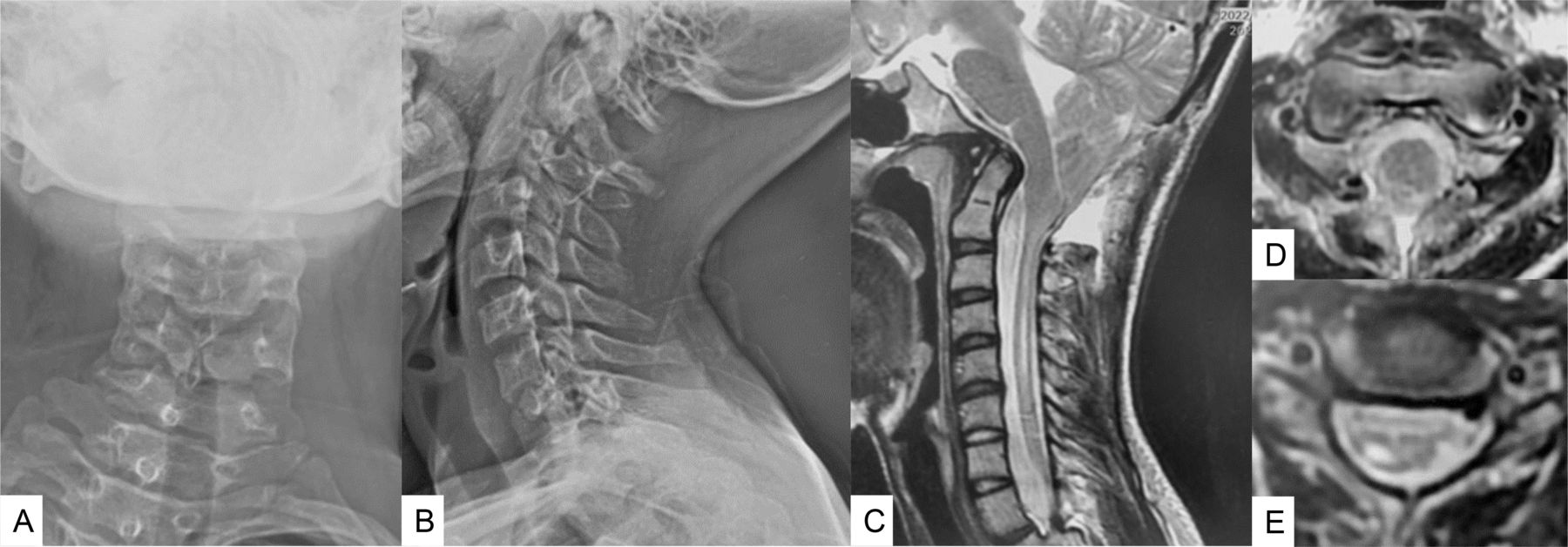
- Figure 1
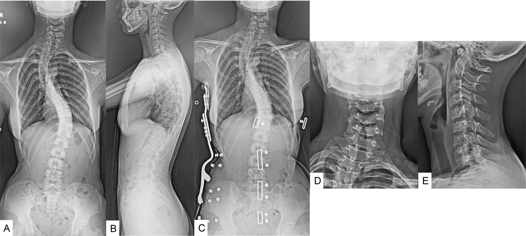
Preoperative radiograms of a 13-year-old girl with Chiari malformation type 1. (A and B) Severe scoliosis was shown, and its Cobb angles were 48° in the upper thoracic and 49° in main thoracic curve. (C) Bracing. (D and E) Cervical spine also had slight scoliosis.
- Figure 2
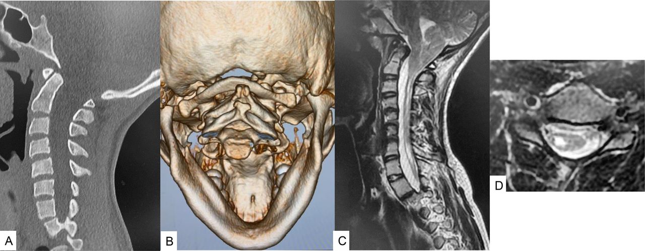
Preoperative images of a 13-year-old girl with Chiari malformation type 1. (A) Sagittal reconstruction computed tomographic (CT) image; (B) 3-D CT image; (C) Mid-sagittal T2-weighted magnetic resonance image (MRI) showed the cerebral tonsile was downward into foramen magnum. (D) Axial T2-weighted MRI at C5 indicated a syrinx in the cervical cord.
- Figure 3
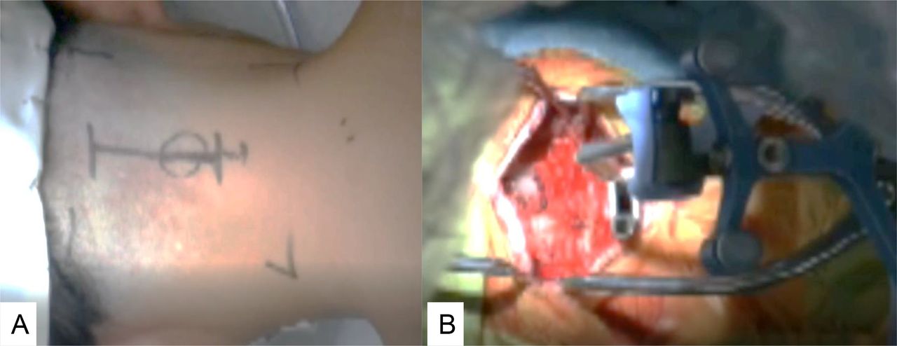
Skin incision and reference frame (A). Approximately 7 cm skin incision was made from greater occipital protuberance to C2 spinous process (B).
- Figure 4
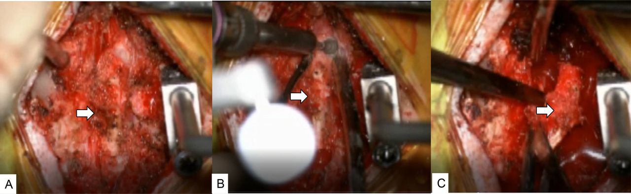
Removal of C1 lamina. (A) Before removal. (B) Cut with a navigated high-speed burr. (C) Removal of C1 lamina.
- Figure 5
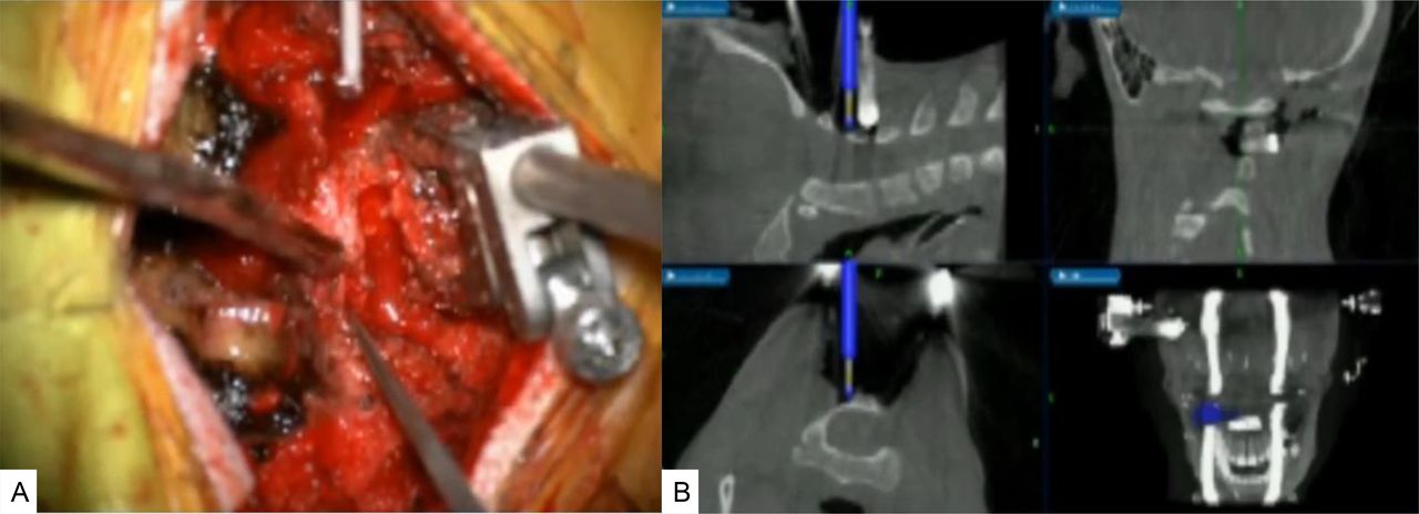
Removal of a half of the C2 lamina. (A) Cut with a navigated high-speed burr. (B) Navigation monitor.
- Figure 6
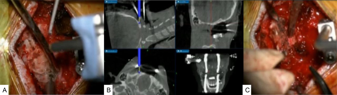
Posterior fossa decompression. (A) Intraoperative image. (B) Navigation monitor. (C) Intraoperative image.
- Figure 7
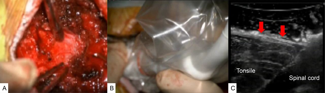
Outer layer dural plasty and intraoperative ultrasonic monitoring. (A) Outer layer dural plasty. (B and C) Intraoperative ultrasonic monitoring. Red arrows show the subarachnoid space between the tonsile and dura.
- Figure 8
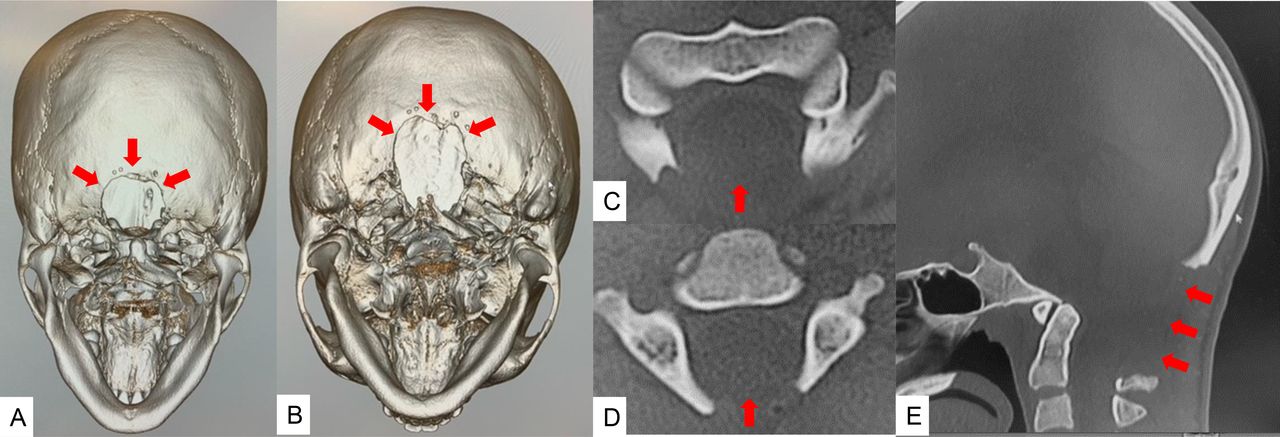
Computed tomographic (CT) images of a 13-year-old girl with Chiari malformation type 1. (A) 45° 3-dimensional (3-D) CT image. (B) 60° 3-D CT image. (C) Axial image at C1. (D) Axial image at C2. (E) Mid-sagittal reconstruction CT image. The foramen magnum was efficiently decompressed (red arrows).
- Figure 9
Follow-up images of a 13-year-old girl with Chiari malformation type 1. (A) Anteroposterior cervical radiogram. (B) Lateral cervical radiogram. (C) Mid-sagittal T2-weighted magnetic resonance image (MRI). (D) Axial T2-weighted MRI at C1. (E) Axial T2-weighted MRI at C5. MRI showed cerebral tonsile was released, and syringomyelia was reduced.


Tables
No. Age, y Gender Preoperative Symptoms Surgical Time, min Blood Loss, mL Complications Clinical Results 1 11 F Scoliosis and headache 106 30 No Excellent 2 11 F Neck pain and hyperreflexia 98 60 No Excellent 3 13 F Scoliosis, hyperreflexia, and muscle weakness 120 50 No Good 4 15 F Scoliosis and headache 116 150 No Good 5 20 F Scoliosis, hyperreflexia, and muscle weakness 131 120 No No change
In this issue


Jump to section
Related Articles
Cited By...
- No citing articles found.