
Main menu

  • Home
  • Content
    • Current Issue
    • Advance Online Publication
    • Archive
  • About Us
    • About ISASS
    • About the Journal
    • Author Instructions
    • Editorial Board
    • Reviewer Guidelines & Publication Criteria
  • More
    • Advertise
    • Subscribe
    • Alerts
    • Feedback
  • Join Us
  • Reprints & Permissions
  • Sponsored Content
  • Other Publications
    • ijss

User menu

  • My alerts

Search

  • Advanced search
International Journal of Spine Surgery
  • My alerts
International Journal of Spine Surgery

Advanced Search

  • Home
  • Content
    • Current Issue
    • Advance Online Publication
    • Archive
  • About Us
    • About ISASS
    • About the Journal
    • Author Instructions
    • Editorial Board
    • Reviewer Guidelines & Publication Criteria
  • More
    • Advertise
    • Subscribe
    • Alerts
    • Feedback
  • Join Us
  • Reprints & Permissions
  • Sponsored Content
  • Follow ijss on Twitter
  • Visit ijss on Facebook
Research ArticleMinimally Invasive Surgery

Two-Year Clinical and Radiographic Outcomes for Percutaneous Lumbar Interbody Fusion With an Expandable Titanium Cage Through Kambin’s Triangle Without Facetectomy

Troy Q. Tabarestani, Timothy Y. Wang, David A.W. Sykes, Vikram A. Mehta, Lexie Z. Yang, Shein-Chung Chow, Christopher I. Shaffrey, Walter F. Wiggins, John H. Chi and Muhammad M. Abd-El-Barr
International Journal of Spine Surgery December 2023, 17 (6) 760-770; DOI: https://doi.org/10.14444/8540
Troy Q. Tabarestani
1 Duke University School of Medicine, Duke University Hospital, Durham, NC, USA
BA
  • Find this author on Google Scholar
  • Find this author on PubMed
  • Search for this author on this site
  • For correspondence: tqt5@duke.edu
Timothy Y. Wang
2 Department of Neurosurgery, Duke University Hospital, Durham, NC, USA
MD
  • Find this author on Google Scholar
  • Find this author on PubMed
  • Search for this author on this site
David A.W. Sykes
1 Duke University School of Medicine, Duke University Hospital, Durham, NC, USA
AB
  • Find this author on Google Scholar
  • Find this author on PubMed
  • Search for this author on this site
Vikram A. Mehta
2 Department of Neurosurgery, Duke University Hospital, Durham, NC, USA
MD, MPH
  • Find this author on Google Scholar
  • Find this author on PubMed
  • Search for this author on this site
Lexie Z. Yang
3 Department of Biostatistics and Bioinformatics, Duke University, Durham, NC, USA
MB
  • Find this author on Google Scholar
  • Find this author on PubMed
  • Search for this author on this site
Shein-Chung Chow
3 Department of Biostatistics and Bioinformatics, Duke University, Durham, NC, USA
PʜD
  • Find this author on Google Scholar
  • Find this author on PubMed
  • Search for this author on this site
Christopher I. Shaffrey
2 Department of Neurosurgery, Duke University Hospital, Durham, NC, USA
MD
  • Find this author on Google Scholar
  • Find this author on PubMed
  • Search for this author on this site
Walter F. Wiggins
4 Department of Radiology, Duke University Hospital, Durham, NC, USA
MD, PʜD
  • Find this author on Google Scholar
  • Find this author on PubMed
  • Search for this author on this site
John H. Chi
5 Department of Neurosurgery, Brigham and Women’s Hospital, Boston, MA, USA
MD, MPH
  • Find this author on Google Scholar
  • Find this author on PubMed
  • Search for this author on this site
Muhammad M. Abd-El-Barr
2 Department of Neurosurgery, Duke University Hospital, Durham, NC, USA
MD, PʜD
  • Find this author on Google Scholar
  • Find this author on PubMed
  • Search for this author on this site
  • Article
  • Figures & Data
  • Info & Metrics
  • PDF
Loading

Article Figures & Data

Figures

  • Tables
  • Figure 1
    • Download figure
    • Open in new tab
    • Download powerpoint
    Figure 1

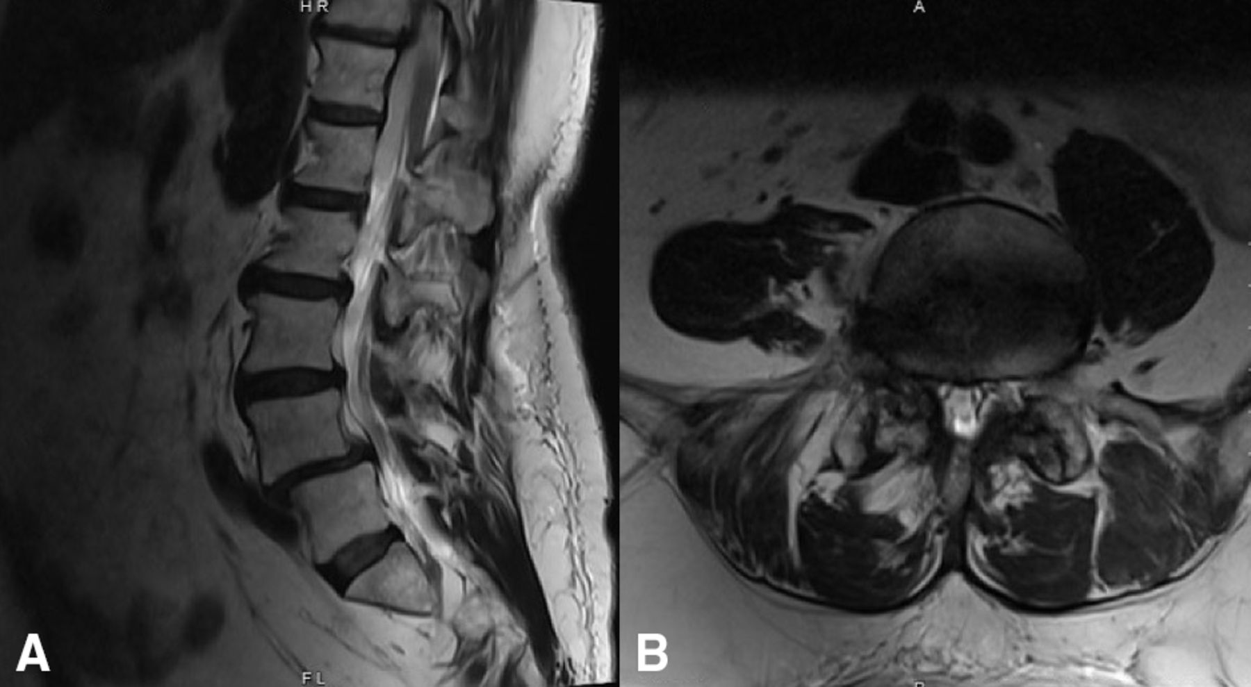
    Preoperative T2 magnetic resonance imaging showing Grade 1 spondylolisthesis of the L4–L5 level on the (a) sagittal view and right-sided foraminal compression of the exiting nerve root on the (b) axial slice.

  • Figure 2
    • Download figure
    • Open in new tab
    • Download powerpoint
    Figure 2

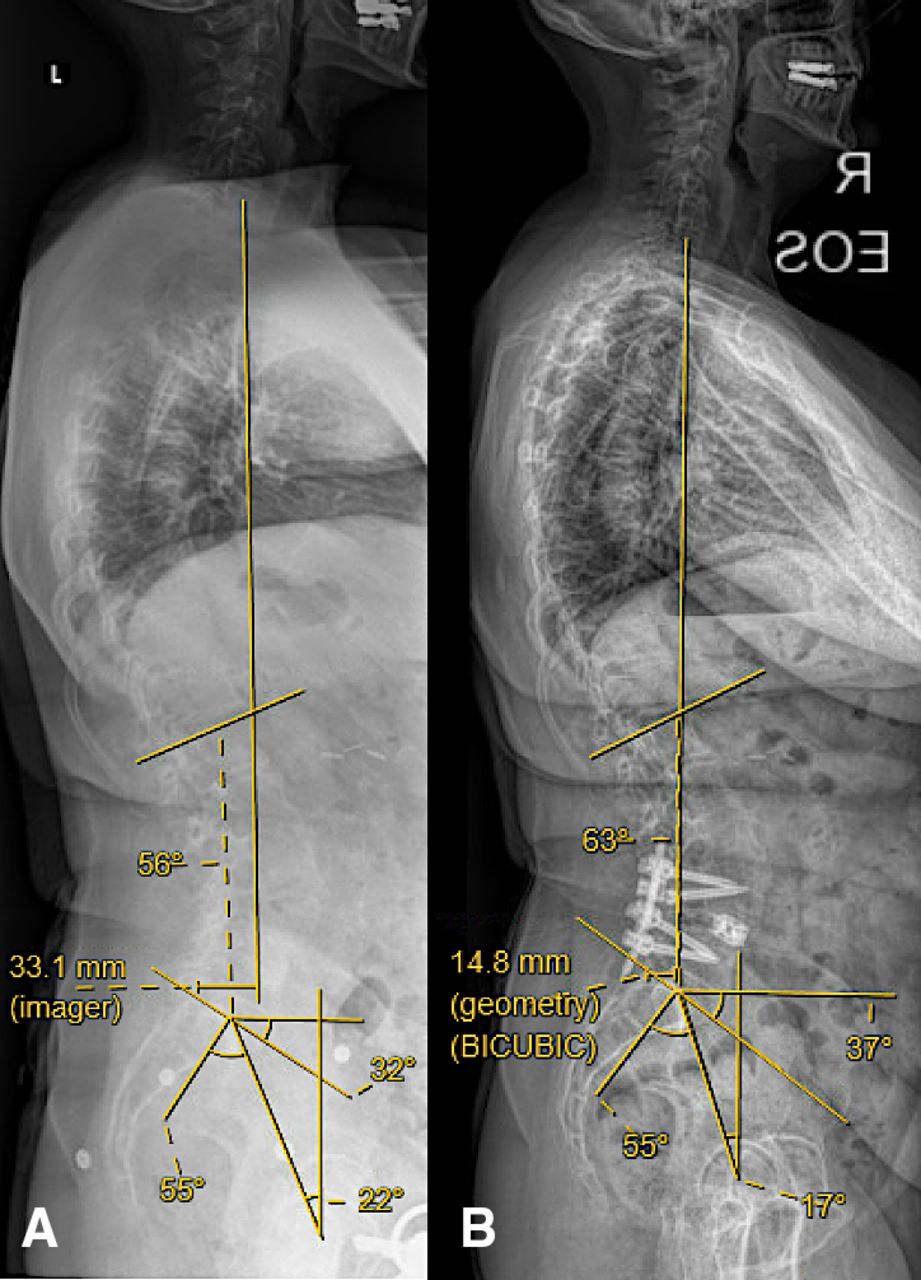
    Full-length standing x-ray images showing the improvement in spinopelvic parameters (lumbar lordosis, sagittal vertical axis, pelvic tilt, pelvic incidence, and sacral slope) comparing the (A) preoperative to (B) 1-year follow-up images.

  • Figure 3
    • Download figure
    • Open in new tab
    • Download powerpoint
    Figure 3

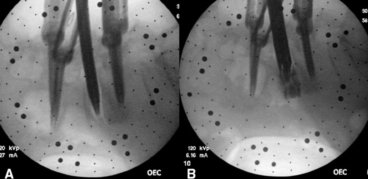
    Sequential fluoroscopic imaging showing (A) a blunt electromyography-guided probe traversing Kambin’s triangle into the disc space. (B) After satisfactory end plate preparation, an introducer is placed at the center of the disc space and loaded with an expandable cage.

  • Figure 4
    • Download figure
    • Open in new tab
    • Download powerpoint
    Figure 4

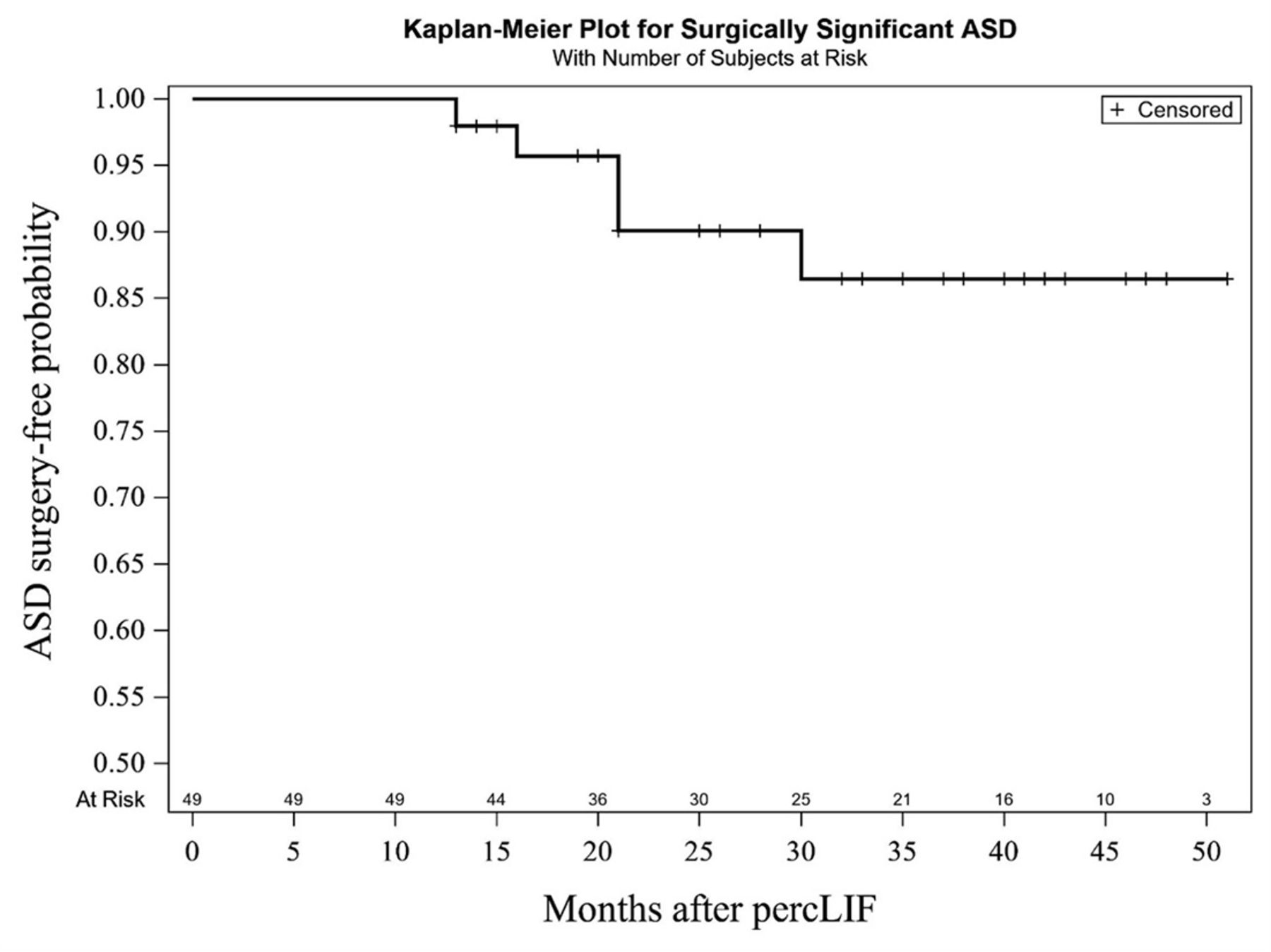
    Survivorship model of surgically significant ASD over time. ASD, adjacent segment disease; percLIF, percutaneous lumbar interbody fusion.
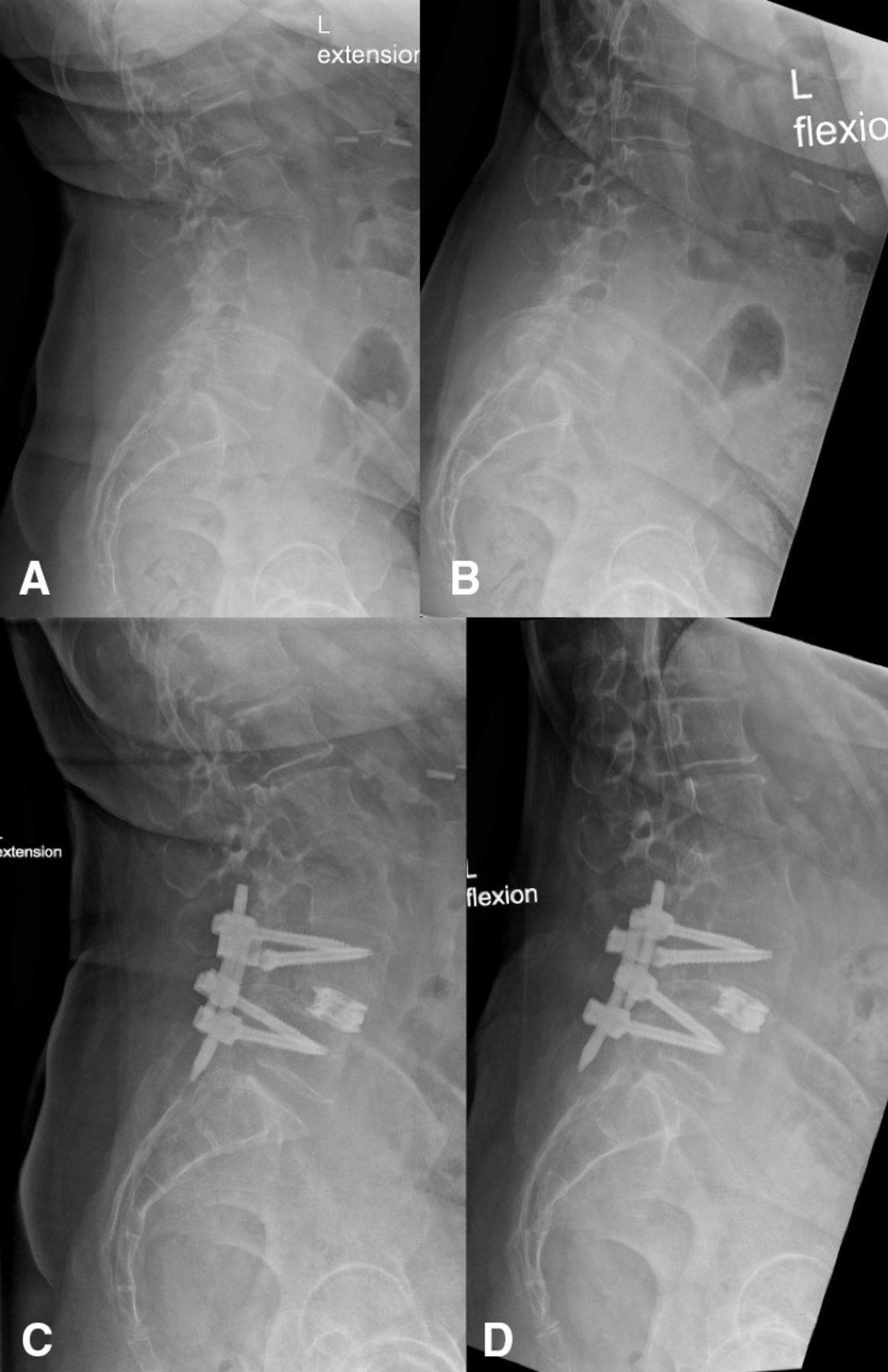
  • Figure 5
    • Download figure
    • Open in new tab
    • Download powerpoint
    Figure 5

    Preoperative (A) extension and (B) flexion standing lumbar x-rays showing reduced anterior and posterior disc space heights at the L4–L5 level. Two-year postoperative (C) extension and (D) flexion films highlighting the maintained long-term increase in disc space heights.
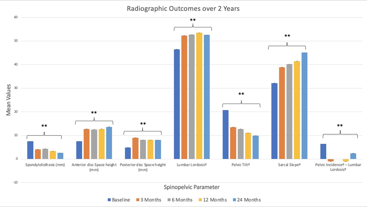
  • Figure 6
    • Download figure
    • Open in new tab
    • Download powerpoint
    Figure 6

    Radiographic outcomes over a 2-year follow-up revealing significant improvement compared with baseline values across each parameter at every recorded postoperative time point. ** P < 0.001.
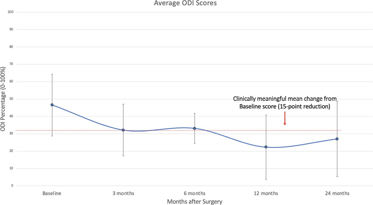
  • Figure 7
    • Download figure
    • Open in new tab
    • Download powerpoint
    Figure 7

    Patient-reported outcomes for the mean Oswestry Disability Index (ODI) through 24 months with the minimal clinically important difference shown (dotted line).
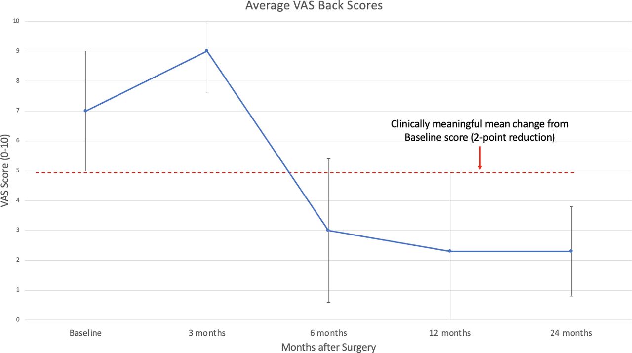
  • Figure 8
    • Download figure
    • Open in new tab
    • Download powerpoint
    Figure 8

    Patient-reported outcomes for the visual analog scale (VAS) back score through 24 months with the minimal clinically important difference shown (dotted line).

Tables

  • Figures
    • View popup
    Table 1

    Demographic data.

    Variable n (%) or Mean (SD)
    N 49 (100%)
    Age, y61.4 (11.4)
    Woman30 (61.2%)
    Body mass index (kg/m2)31.3 (5.1)
    Caucasian38 (77.6%)
    Not Hispanic48 (98.0%)
    Operative level
     L1–L20 (0%)
     L2–L33 (6.1%)
     L3–L410 (20.4%)
     L4–L527 (55.1%)
     L5–S19 (18.4%)
    • View popup
    Table 2

    Hospitalization and operative data.

    Variable n (%) or Mean (SD)
    Operative time, min190.4 (73.9)
    Estimated blood loss, mL70.3 (82.7)
    Length of stay, nights3.4 (2.9)
    Readmissions3 (6.12%)
    Operations for adjacent segment disease5 (10.2%)
    • View popup
    Table 3

    Annual incidence of surgery for ASD.

    Years After Initial FusionNumber of New ASD SurgeryNumber CensoredEffective Sample SizeAnnual Incidence of ASD
    00049.00
    10049.00
    241541.50.0964
    311124.50.0408
    401311.50
    5052.50
    • Abbreviation: ASD, adjacent segment disease.

    • View popup
    Table 4

    Radiographic and spinopelvic parameters data.

    VariableMean (SD) P Valuea
    Baseline3 Mo6 Mo12 Mo24 Mo
    Spondylolisthesis, mm7.5 (3.7)4.1 (2.1)4.3 (2.9)3.4 (2.5)2.6 (2.8) <0.001 at 3, 6, 12, and 24 mo
    Anterior disc space height, mm7.5 (3.2)12.7 (2.8)12.5 (2.8)12.7 (3.0)13.6 (3.2) <0.001 at 3, 6, 12, and 24 mo
    Posterior disc space height, mm4.9 (2.2)9.0 (2.8)8.1 (2.5)8.1 (3.0)8.1 (2.1) <0.001 at 3, 6, 12, and 24 mo
    LL°46.5 (12.9)52.3 (12.9)52.7 (11.5)53.5 (12.5)52.6 (12.6) <0.001 at 3, 6, 12, and 24 mo
    Sagittal vertical axis, mm58.4 (24.2)33.5 (14.4)49.1 (21.0)45.8 (32.3)56.4 (38.4)3 mo: P < 0.001
    6 mo: P = 0.074
    12 mo: P = 0.087
    24 mo: P = 0.948
    Pelvic tilt°20.7 (4.8)13.5 (9.1)12.7 (8.8)11.1 (8.1)9.9 (9.7) <0.001 at 3, 6, 12, and 24 mo
    Sacral slope°32.2 (8.9)38.9 (9.3)40.2 (9.4)41.5 (9.9)45.1 (8.4) <0.001 at 3, 6, 12, and 24 mo
    Pelvic incidence°–LL°6.4 (10.5)-1.0 (15.9)0.0 (11.7)−1.0 (11.2)2.4 (13.2) <0.001 at 3, 6, 12, and 24 mo
    • Abbreviation: LL, lumbar lordosis.

    • ↵a Significance was determined if the P < 0.05.

PreviousNext
Back to top

In this issue

International Journal of Spine Surgery
Vol. 17, Issue 6
1 Dec 2023
  • Table of Contents
  • Index by author

Print
Download PDF
Article Alerts
Sign In to Email Alerts with your Email Address
Email Article

Thank you for your interest in spreading the word on International Journal of Spine Surgery.

NOTE: We only request your email address so that the person you are recommending the page to knows that you wanted them to see it, and that it is not junk mail. We do not capture any email address.

Enter multiple addresses on separate lines or separate them with commas.
Two-Year Clinical and Radiographic Outcomes for Percutaneous Lumbar Interbody Fusion With an Expandable Titanium Cage Through Kambin’s Triangle Without Facetectomy
(Your Name) has sent you a message from International Journal of Spine Surgery
(Your Name) thought you would like to see the International Journal of Spine Surgery web site.
CAPTCHA
This question is for testing whether or not you are a human visitor and to prevent automated spam submissions.
Citation Tools
Two-Year Clinical and Radiographic Outcomes for Percutaneous Lumbar Interbody Fusion With an Expandable Titanium Cage Through Kambin’s Triangle Without Facetectomy
Troy Q. Tabarestani, Timothy Y. Wang, David A.W. Sykes, Vikram A. Mehta, Lexie Z. Yang, Shein-Chung Chow, Christopher I. Shaffrey, Walter F. Wiggins, John H. Chi, Muhammad M. Abd-El-Barr
International Journal of Spine Surgery Dec 2023, 17 (6) 760-770; DOI: 10.14444/8540

Citation Manager Formats

  • BibTeX
  • Bookends
  • EasyBib
  • EndNote (tagged)
  • EndNote 8 (xml)
  • Medlars
  • Mendeley
  • Papers
  • RefWorks Tagged
  • Ref Manager
  • RIS
  • Zotero
Share
Two-Year Clinical and Radiographic Outcomes for Percutaneous Lumbar Interbody Fusion With an Expandable Titanium Cage Through Kambin’s Triangle Without Facetectomy
Troy Q. Tabarestani, Timothy Y. Wang, David A.W. Sykes, Vikram A. Mehta, Lexie Z. Yang, Shein-Chung Chow, Christopher I. Shaffrey, Walter F. Wiggins, John H. Chi, Muhammad M. Abd-El-Barr
International Journal of Spine Surgery Dec 2023, 17 (6) 760-770; DOI: 10.14444/8540
Twitter logo Facebook logo Mendeley logo
  • Tweet Widget
  • Facebook Like
  • Google Plus One

Jump to section

  • Article
    • Abstract
    • INTRODUCTION
    • METHODS
    • RESULTS
    • DISCUSSION
    • CONCLUSION
    • Footnotes
    • References
  • Figures & Data
  • Info & Metrics
  • PDF

Related Articles

  • No related articles found.
  • PubMed
  • Google Scholar

Cited By...

  • No citing articles found.
  • Google Scholar

More in this TOC Section

  • Comparative Review of Lateral and Oblique Lumbar Interbody Fusion: Technique, Outcomes, and Complications
  • Key Considerations in Surgical Decision-Making on the Side of Approach for Lumbar Lateral Transpsoas Interbody Fusion Techniques
  • A Spine Surgeon’s Learning Curve With the Minimally Invasive L5 to S1 Lateral ALIF Surgical Approach: Perioperative Outcomes and Technical Considerations
Show more Minimally Invasive Surgery

Similar Articles

Keywords

  • expandable cage
  • Kambin’s triangle
  • lumbar interbody fusion
  • minimally invasive surgery
  • percutaneous spinal fusion
  • patient-reported outcomes
  • spinopelvic parameters

Content

  • Current Issue
  • Latest Content
  • Archive

More Information

  • About IJSS
  • About ISASS
  • Privacy Policy

More

  • Subscribe
  • Alerts
  • Feedback

Other Services

  • Author Instructions
  • Join ISASS
  • Reprints & Permissions

© 2025 International Journal of Spine Surgery

International Journal of Spine Surgery Online ISSN: 2211-4599

Powered by HighWire