


Article Figures & Data
Figures
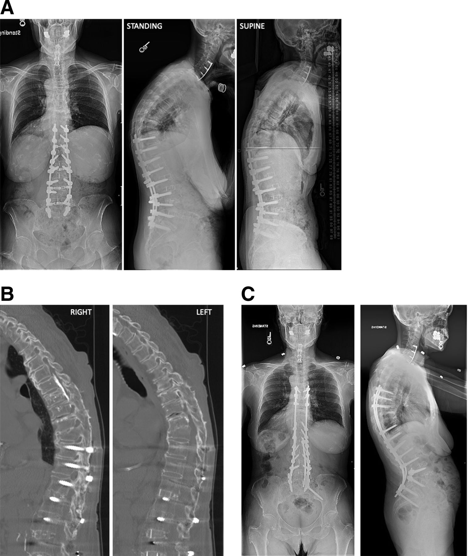
- Figure 1
(A). Preoperative x-ray images showed lucencies around pelvic instrumentation and pedicle screw pullout/loosening at T4-T6 with spondylolisthesis at T3-T4. Alignment parameters showed a low-grade PI, no significant PI−LL mismatch, and elevated pelvic tilt: PI 37°, LL 28°, PI−LL 9°, PT 27°, TK 63°, and SVA 23 mm. (B) Computed tomography images showed lucencies around pedicle screws T4-T6 and pedicle screw pullout with spondylolisthesis T3-T4 and L5-S1 pseudarthrosis. (C) 6-month postoperative x-ray images after revision posterior spinal fusion with instrumentation T2-pelvis and L3 pedicle subtraction osteotomy. Alignment parameters show that the pelvic tilt has normalized. PI 37°, LL 53°, PI−LL −16°, PT 14°, TK 63°, and SVA −44 mm. PI, pelvic incidence; LL, lumbar lordosis; PI−LL, PI LL mismatch; PT, pelvic tilt; TK, thoracic kyphosis; SVA, sagittal vertical axis
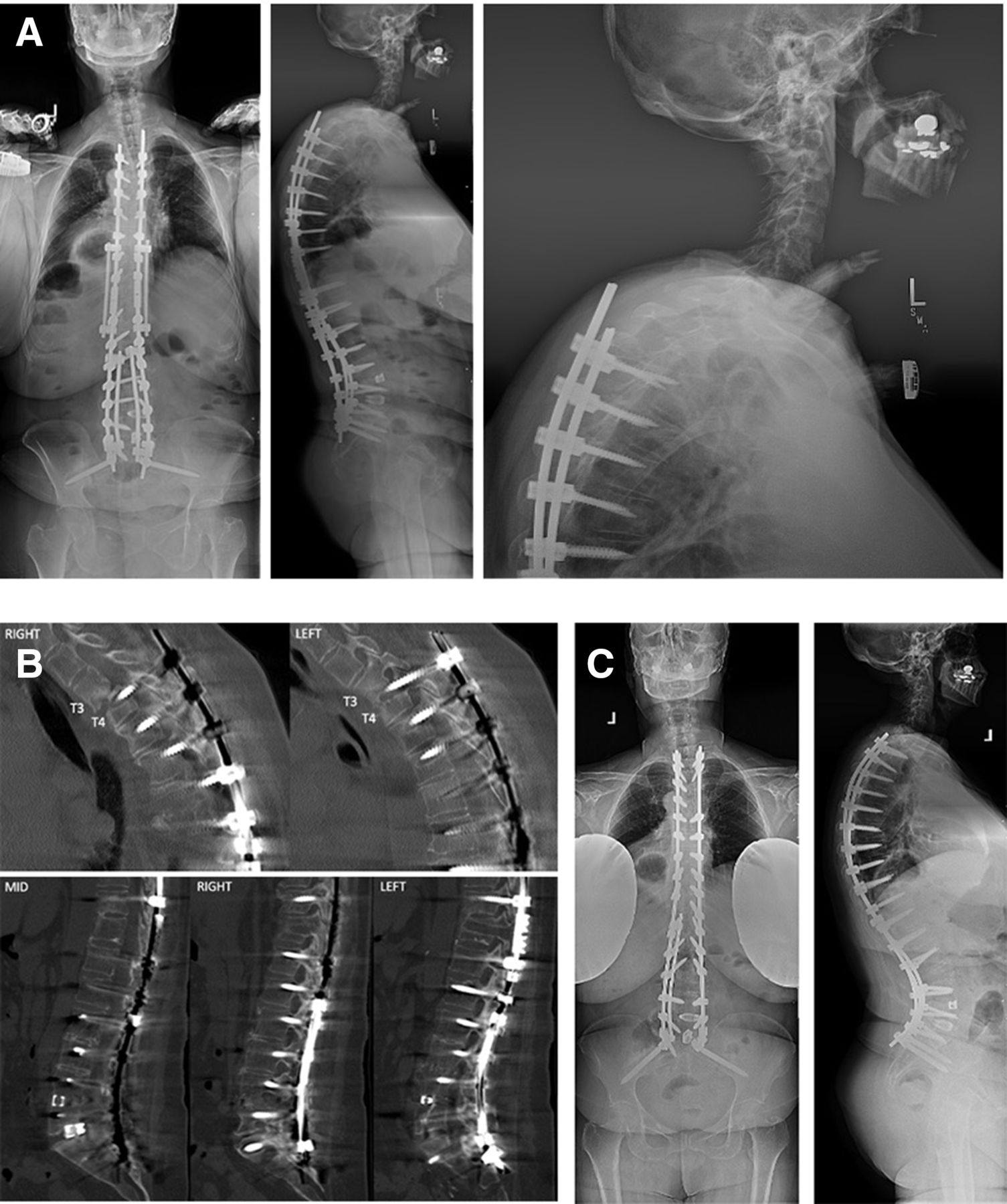
- Figure 2
(A) Preoperative x-ray images show instrumentation from prior T10-S1 fusion with interbody fusion T12-L5. There is a positive sagittal imbalance secondary to flatback deformity with a healed T9 compression fracture. PI 74°, LL 57°, PI−LL 17°, PT 34°, TK 66° (50° supine), and SVA 83 mm. (B) Computed tomography (CT) image showed an intact fusion mass T6-S1 and hyperostosis T3-T5 resulting in a spine that is functionally fused T3-S1 except for vacuum phenomena at T6-T7 and T7-T8 indicating motion at these levels (with a decrease in kyphosis on supine radiographs). Also noted are screw tracts from prior T6-T9 fixation and cement augmentation in the midthoracic spine. Full lumbar CT image not shown. (C) Postoperative day 5 x-ray images after revision posterior spinal fusion with instrumentation T5-pelvis and L4 pedicle subtraction osteotomy. PI 74°, LL 86°, PI−LL −12°, PT 18°, and SVA −37 mm. PI, pelvic incidence; LL, lumbar lordosis; PT, pelvic tilt; TK, thoracic kyphosis; SVA; sagittal vertical axis.
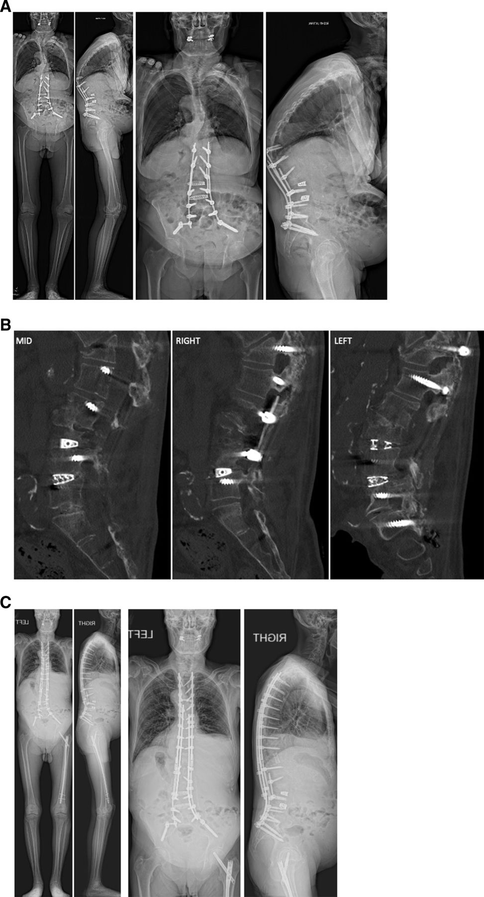
- Figure 3
(A) Preoperative x-ray images show prior T12-S1/pelvis fusion with proximal junctional failure (PJF). There is a positive sagittal imbalance secondary to PJF. PI 35°, LL 27°, PI−LL 8°, PT 30°, SVA 182 mm, and PJA 50°. (B) Lumbar computed tomography image showed a T12 compression fracture, an L5-S1 pseudarthrosis, and an otherwise solid fusion mass T12-L5. (C) 2.5-year postoperative x-ray images after revision posterior spinal fusion with instrumentation T4-S1/pelvis and T12 VCR. Alignment parameters show that the pelvic tilt has normalized, and global sagittal alignment has improved without evidence of recurrent proximal junctional kyphosis. PI 35°, LL 27°, PI−LL 8°, PT 12°, and SVA 107 mm. PI, pelvic incidence; LL, lumbar lordosis; PT, pelvic tilt; TK, thoracic kyphosis; SVA, sagittal vertical axis; PJA, proximal junctional angle.


Tables
Risk Factors Prevention Techniques Surgical Disruption of posterior soft tissues Meticulous dissection at UIV and care to protect facet capsule of the level above Rigidity of instrumentation Use of hooks vs screws at proximal level, not engaging all screw threads proximally, use of transition rods and tethers Choice of vertebral levels UIV in the lower thoracic spine increases the risk of failure and vertebral fracture, UIV in the upper thoracic spine may increase the risk of junctional kyphosis, and lower instrumented vertebra to the sacrum/pelvis may increase the risk of PJK/PJF Choice of approach Avoid combined anterior-posterior approaches if feasible Degree of correction: high SVA correction, increased correction of lumbar lordosis Optimize global sagittal alignment, SVA of 0 cm may not be optimal for all patients, and PJK may be a compensatory mechanism for overcorrection; consider age-adjusted alignment targets, “ideal” Roussouly type, Global Alignment and Proportion Score UIV loading Under-loading of the UIV (decreased bending moment) associated with PJK/PJF Radiographic Increased preoperative thoracic kyphosis Nonmodifiable Increased preoperative proximal junctional angle (>5°) Ensure the construct includes any levels with junctional kyphosis >5° Patient Specific Advanced age (>55 y) Nonmodifiable Body mass index Encourage weight loss and nutrition counseling Osteopenia/osteoporosis Vertebral augmentation and preoperative optimization (consider interventions such as intermittent teriparatide treatment) Lower muscularity and fatty degeneration in the thoracolumbar region Consider UIV in the upper rather than lower thoracic in these patients Higher preoperative thoracic spine flexibility associated with PJK Obtain preoperative supine radiographs to identify patients at risk of thoracic spine flattening during positioning Abbreviations: PJF, proximal junctional failure; PJK, proximal junctional kyphosis; SVA, sagittal vertical axis; UIV, upper instrumented vertebra.
Note: Modified from: Kim HJ, Iyer S. Proximal junctional kyphosis. J Am Acad Orthop Surg. 2016;24(5):318–326. doi:10.5435/JAAOS-D-14-00393.
In this issue


