


Article Figures & Data
Figures
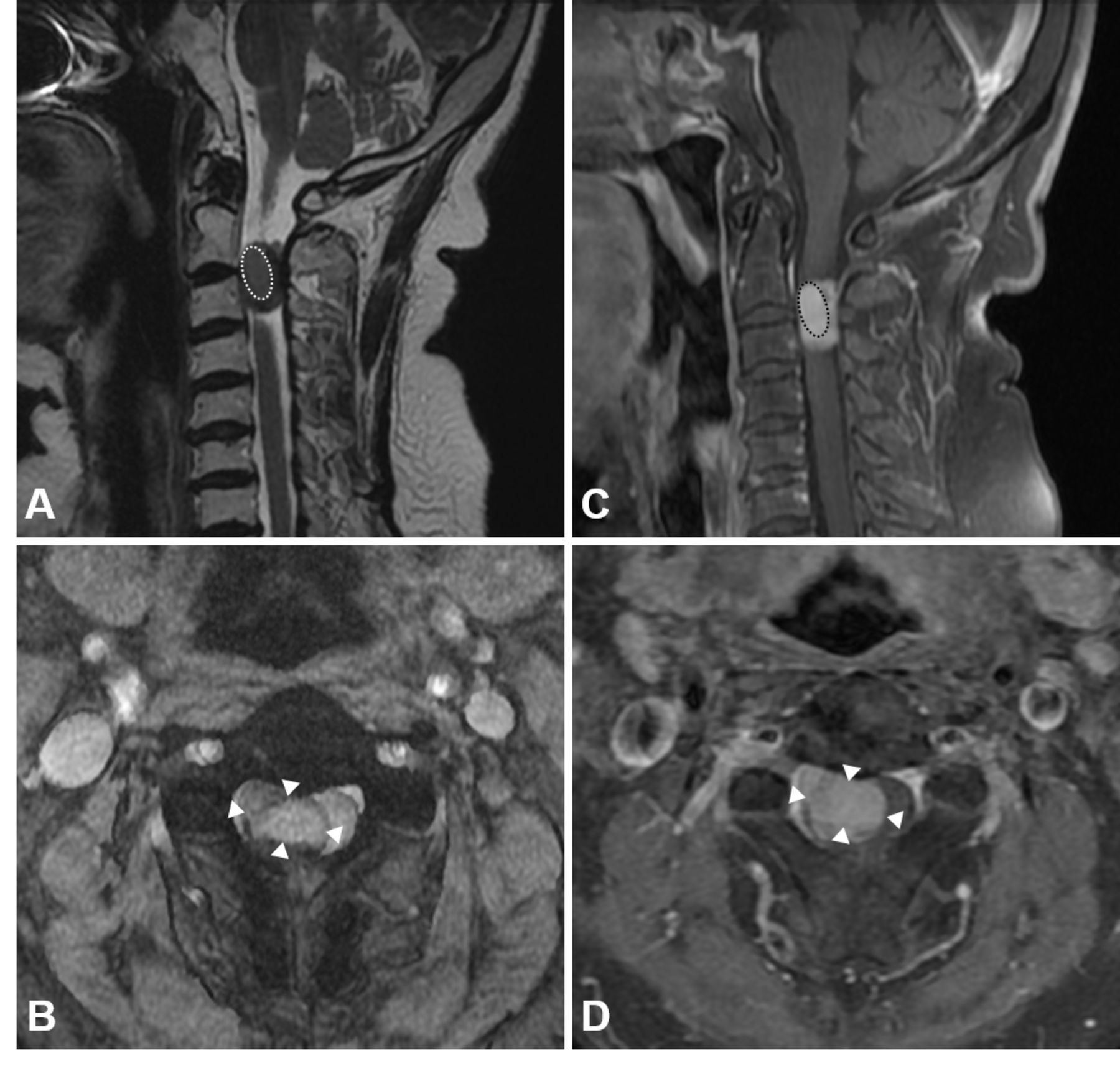
- Figure 1
The intradural extramedullary mass at the level of C2–C3 vertebrae. An oval, well-demarcated, isointensity mass (dotted line) was positioned anteriorly in the central canal on the sagittal T2-weighted image (A). The mass had a heterogeneous gadolinium enhancement on the sagittal T1-weighted image (C). The contrast-enhanced mass (white arrowhead) was positioned in the dura mater and deviated from the adjacent spinal cord on the axial images (T2-weighted image on B and T1-weighted images on D).
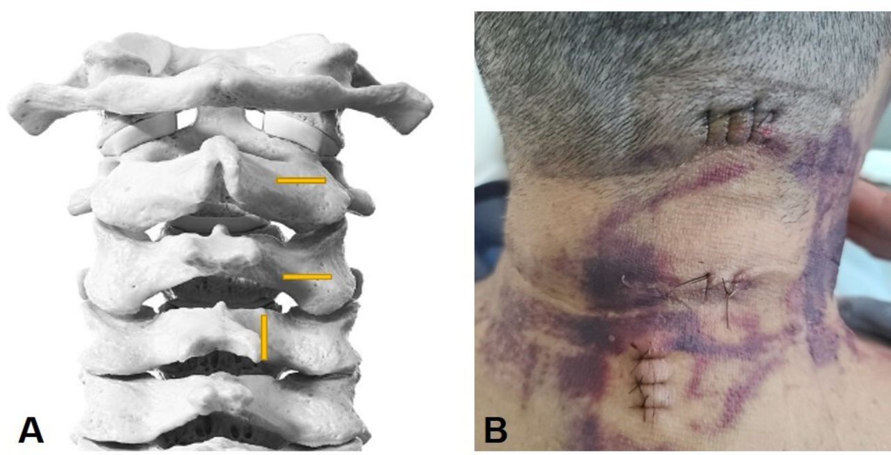
- Figure 2
(A) Schematic illustration. A cranial portal was made on the right side of the midline of the C2 spinous process. A caudal portal, positioned 2–3 cm inferior to the first, was also created. A third portal was established mediocaudally to the caudal portal. (B) Sutured state of the portals after surgery.
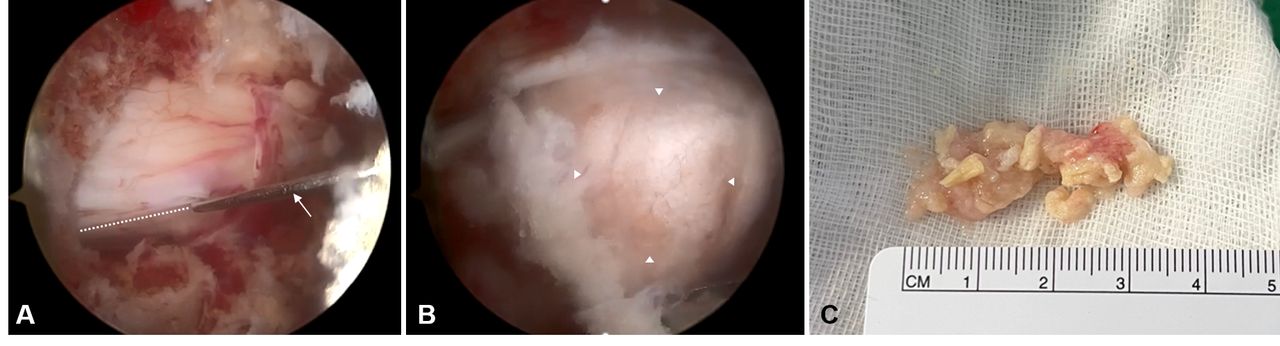
- Figure 3
(A) Endoscopic view of performing a longitudinal incision (dotted line) following dura mater exposure (sharp probe, arrow). (B) A substantial grayish-white mass (white arrowhead) was being excised through the incised dural margins, with the microforceps positioned inferiorly. (C) Gross appearance of the excised tissues.
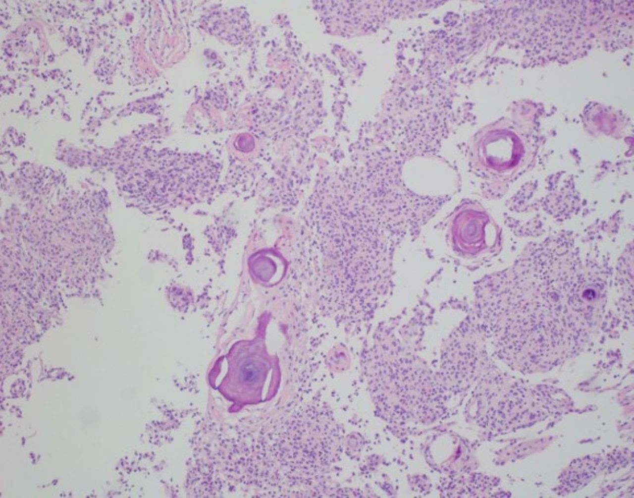
- Figure 4
Photomicrograph of the resected tumor. The tumor cells exhibited lobules and whorl formation of spindle-shaped cells. Psammoma bodies were also visible (hematoxylin and eosin staining, ×100 magnification).
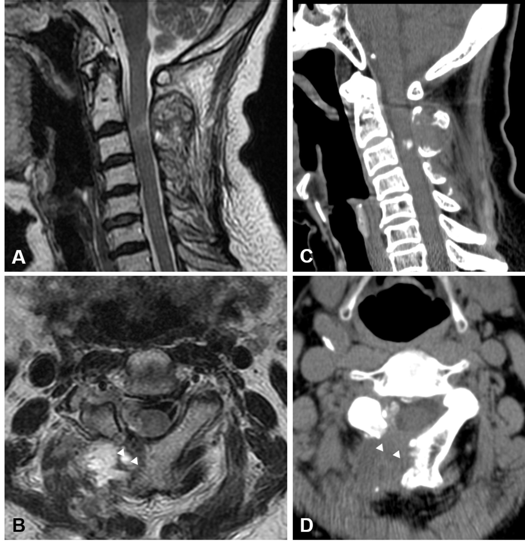
- Figure 5
Postoperative magnetic resonance imaging (sagittal T1-weighted image on A and axial T2-weighted image on B) and computed tomographic scans (sagittal view on C and axial view on D) confirmed that the mass was nearly completely removed, and the right-sided laminectomy (white arrowhead) was clearly visible.
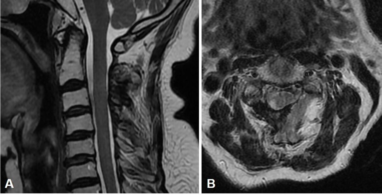
- Figure 6
Magnetic resonance imaging 18 months after surgery (sagittal view on A and axial view on B) demonstrated a clear visualization of the spinal cord without evidence of residual mass, absence of tumor recurrence, and adequate central canal space.


In this issue


Jump to section
Related Articles
Cited By...
- No citing articles found.