


Article Figures & Data
Figures
- Fig. 1
White light interferometry images of an unworn (left) and worn (right) polyethylene implant surface.
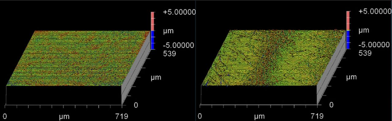
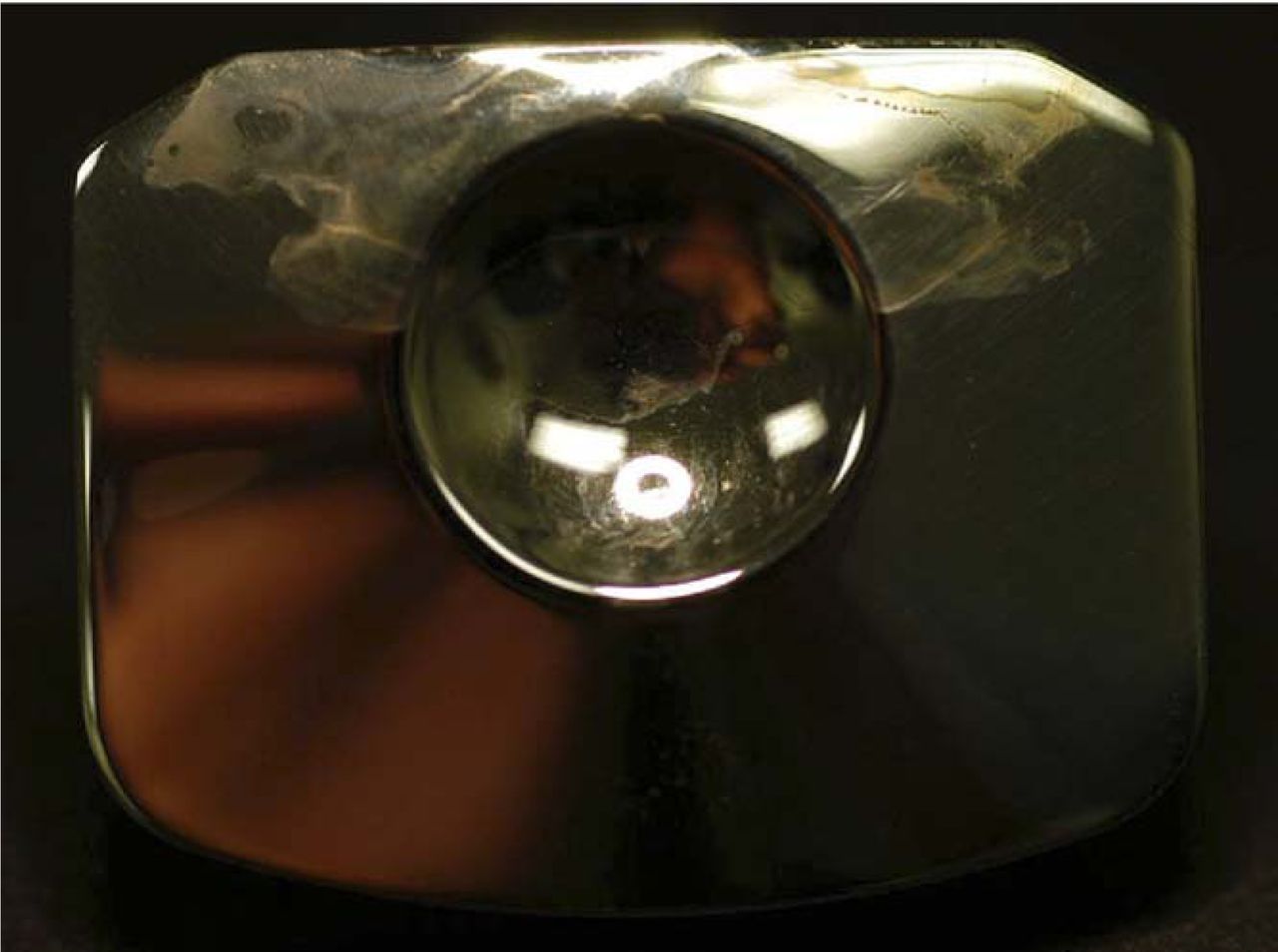
- Fig. 2
White light interferometry images of an unworn (left) and worn (right) metal-on-metal implant surface.
- Fig. 3
Abrasive wear observed on polymeric components from retrieved Dynesys systems, implanted 1.1 (left) and 1 (right) years.
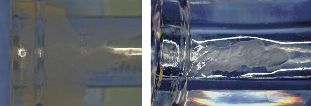
- Fig. 4
Microscopic, multidirectional scratches and crisscrossing wear paths at the dome of a retrieved polyethylene total disc replacement that was implanted 6.2 years.
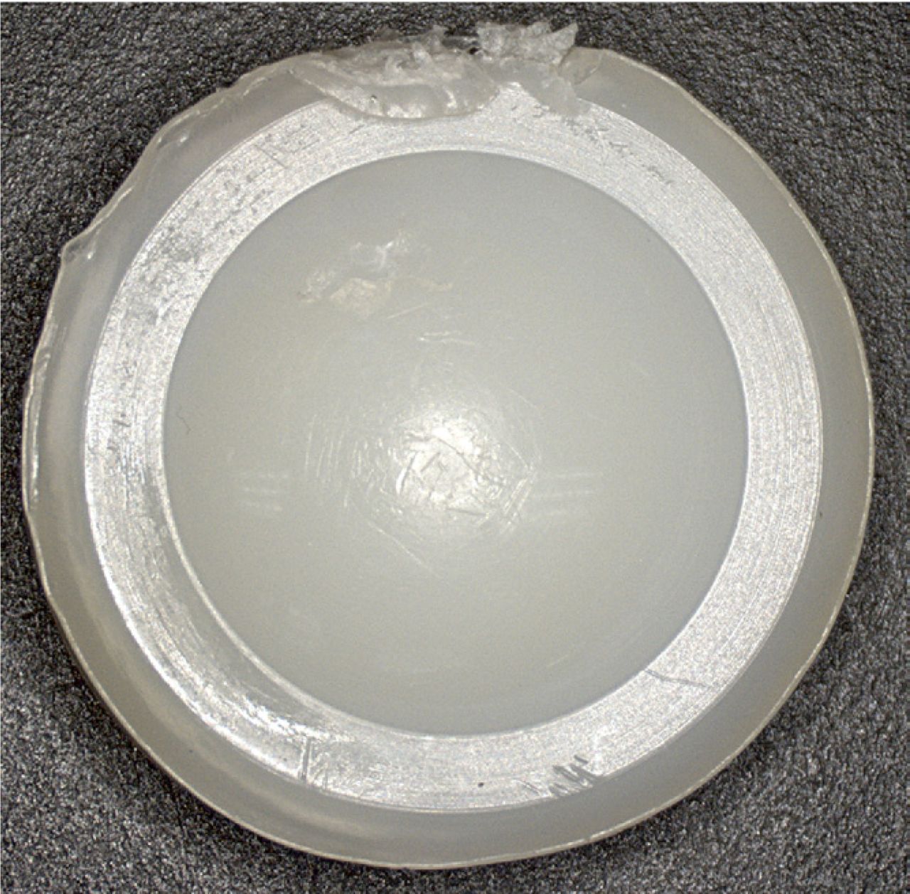
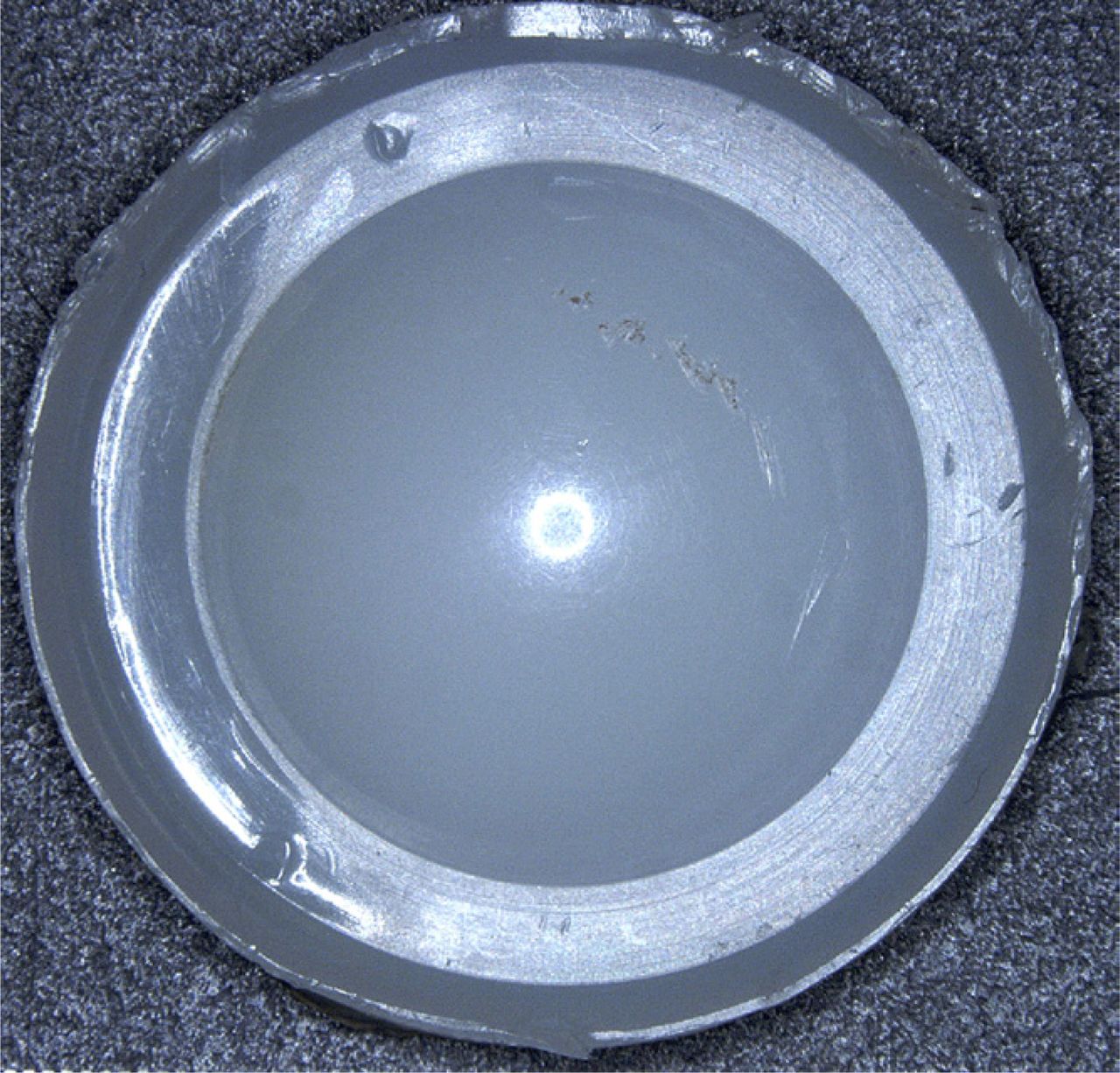
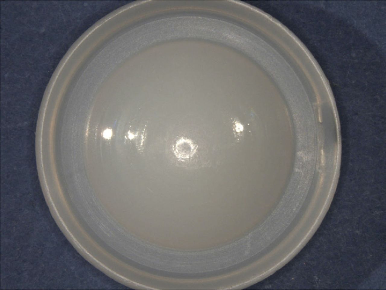
- Fig. 5
Burnishing observed on the dome of the polyethylene component of a retrieved total disc replacement.
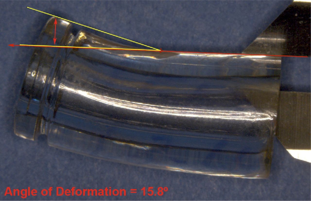
- Fig. 6
Polycarbonate urethane component of a retrieved Dynesys system that was implanted for 1 year, demonstrating evidence of permanent bending along the length in response to off-axis compressive loading.
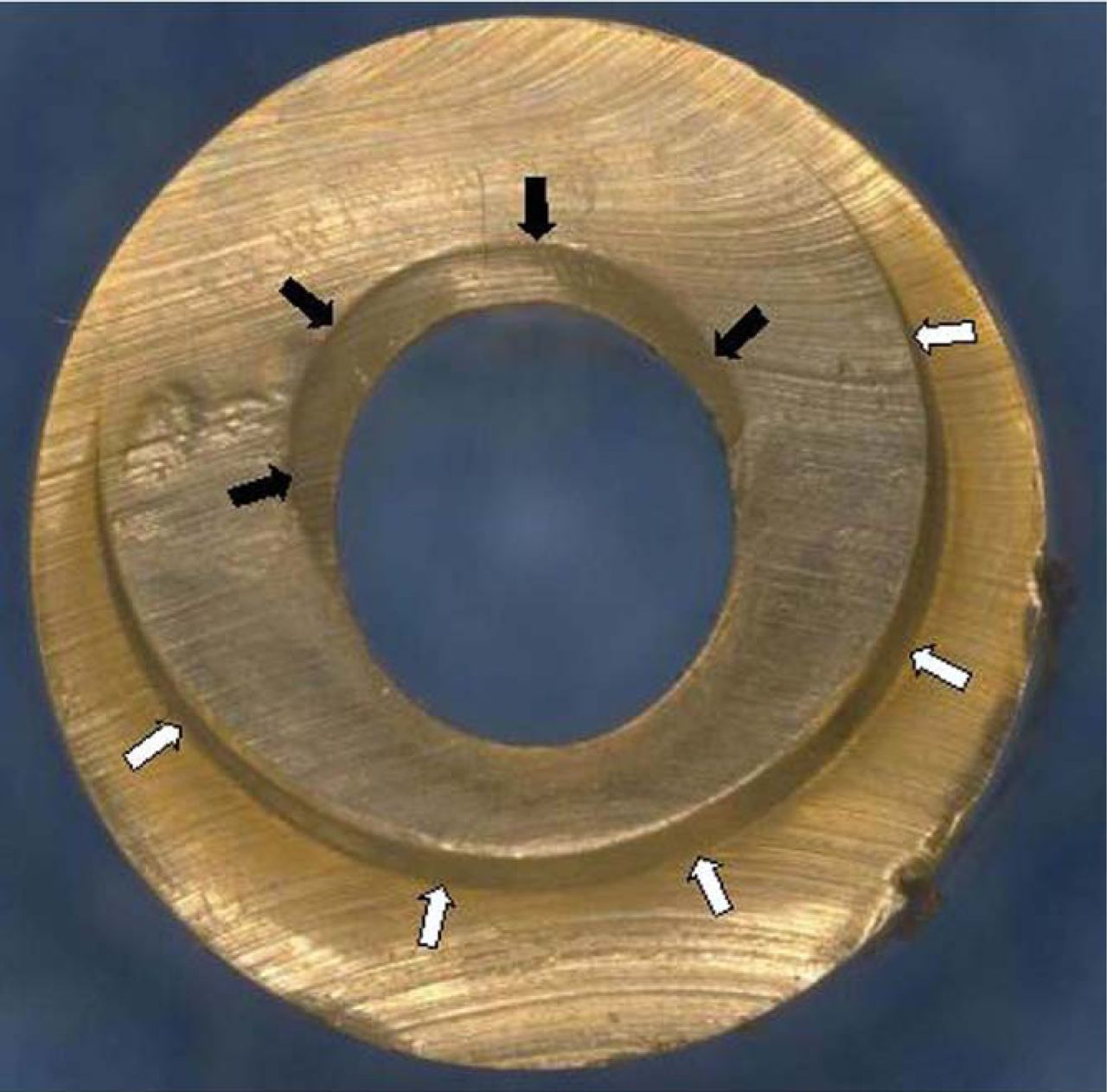
- Fig. 7
Indentations observed in the spacer component from a retrieved Dynesys system that was implanted for 1.9 years. Black arrows denote deformation from the cord, while white arrows indicate deformation from the supporting pedicle screw.
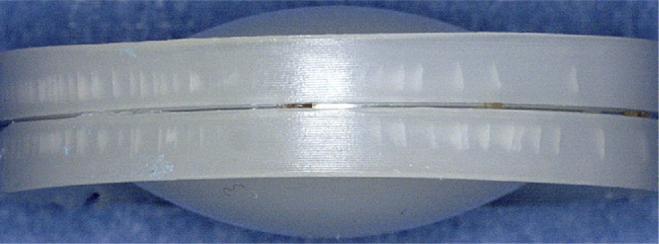
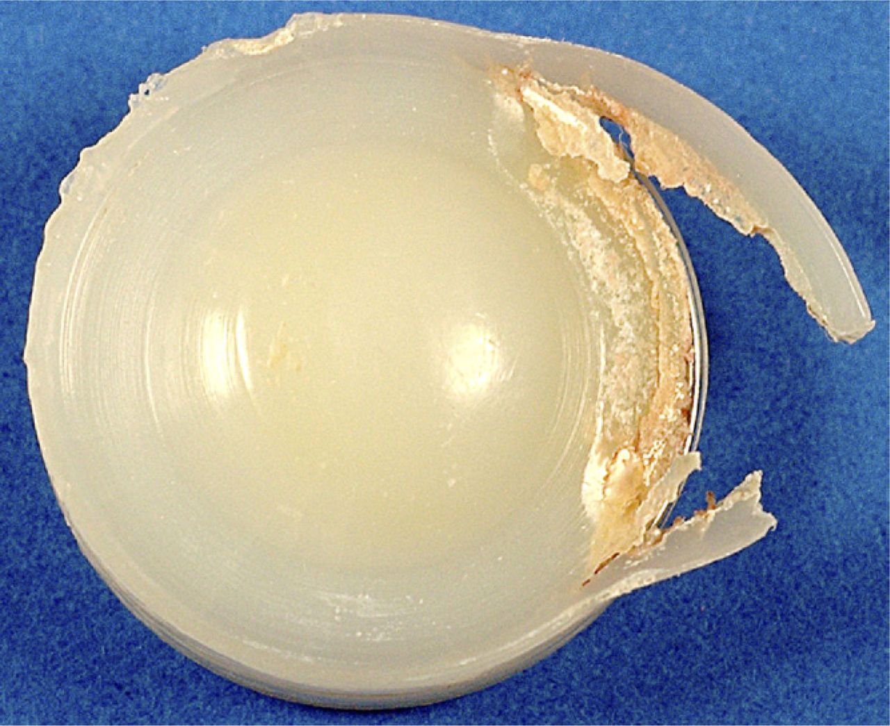
- Fig. 8
Fatigue-related full-thickness rim fracture observed in a retrieved Charité implant that was implanted for 16.1 years.
- Fig. 9
Fatigue-related radial rim cracking observed in a retrieved Charité prosthesis that was implanted for 5.3 years.
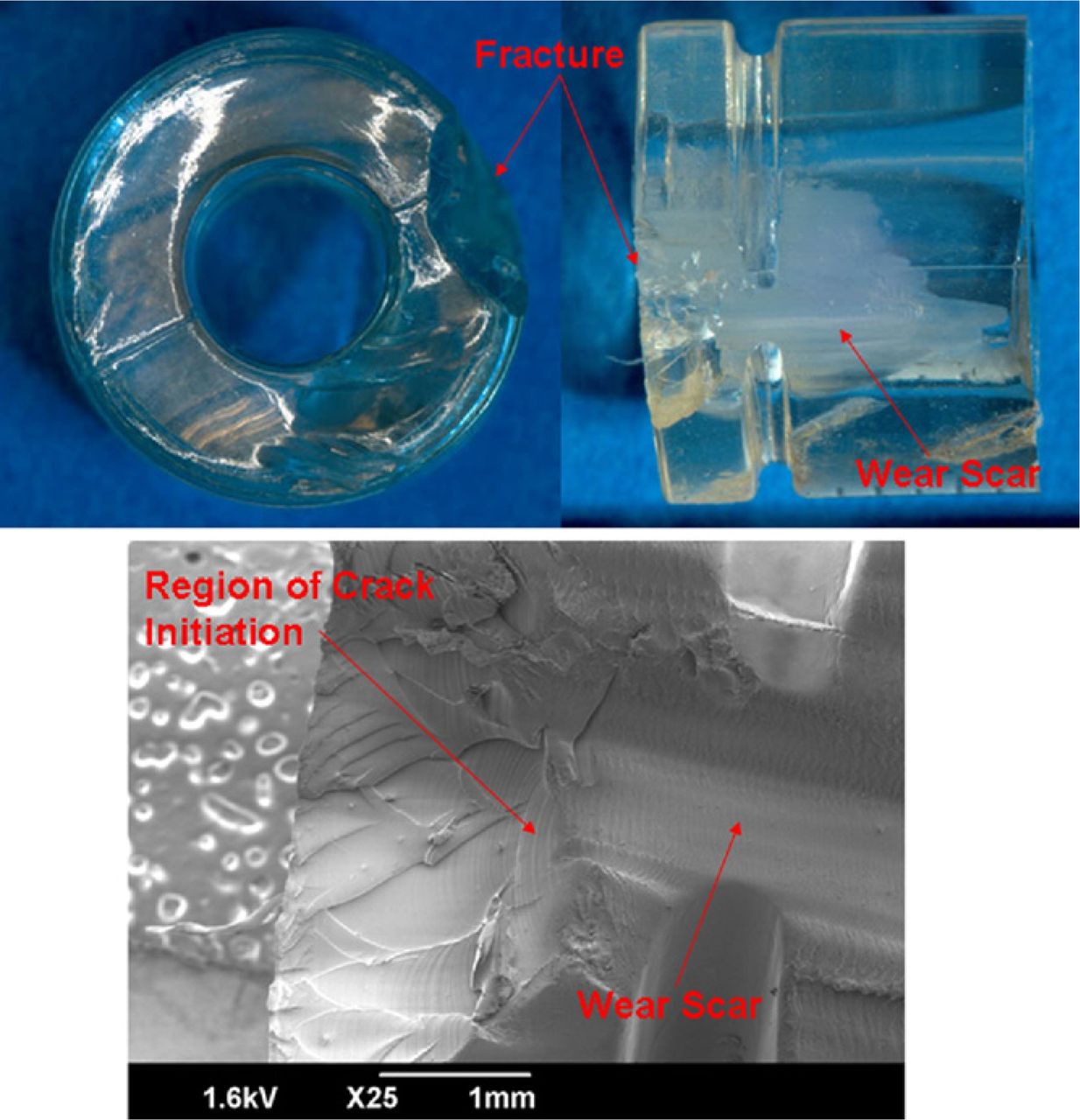
- Fig. 10
Optical microscopy and SEM analysis of a fatigue-fractured spacer from a retrieved Dynesys system that was implanted for 1.1 years.
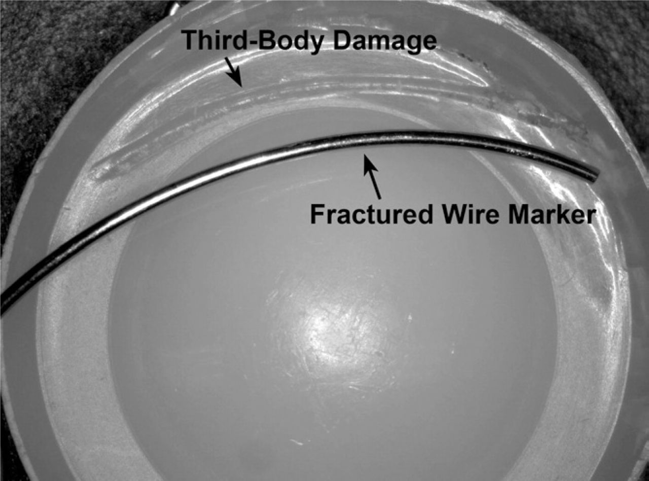
- Fig. 11
Third-body damage caused by a fractured radiographic wire marker in a 12.7 year implanted component, which depicts rim damage in the polyethylene core.
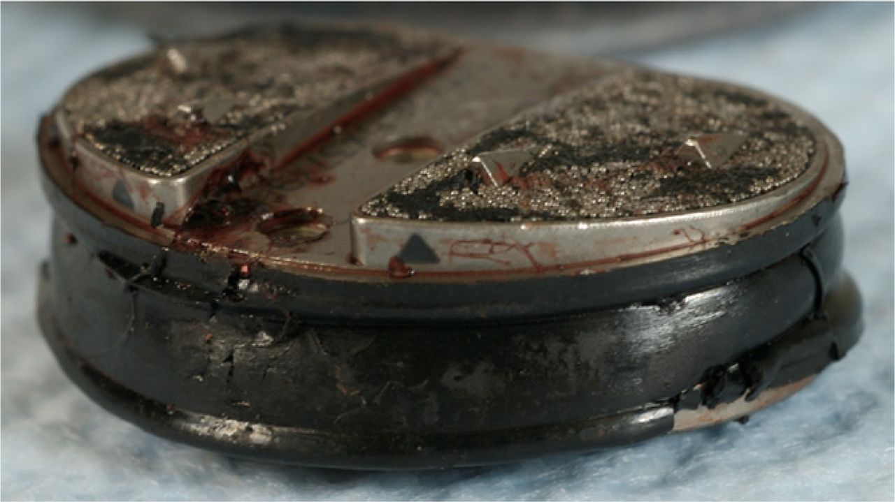
- Fig. 12
Biofilm observed in CoCr alloy, metal-on-metal total disc replacement.
- Fig. 13
Retrieved Acroflex.
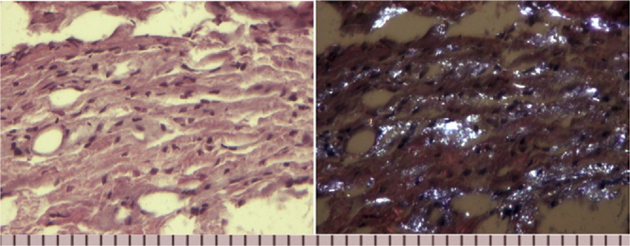
- Fig. 14
TDR tissue sections stained with hematoxylin and eosin and imaged with brightfield (left) and polarized light (right) microscopy. The polarized image is of tissue retrieved at the time of revision surgery (9.2 yr) from a patient who received a pre-1998 Charité TDR implant. The PE debris is white and the score is 3 (range, 0–3). Scale interval represents 0.01 mm.
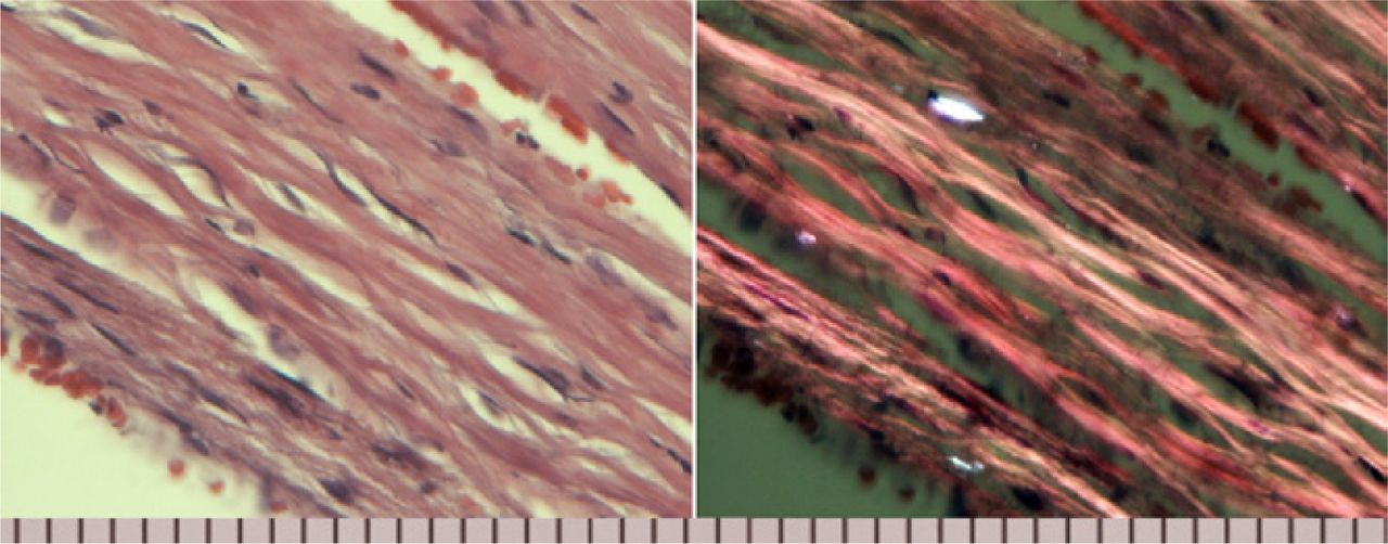
- Fig. 15
TDR tissue sections stained with hematoxylin and eosin and imaged with brightfield (left) and polarized light (right) microscopy. The polarized image is of tissue retrieved at the time of revision surgery (2.2 yr) from a patient who received a post-1998 Charité TDR implant. The PE debris is white and the score is 1 (range, 0–3). Scale interval represents 0.01 mm.
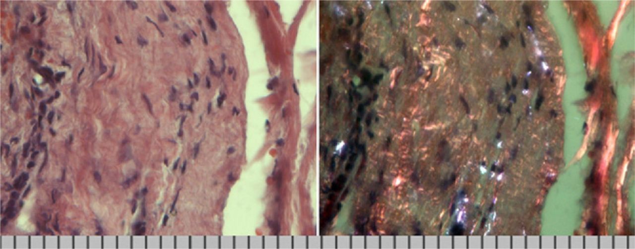
- Fig. 16
TDR tissue sections stained with hematoxylin and eosin and imaged with brightfield (left) and polarized light (right) microscopy. The polarized image is of tissue retrieved at the time of revision surgery (2.2 yr) from the same patient (Fig. 15) who received a post-1998 Charité TDR implant. The PE debris is white and the score is 2 (range, 0–3). Scale interval represents 0.01 mm.
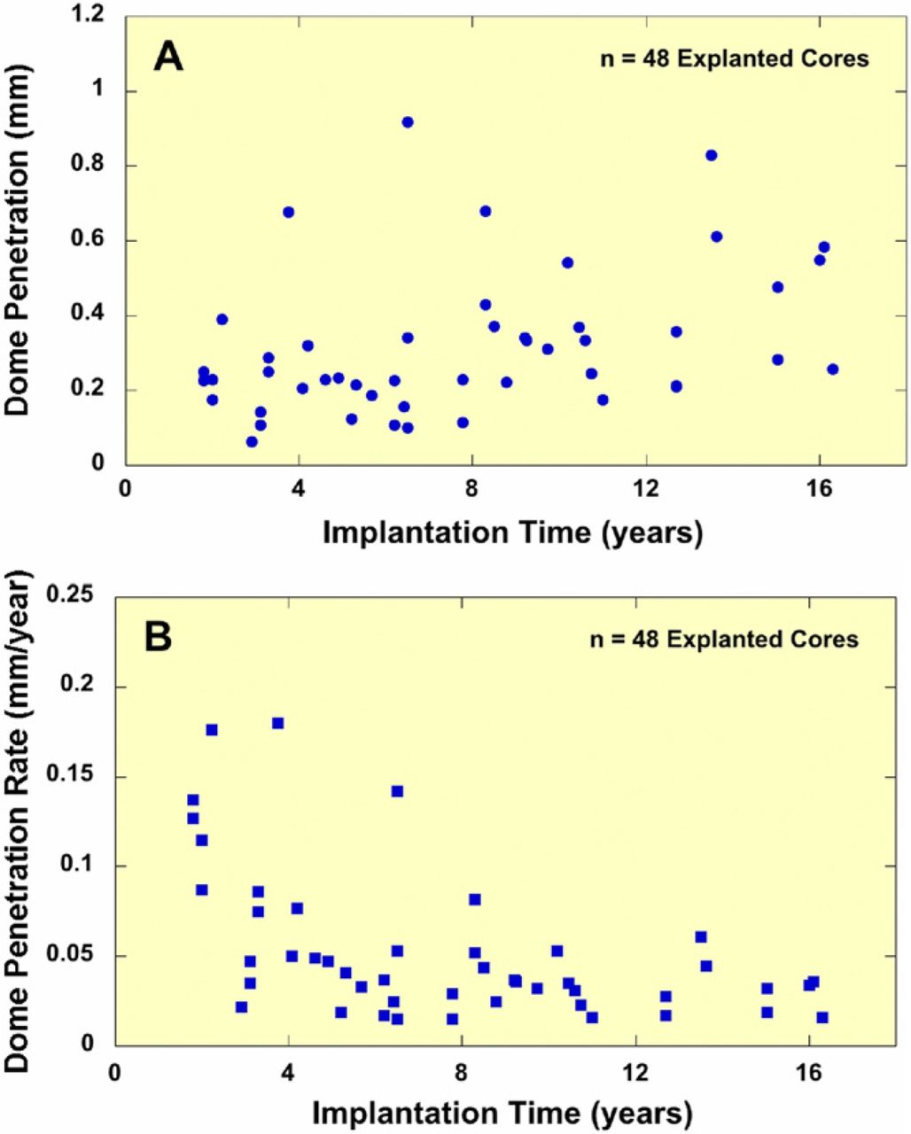
- Fig. 17
A significant correlation was observed between implantation time and (A) penetration (Spearman's Rho = 0.42, p = 0.003) and (B) penetration rate (Spearman's Rho = −0.53, p = 0.0001) in retrieved Charité implants. (Adapted with permission.15)
- Fig. 18
Charité components tested using ISO wear testing protocols incorporating coupled motions exhibited regional burnishing and wear at a rate of 0.124 mm/Mcycles.


In this issue


Jump to section
- Article
- Abstract
- Practical aspects of retrieval analysis
- Wear and damage assessment
- MicroCT analysis
- White light interferometry
- Wear and damage mechanisms
- Analysis of retrieved tissues and particles
- Histological assessment
- Wear particle assessment
- Review of the literature on retrieval analysis
- Cervical spine TDRs
- Lumbar spine TDRs
- Dynamic motion preservation studies
- Recommendations for future testing and research
- Acknowledgments
- References
- Figures & Data
- Info & Metrics
Related Articles
Cited By...
- No citing articles found.