
Main menu

  • Home
  • Content
    • Current Issue
    • Advance Online Publication
    • Archive
  • About Us
    • About ISASS
    • About the Journal
    • Author Instructions
    • Editorial Board
    • Reviewer Guidelines & Publication Criteria
  • More
    • Advertise
    • Subscribe
    • Alerts
    • Feedback
  • Join Us
  • Reprints & Permissions
  • Sponsored Content
  • Other Publications
    • ijss

User menu

  • My alerts

Search

  • Advanced search
International Journal of Spine Surgery
  • My alerts
International Journal of Spine Surgery

Advanced Search

  • Home
  • Content
    • Current Issue
    • Advance Online Publication
    • Archive
  • About Us
    • About ISASS
    • About the Journal
    • Author Instructions
    • Editorial Board
    • Reviewer Guidelines & Publication Criteria
  • More
    • Advertise
    • Subscribe
    • Alerts
    • Feedback
  • Join Us
  • Reprints & Permissions
  • Sponsored Content
  • Follow ijss on Twitter
  • Visit ijss on Facebook
Research ArticleFull Length Article
Open Access

Presacral retroperitoneal approach to axial lumbar interbody fusion: a new, minimally invasive technique at L5-S1: Clinical outcomes, complications, and fusion rates in 50 patients at 1-year follow-up

Robert J. Bohinski, Viral V. Jain and William D. Tobler
International Journal of Spine Surgery January 2010, 4 (2) 54-62; DOI: https://doi.org/10.1016/j.esas.2010.03.003
Robert J. Bohinski
aDepartment of Neurosurgery, University of Cincinnati (UC) Neuroscience Institute, UC College of Medicine, Mayfield Clinic and Spine Institute, and The Christ Hospital, Cincinnati, OH
MD, PhD
  • Find this author on Google Scholar
  • Find this author on PubMed
  • Search for this author on this site
Viral V. Jain
bDepartment of Orthopedic Surgery, Cincinnati Children's Hospital Medical Center, Cincinnati, OH
MD, PhD
  • Find this author on Google Scholar
  • Find this author on PubMed
  • Search for this author on this site
William D. Tobler
aDepartment of Neurosurgery, University of Cincinnati (UC) Neuroscience Institute, UC College of Medicine, Mayfield Clinic and Spine Institute, and The Christ Hospital, Cincinnati, OH
MD
  • Find this author on Google Scholar
  • Find this author on PubMed
  • Search for this author on this site
  • For correspondence: editor@mayfieldclinic.com
  • Article
  • Figures & Data
  • Info & Metrics
  • PDF
Loading

Article Figures & Data

Figures

  • Tables
  • Fig. 1
    • Download figure
    • Open in new tab
    • Download powerpoint
    Fig. 1

    Preoperative evaluation of the sacrum and coccyx for the presacral ALIF procedure. To assess the feasibility of the approach, a line is drawn on the midsagittal MR scan from the probe entry point below the sacrococcygeal joint (red arrow) to the midpoint of the S1 endplate (red line). The optimal trajectory zone (green shaded area) for placement of the implant should be perpendicular to and centered at the midpoint of the superior endplate of S1. MR scan (A) and illustration (B) shows an ideal trajectory for insertion of the screw into the L5-S1 vertebrae. Illustration of the posterior view of the sacrum shows the probe entry point lateral to the coccyx and inferior to the attachments of the sacrospinous and sacrotuberous ligaments. MR scan (C) shows an excessive curvature of the sacrum and coccyx that would result in screw placement at an acute angle to the superior endplate of S1, which is biomechanically suboptimal, and in violation of the spinal canal. This procedure is contraindicated in these patients (with permission from Mayfield Clinic).

  • Fig. 2
    • Download figure
    • Open in new tab
    • Download powerpoint
    Fig. 2

    Bar graphs of the pre- and postoperative Visual Analog Scale (A) and Oswestry Disability Index (B) scores. At 1-year follow up, VAS improved 49% and ODI improved 50% (statistically significant P < .0001) (with permission from Mayfield Clinic).
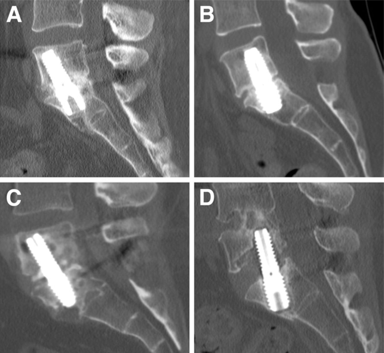
  • Fig. 3
    • Download figure
    • Open in new tab
    • Download powerpoint
    Fig. 3

    Postoperative fusion grades for the procedure depicted on 1-year postoperative CT scans. (A) Grade I shows continuous bridging bone extending from the L5 to the S1 endplate and occupying >50% of available space for fusion. (B) Grade II has bridging bone extending from the L5 to the S1 endplate and occupying < 50% of available space for fusion. (C) Grade III has developing bone with presence of bone connected to either L5 or S1 endplate without bridging. (D) Grade V is a pseudoarthrosis (with permission from Mayfield Clinic).

Tables

  • Figures
    • View popup
    Table 1

    Fusion criteria on 3-mm high-resolution CT scan reconstructions in coronal and sagittal planes (with permission from Mayfield Clinic)

    GradeTime periodDefinition
    IApplicable at any follow-upBridging bone, advanced fusion. Presence of continuous bridging bone extending
    from the L5 to the S1 endplate on the reconstructed images, occupying >50%
    of available space for fusion.
    IIApplicable at any follow-upBridging bone, fusion. Bridging bone filling <50% of available space between
    vertebral bodies.
    IIIEarly: 6 monthsEarly: Presence of developing bone connected to either end plate without bridging.
    Late: 12,18, 24 monthsLate: Presence of developing bone connected to either endplate without bridging
    with fusion mass greater than previous scan.
    IV6 monthsNo bridging or developing bone connected to either endplate.
    VApplicable at 12, 18, 24 monthsNo bridging or developing bone. Decrease or nonprogression of developing bone since previous visit.
    • View popup
    Table 2

    Mean intraoperative blood loss and operative times as noted from anesthetist's chart were significantly lower in stand-alone procedures when compared with procedures supplemented by either pedicle screw fixation or TLIF procedure at another level. Although few patients underwent the procedure alone, morbidity associated with this procedure was substantially lower

    TotalStand-aloneWith posterior procedurePaired t test
    Criteria(N = 50)(n = 5)(n = 45)P value
    Estimated blood loss (ml)21829234.02
    Operative time (minutes)19455207.0001
PreviousNext
Back to top

In this issue

International Journal of Spine Surgery
Vol. 4, Issue 2
1 Jan 2010
  • Table of Contents
  • Index by author

Print
Download PDF
Article Alerts
Sign In to Email Alerts with your Email Address
Email Article

Thank you for your interest in spreading the word on International Journal of Spine Surgery.

NOTE: We only request your email address so that the person you are recommending the page to knows that you wanted them to see it, and that it is not junk mail. We do not capture any email address.

Enter multiple addresses on separate lines or separate them with commas.
Presacral retroperitoneal approach to axial lumbar interbody fusion: a new, minimally invasive technique at L5-S1: Clinical outcomes, complications, and fusion rates in 50 patients at 1-year follow-up
(Your Name) has sent you a message from International Journal of Spine Surgery
(Your Name) thought you would like to see the International Journal of Spine Surgery web site.
CAPTCHA
This question is for testing whether or not you are a human visitor and to prevent automated spam submissions.
Citation Tools
Presacral retroperitoneal approach to axial lumbar interbody fusion: a new, minimally invasive technique at L5-S1: Clinical outcomes, complications, and fusion rates in 50 patients at 1-year follow-up
Robert J. Bohinski, Viral V. Jain, William D. Tobler
International Journal of Spine Surgery Jan 2010, 4 (2) 54-62; DOI: 10.1016/j.esas.2010.03.003

Citation Manager Formats

  • BibTeX
  • Bookends
  • EasyBib
  • EndNote (tagged)
  • EndNote 8 (xml)
  • Medlars
  • Mendeley
  • Papers
  • RefWorks Tagged
  • Ref Manager
  • RIS
  • Zotero
Share
Presacral retroperitoneal approach to axial lumbar interbody fusion: a new, minimally invasive technique at L5-S1: Clinical outcomes, complications, and fusion rates in 50 patients at 1-year follow-up
Robert J. Bohinski, Viral V. Jain, William D. Tobler
International Journal of Spine Surgery Jan 2010, 4 (2) 54-62; DOI: 10.1016/j.esas.2010.03.003
Twitter logo Facebook logo Mendeley logo
  • Tweet Widget
  • Facebook Like
  • Google Plus One

Jump to section

  • Article
    • Abstract
    • Materials and methods
    • Results
    • Discussion
    • Conclusions
    • Disclosure
    • References
  • Figures & Data
  • Info & Metrics
  • PDF

Related Articles

  • No related articles found.
  • Google Scholar

Cited By...

  • No citing articles found.
  • Google Scholar

More in this TOC Section

  • Instrumented reduction of a fixed C1–2 subluxation using occipital and C2/C3 fixation: A case report
  • Preoperative laryngoscopic examination in patients undergoing repeat anterior cervical discectomy and fusion
  • Kinematic analysis following implantation of the PRESTIGE LP
Show more Full Length Article

Similar Articles

Keywords

  • AxiaLIF
  • Percutaneous fusion
  • Minimally invasive fusion
  • interbody fusion

Content

  • Current Issue
  • Latest Content
  • Archive

More Information

  • About IJSS
  • About ISASS
  • Privacy Policy

More

  • Subscribe
  • Alerts
  • Feedback

Other Services

  • Author Instructions
  • Join ISASS
  • Reprints & Permissions

© 2025 International Journal of Spine Surgery

International Journal of Spine Surgery Online ISSN: 2211-4599

Powered by HighWire