


Article Figures & Data
Figures
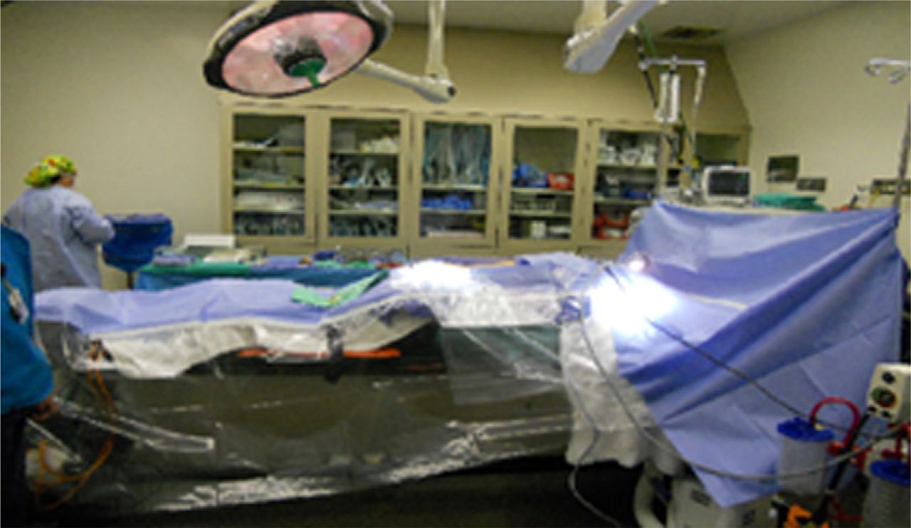
- Fig. 1
Patient prone on open frame, transparent drape is used to maintain visual control of foot pedals, and lateral position of fluoroscope.
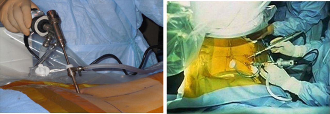
- Fig. 2
Uniportal, and biportal transforaminal approaches to lumbar spine.
- Fig. 3
Illustration of the path of posterolateral transforaminal instrumentation of the lumbar disc. Note the traversing and exiting nerves, seen end-on.
- Fig. 4
Demonstration of sequence of transforaminal instrumentation of lumbar disc for endoscopy. Instrumentation begins with long spine needle, followed by guide-wire, obturator, and cannula, in that order. The middle of the lower row of photographs shows percutaneous removal of the disc nucleus, seen on the last of the photographs.
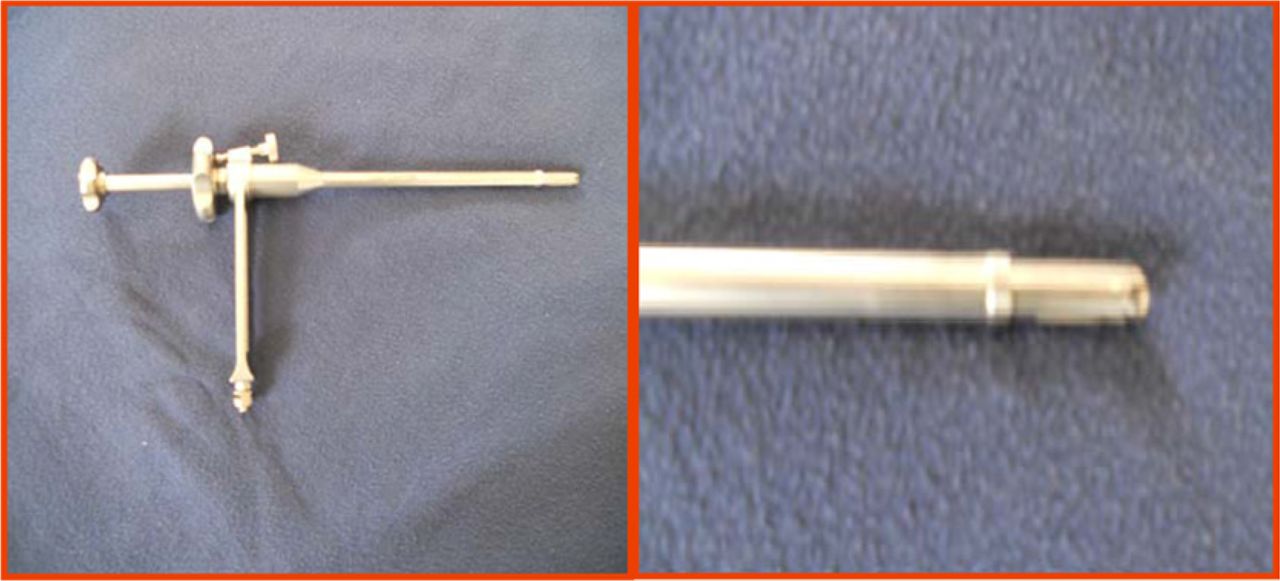
- Fig. 5
Expandable reamer in un-expanded and expanded state. Note the distal end of the reamer cannula is threaded so that it locks into the endplates, thus preventing deep penetration and backing out of the reamer.
- Fig. 6
The photograph on the left shows expandable disc reamer in place. The middle image shows expanded reamer in the disc space. The picture on the right shows bilateral, bi-portal approach with the arthroscope (on the left) visualizing endplate preparation with expandable reamer in the contralateral portal.
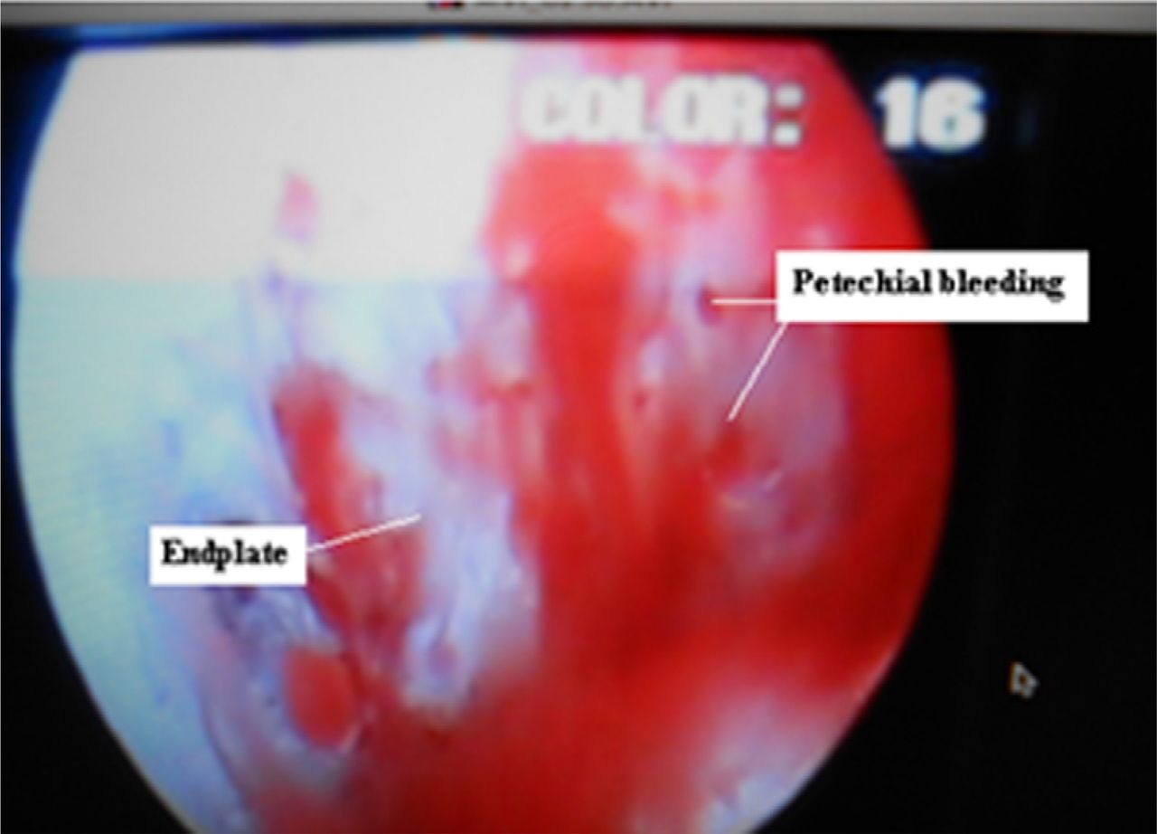
- Fig. 7
Arthroscopic visualization of the prepared endplate. Note the petechial bleeding and the shiny subchondral bone which has not been breached during the reaming.
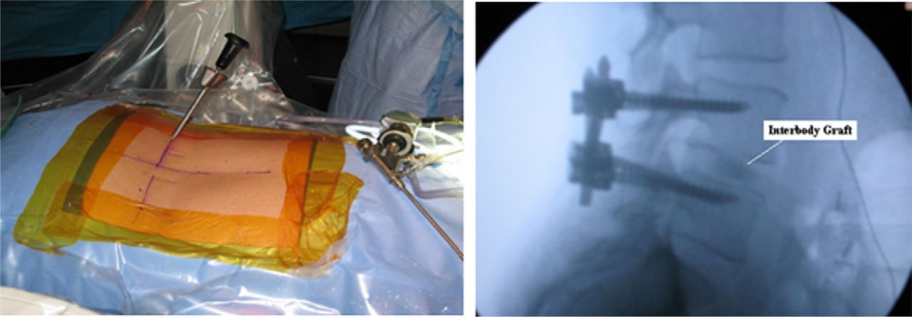
- Fig. 8
The photograph illustrates the insertion of the graft in the intervertebral space, and the lateral fluoroscopic view shows interbody graft in place.
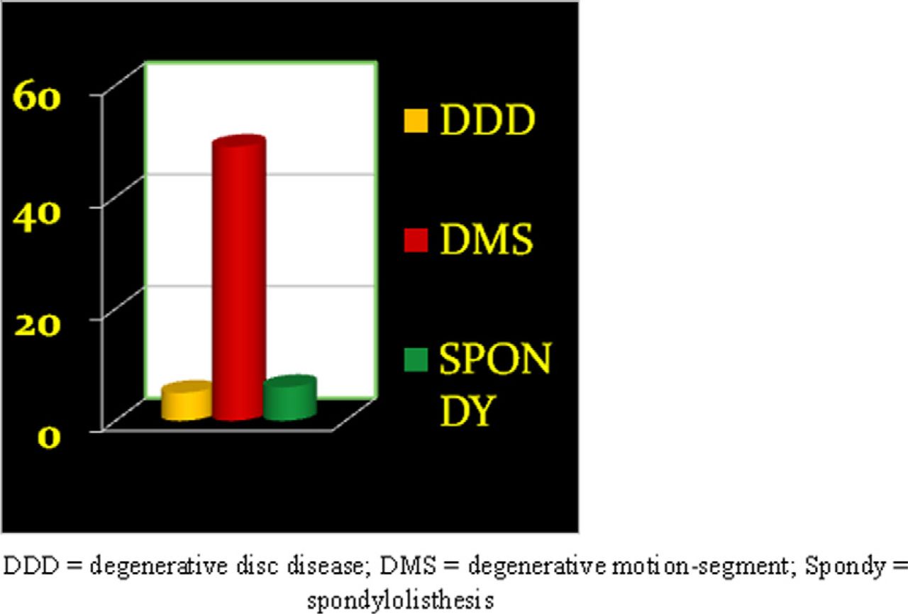
- Fig. 9
Illustrates the diagnoses in this series: DDD = degenerative disc disease; DMS = degenerative motion-segment; and Spondylolisthesis.
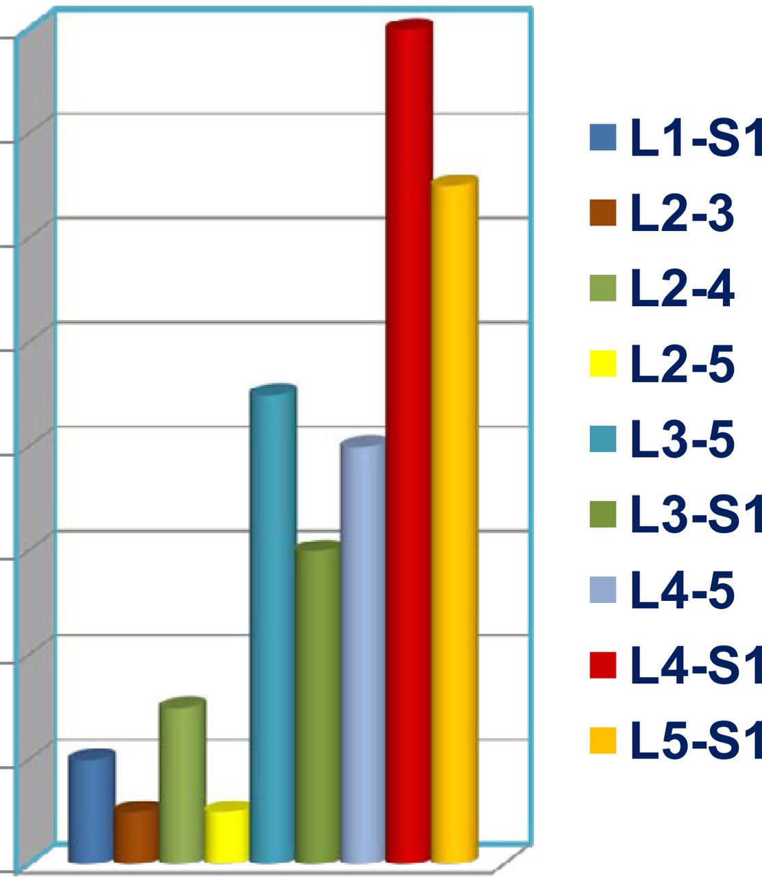
- Fig. 10
Illustrates the frequency of motion-segments treated.
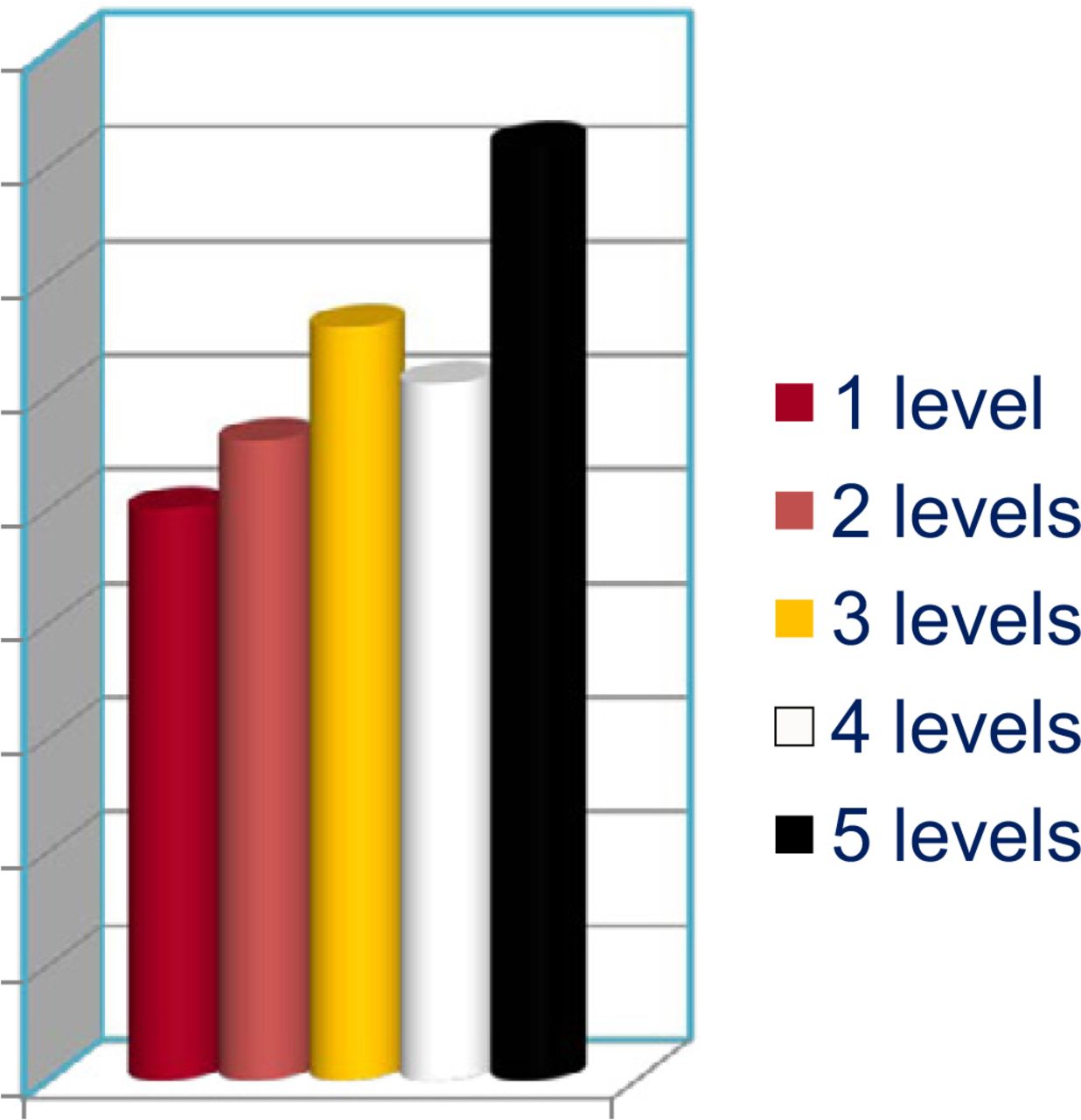
- Fig. 11
Illustrates operating time according to levels fused.
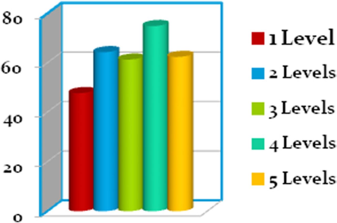
- Fig. 12
Illustrates estimated blood loss according to levels fused.
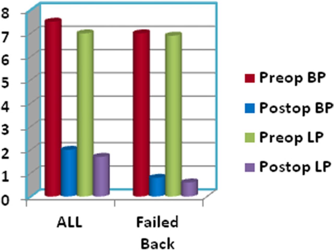
- Fig. 13
a. Pre- and post-operative back pain; b. pre- and post-operative leg pain.
- Fig. 14
Graph compares outcomes of all cases undergoing fusion versus those who had prior spine surgery.
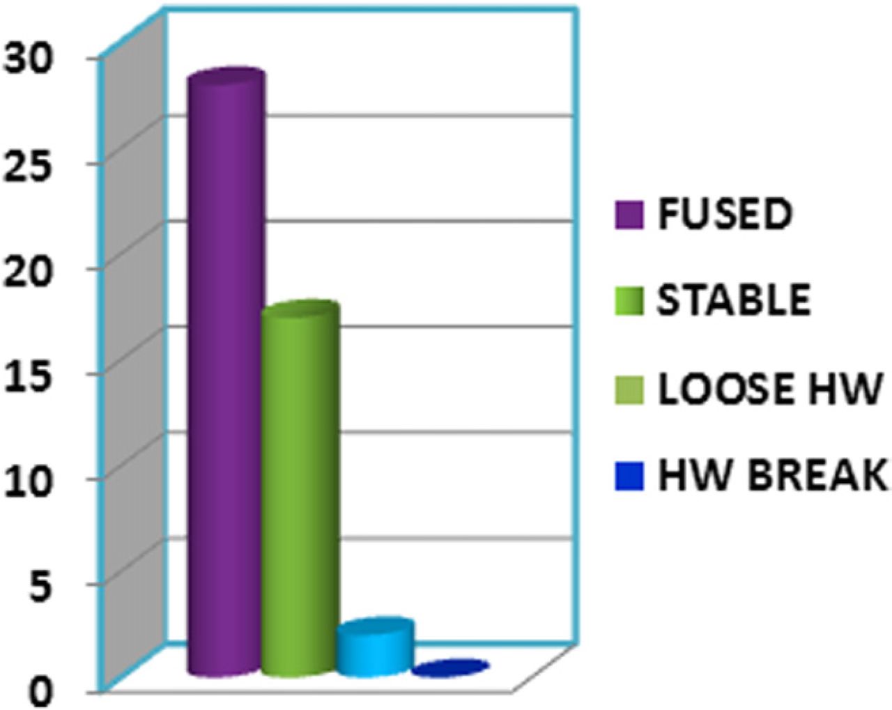
- Fig. 15
Radiographic results following lumbar fusion.
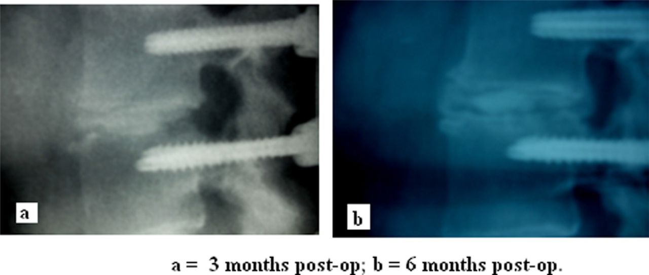
- Fig. 16
Lateral views of a fused segment at 3 months and 6 months post-operatively.


In this issue


Jump to section
Related Articles
Cited By...
- A Narrative Review of Uniportal Endoscopic Lumbar Interbody Fusion: Comparison of Uniportal Facet-Preserving Trans-Kambin Endoscopic Fusion and Uniportal Facet-Sacrificing Posterolateral Transforaminal Lumbar Interbody Fusion
- Full-Endoscopic Oblique Lateral Lumbar Interbody Fusion: A Technical Note With 1-Year Follow-Up
- Full Endoscopic Lumbar Transforaminal Interbody Fusion in DDD Lumbar Degenerative Disc Disease: A Latest Technique
- Feasibility of Endoscopic Inspection of Pedicle Wall Integrity in a Live Surgery Model