


Article Figures & Data
Figures
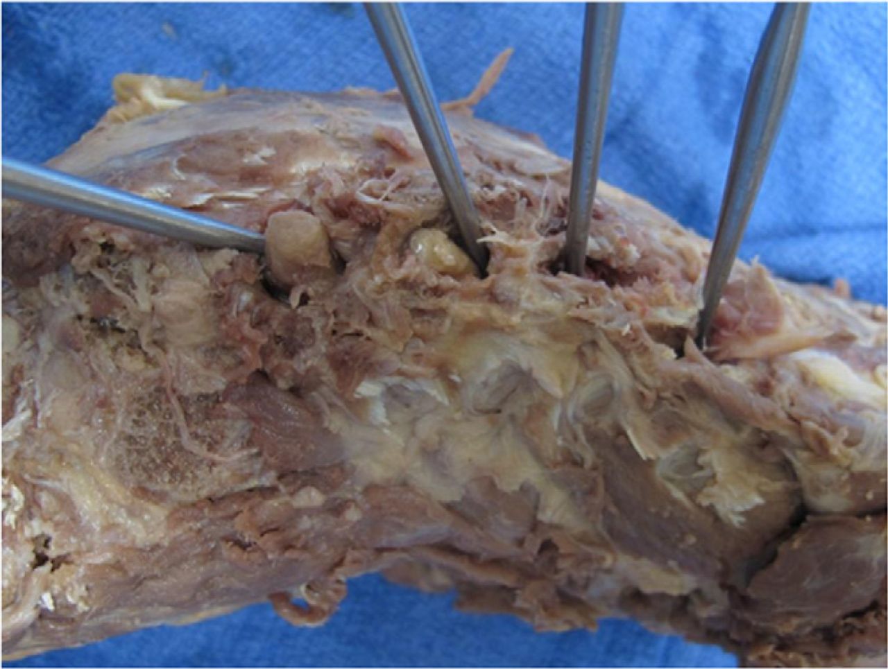
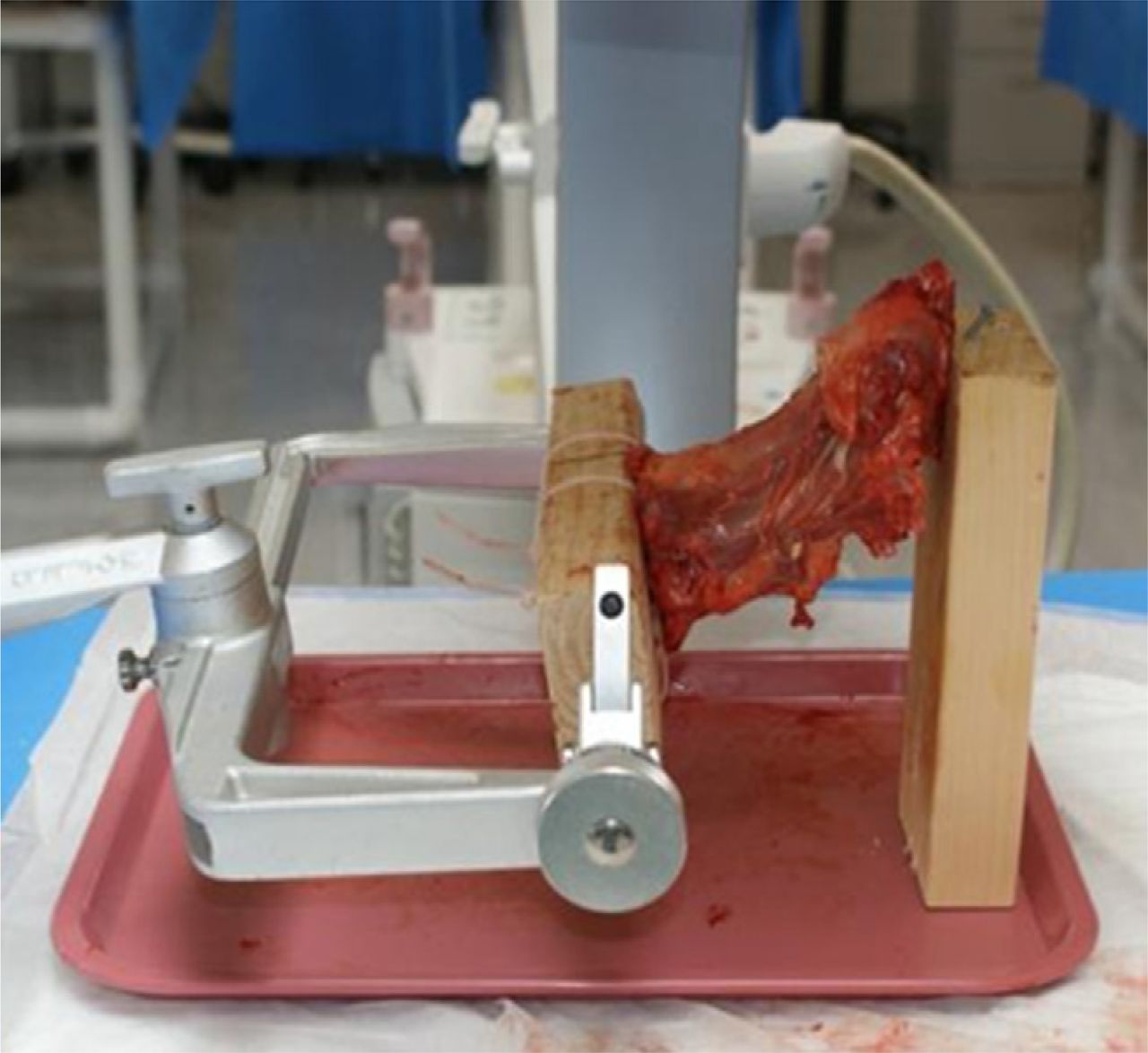
- Fig. 1
Specimen mounted for instrumentation.
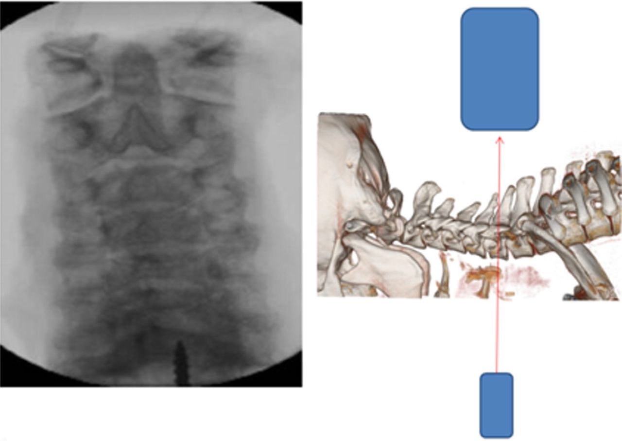
- Fig. 2
Standard AP view. The beam is angled parallel to the disc spaces, providing an oblique view of the joints.
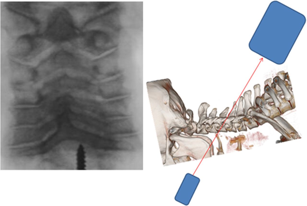
- Fig. 3
Facet AP view. The patient is placed in the Trendelenburg position, and the C-arm is angled such that the beam is parallel to the facet joint being instrumented (in this case, C4-5). The joint appears as a clear space on the monitor.
- Fig. 4
Guide placed perpendicular to C-arm beam, confirmed by targeting hole.
- Fig. 5
Radiographic targeting device. The 18-gauge needle passing through the radiopaque section perpendicular to the longitudinal access should be noted. Under the facet AP view, when this hole is visible on the fluoroscopic image, the guide will be perpendicular to the beam and therefore perpendicular to the facet joint.
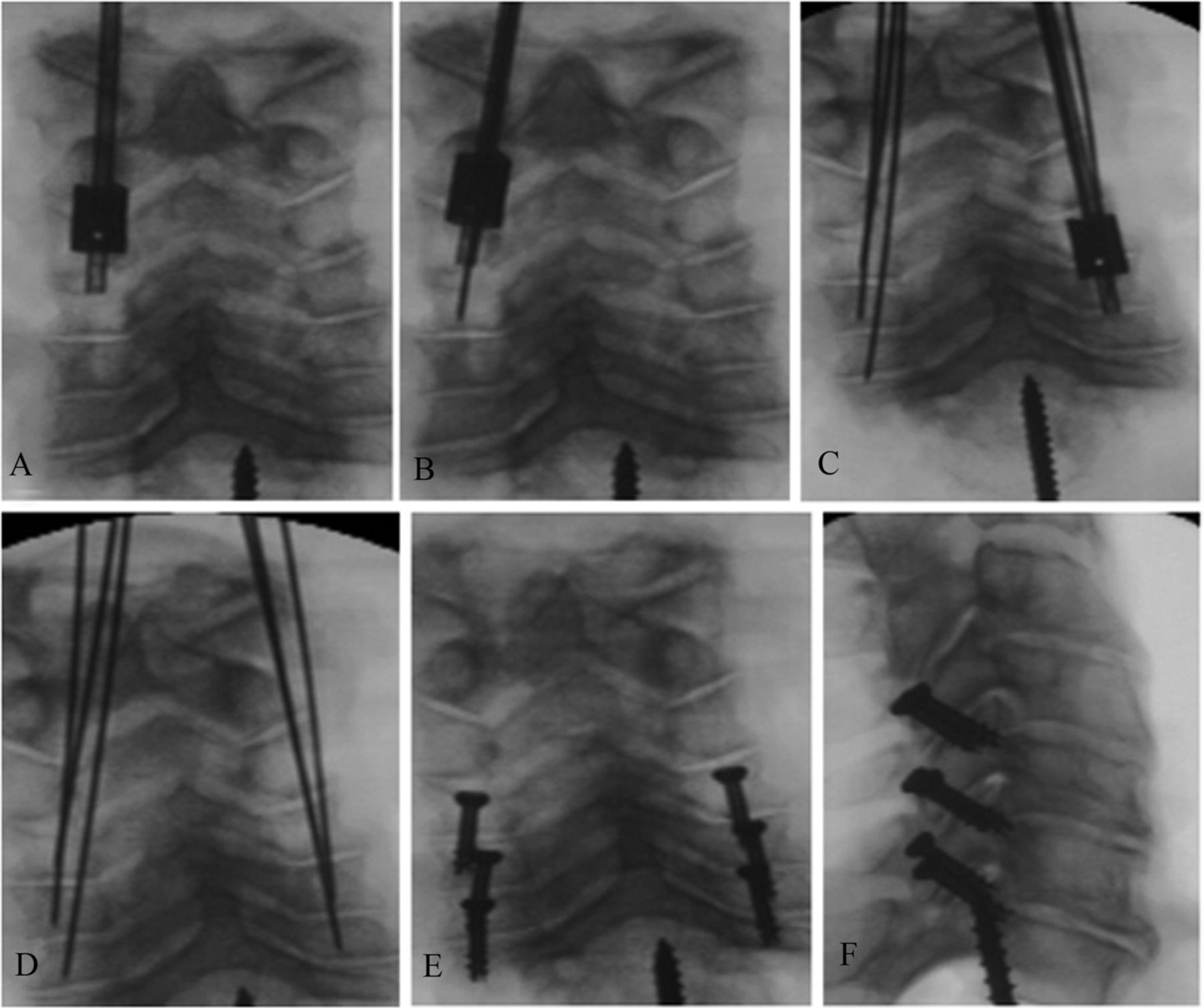
- Fig. 6
Screw placement technique. (A) The guide was placed at the starting point, and cranial-caudal angulation was adjusted until the targeting hole came into view. (B) A K-wire was placed down the guide and advanced to the level of the joint. (C) This was repeated for all levels instrumented, with the C-arm being adjusted at each level to ensure an accurate facet AP view. (D) All K-wires were placed. (E) Self-drilling or self-tapping 14 × 4–mm cannulated screws were placed over the wires, and the wires were removed. (F) The lateral view confirms screw placement.
- Fig. 7
Acceptable screw placement confirmed by CT.
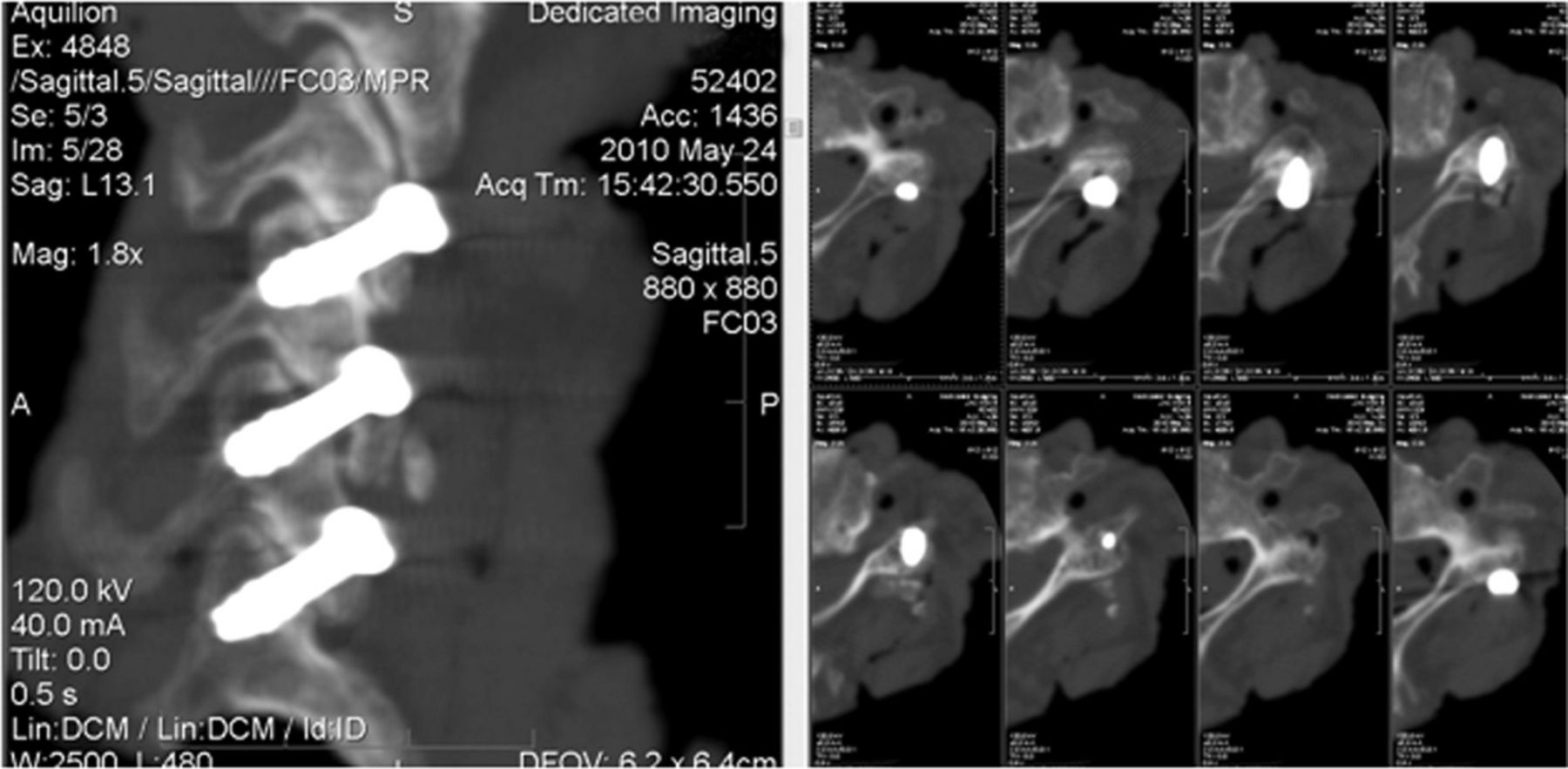
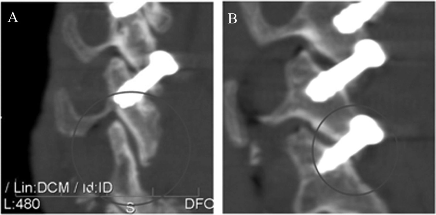
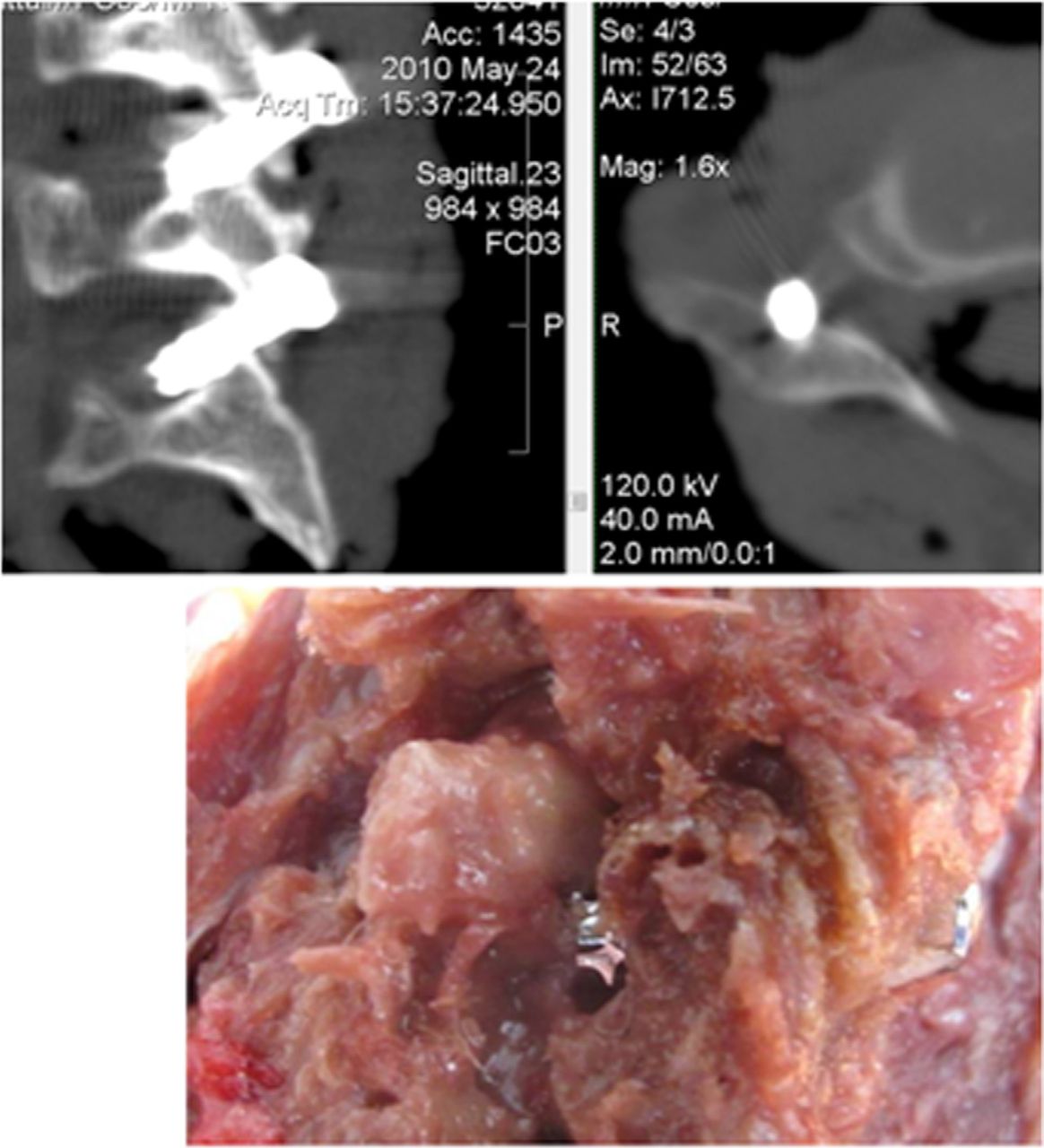
- Fig. 8
Direct nerve root dissection.
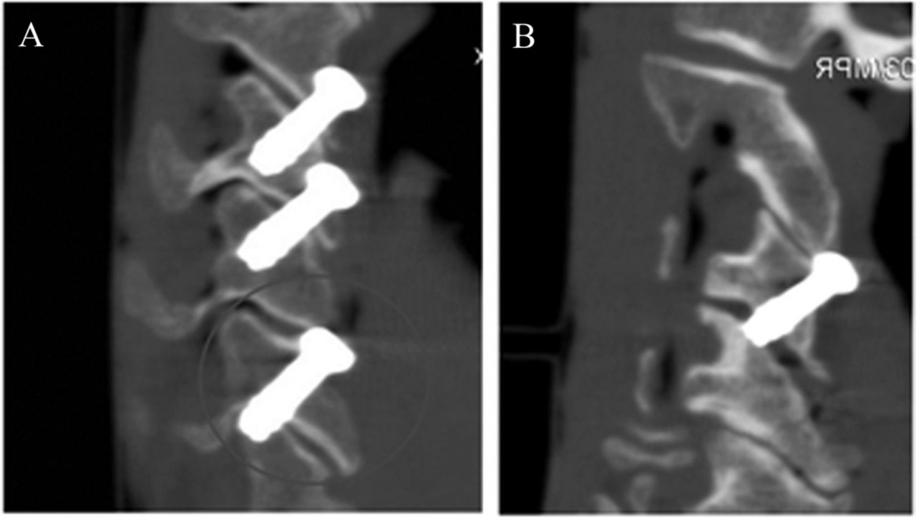
- Fig. 9
Breaches. (A) Facet fracture (bottom screw). (B) Distraction.
- Fig. 10
C6-7 nerve root injury. In a specimen with a small C7 superior articular process, the screw is seen passing through the bone and into the neural foramen. Direct dissection shows that the screw is in the neural foramen, displacing the C7 nerve root superiorly and anteriorly.
- Fig. 11
Variable C7 superior articular facet anatomy, as noted in our study. (A) A thin, vertical C7 superior articular process that is not appropriate for screw placement. (B) A C7 superior articular process that is ideal for screw placement.


Tables
Level No. of potential screws No. of screws Placed Acceptable Placement Breach C3-4 14 10 9 1 C4-5 14 14 14 0 C5-6 14 14 13 1 C6-7 14 10 9 1 Total 56 48 45 3
In this issue


Jump to section
Related Articles
Cited By...
- No citing articles found.