


Article Figures & Data
Figures
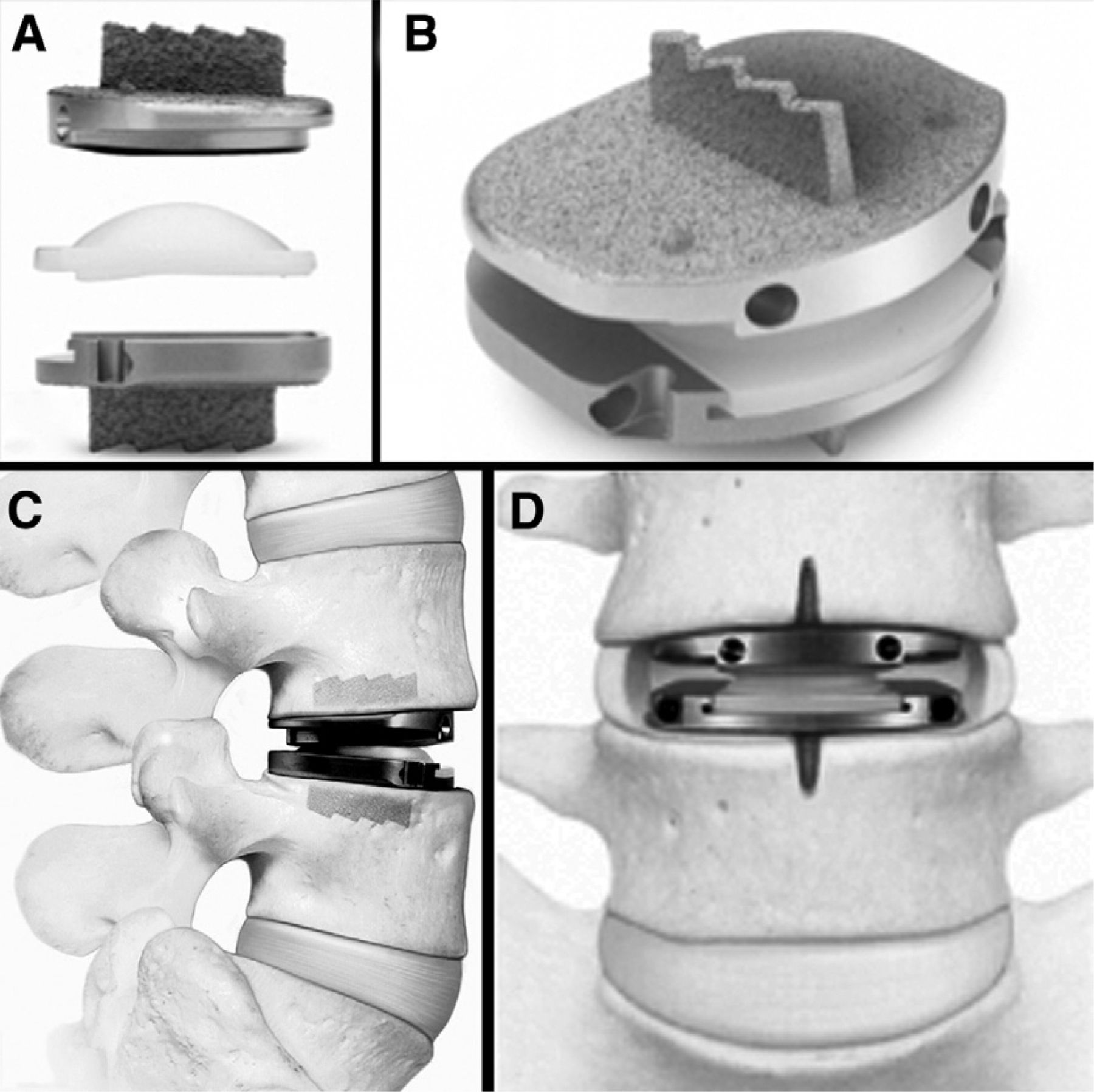
- Fig. 1
PD-L components (A). PD-L device assembled (B). PD-L device implanted: lateral view (C) and anteroposterior view (D). One should note the keels and associated chisel cuts. (Images reproduced with permission of Synthes Spine, Inc.)
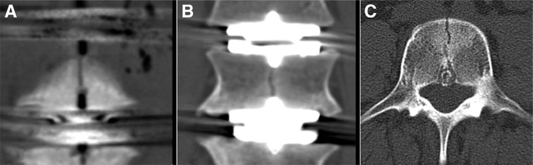
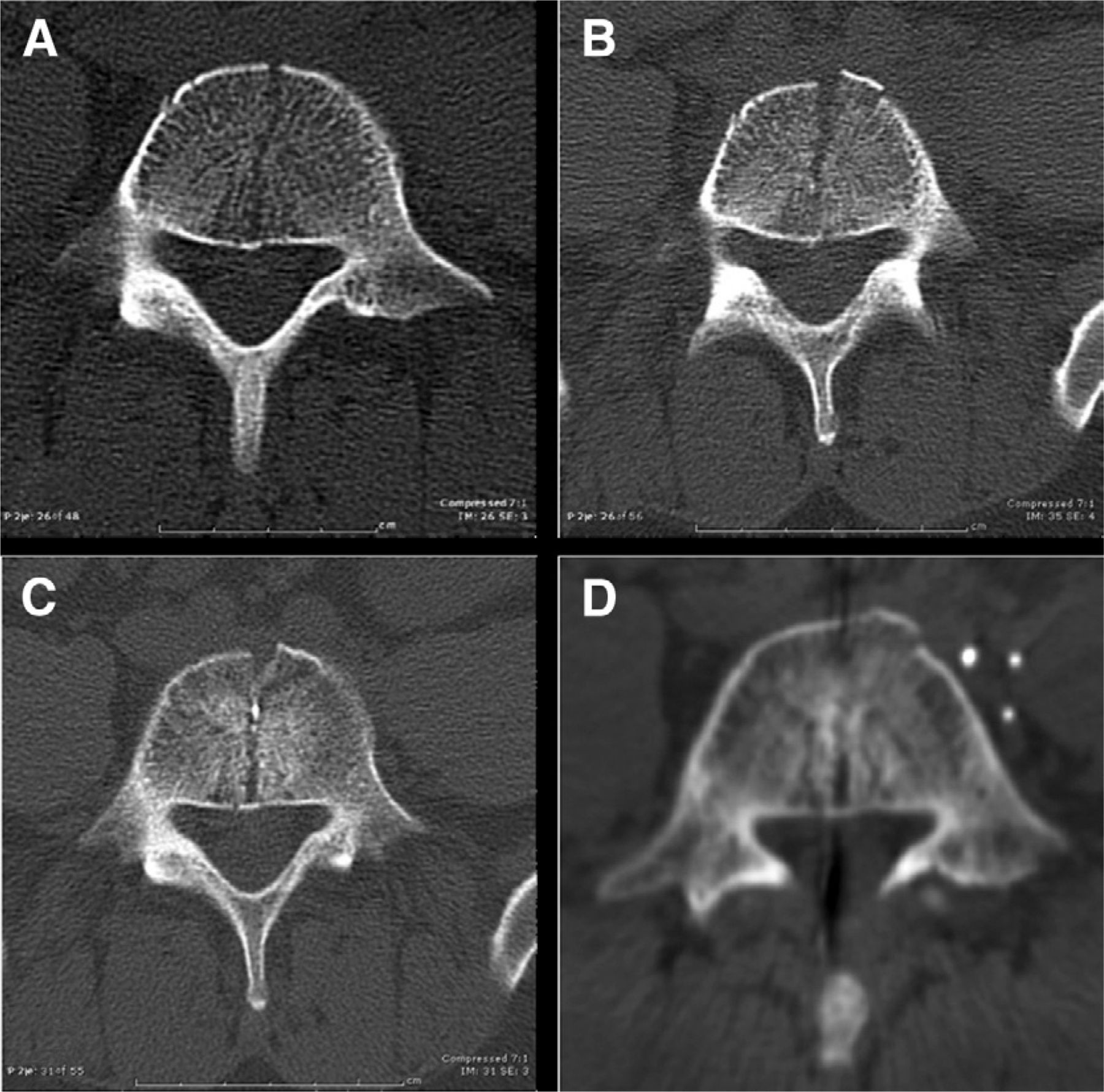
- Fig. 2
Typical appearance of a VB-SF (patient 6, PD-L implants at L3-4, L4-5, and L5-S1). Coronal CT reconstruction of L4. The fracture connects the keel cuts in L4 (A). Coronal CT reconstruction through the midbody of L4 (B). The fracture connects the keels of adjacent PD-L implants. Axial CT reconstruction of L4 (C). The fracture extends from the anterior cortex to the posterior venous drainage.
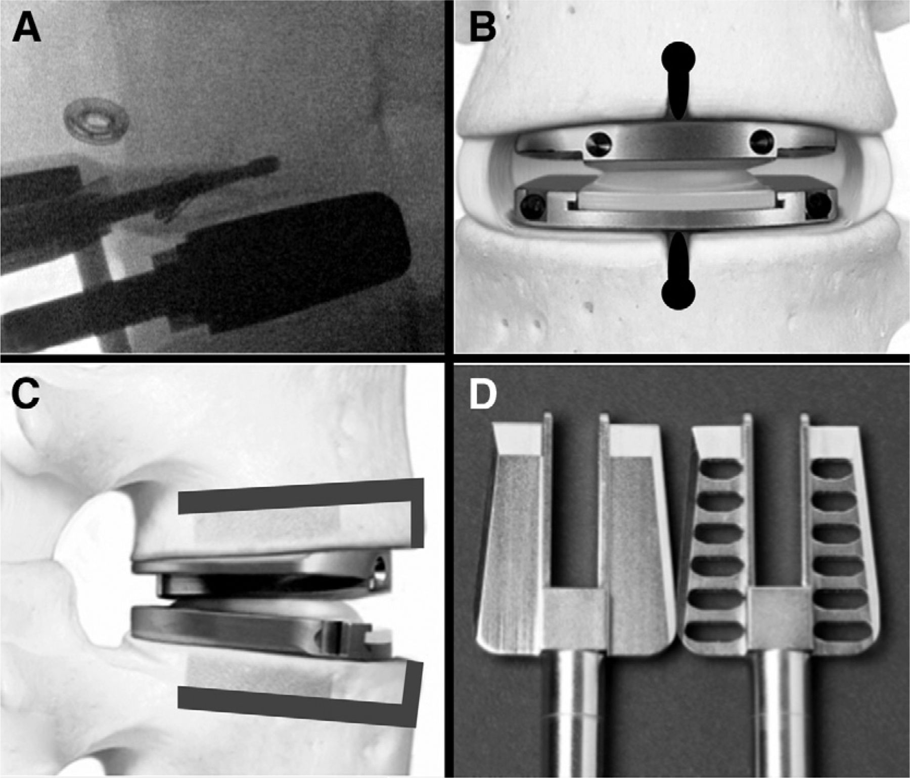
- Fig. 3
PH/CR/FC technique for PD-L device implantation. Drilling of pilot hole in superior vertebral body as seen on intraoperative lateral fluoroscopy (A). Anteroposterior diagram of implanted PD-L device with location of pilot holes and anterior cortex removal in heavy black overlay (B). Lateral diagram of implanted PD-L device with gray overlay depicting the location of pilot holes and anterior cortex removal (C). Comparison of standard US chisel (left) with fenestrated chisel (right) used in modified surgical technique (D). The reverse-cutting horizontal surfaces in the fenestrated chisel should be noted. (B and C, excluding the overlays, are reproduced with permission of Synthes Spine, Inc.)
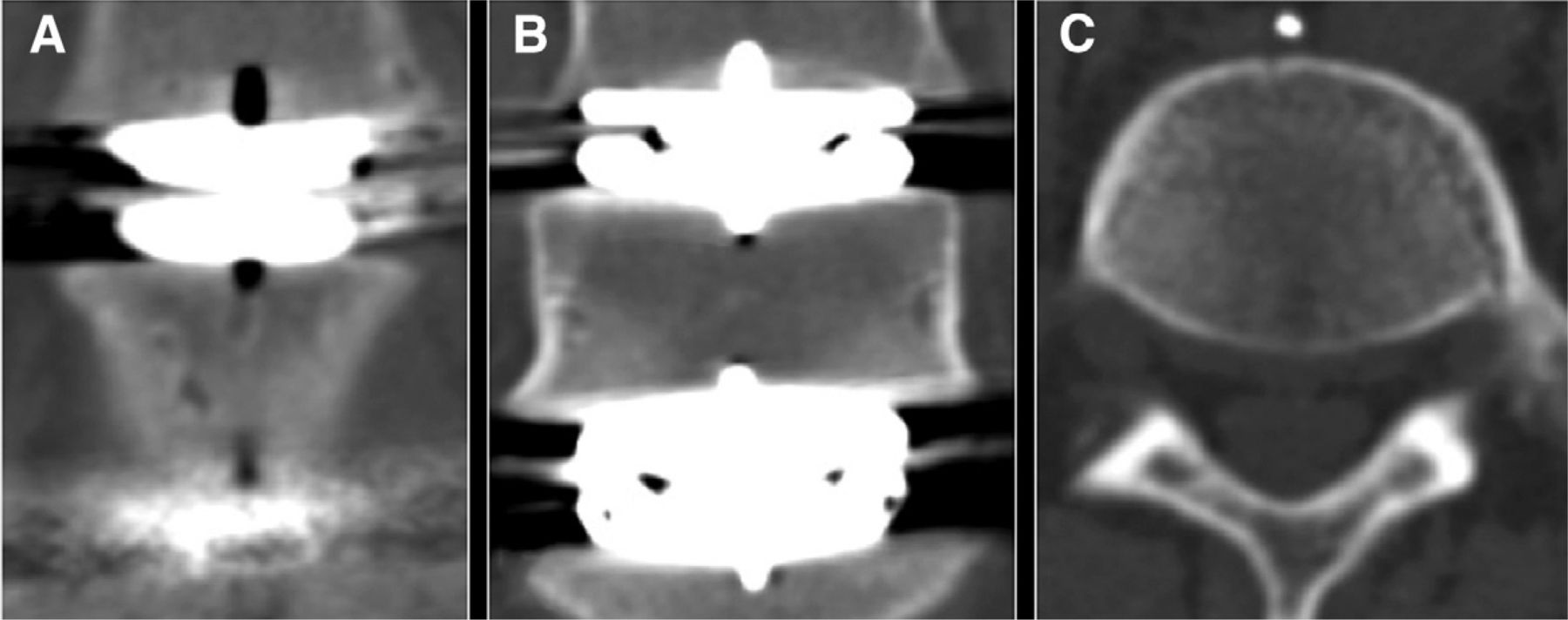
- Fig. 4
Patient 4, a 2-level PD-L case with a cranial-caudal anterior keel cut–to–anterior keel cut fracture. Intraoperative photograph taken after removal of the anterior periosteum between the keel cuts, showing a fracture connecting adjacent keel cuts (A). Axial (B) and coronal (C) CT reconstructions showing no evidence of fracture within the L5 vertebral body.
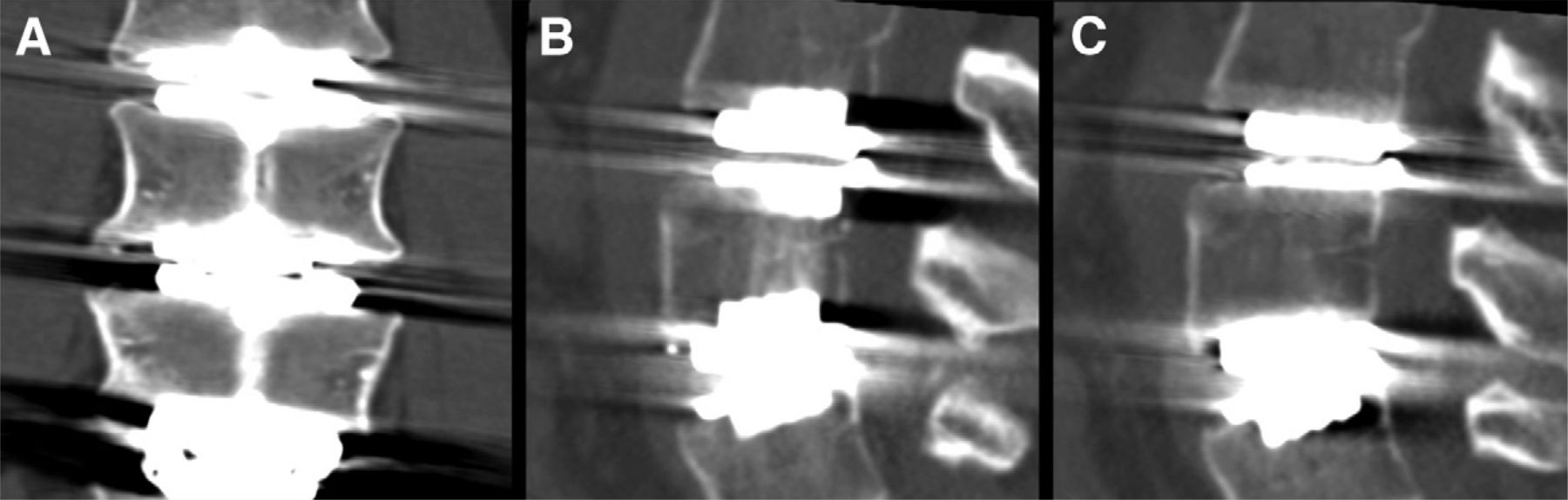
- Fig. 5
Axial CT reconstruction of the L5 vertebral body from patient 5, who may have had an adverse outcome related to a VB-SF. Postoperative axial CT showing the VB-SF (A). Axial CT scan 10 days postoperatively, immediately after an accident with extreme vertical and rotational loading of the spine (B). One should note the fracture separation and the anterior wedge of bone that has been extruded. Axial CT scan 4 weeks after injury showing anterior healing or fragment migration with posterior fracture persistence (C). Axial CT scan 8 months after injury showing continued anterior fracture healing with sclerosis around the posterior portion of the fracture, which remains open (D).
- Fig. 6
Postoperative CT scans of patient 7 centered on L5 with no evidence of fracture. Anterior coronal cut (A). Midcoronal cut (B). Midbody axial cut (C).
- Fig. 7
Twelve-month follow-up CT scan of patient 6, a 3-level PD-L case operated on by the PHO technique, showing late formation of a vertical sclerotic band in the nonfractured L5 vertebral body on axial CT reconstruction (A). One should note the similarities between the sclerosis at this level and what is seen in the fractured L4 vertebral body at the level above. Twelve-month follow-up CT scan of patient 1 (B). One should note that the sclerotic development is seen only between 2 adjacent keels. Off-midline sagittal CT cut depicting absence of sclerotic band (C).
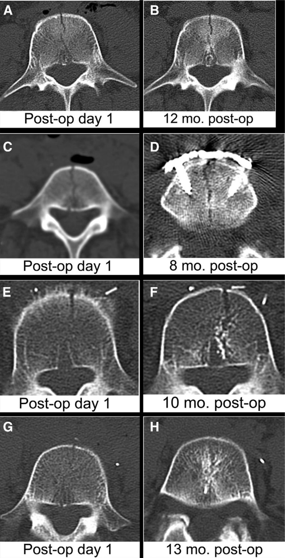
- Fig. 8
Initial and final follow-up axial CT images of the multilevel PD-L cases with VB-SFs without clinical sequelae. In patients 6 (A and B), 3 (C and D), and 2 (E and F) with 12, 8, and 10 months’ follow-up, respectively, the fractures remain open and show sclerotic margins without evidence of bridging of the fracture. One should note the unhealed fracture in D, where a plate was placed across the fracture on the second postoperative day. On these images, the fractures are more obvious at late follow-up. In patient 1 (G and H), a 13-month follow-up axial CT reconstruction shows cortical bridging at the fracture and sclerosis around the fracture path.


Tables
- Table 1
Patient-specific surgical and fracture details for group I, transitional, and group II patients
Patient No. No. of levels Fusion PD-L Technique Site Primary/assisting surgeon Fracture type, level Group I 1 2 — L4-5, L5-S1 United States United States H.G.S. VB-SF, L5 2 2 — L3-4, L4-5 United States United States H.G.S. VB-SF, L4 3 2 — L4-5, L5-S1 United States United States H.G.S. VB-SF, L5* 4 2 — L4-5, L5-S1 United States United States H.G.S. Anterior keel cut–to–anterior keel cut†, L5 5 2 — L4-5, L5-S1 United States United States H.G.S. VB-SF, L5 T 6 3 — L3-4, L4-5, L5-S1 PHO United States R.B./H.G.S. VB-SF, L4 Group II 7 2 — L4-5, L5-S1 PH/CR/FC United States H.G.S. — 8 2 — L4-5, L5-S1 PH/CR/FC United States H.G.S. — 9 2 L5-S1 L3-4, L4-5 PH/CR/FC United States H.G.S. — 10 2 — L4-5, L5-S1 PH/CR/FC United States H.G.S. — 11 2 — L3-4, L4-5 PH/CR/FC Germany R.B. — 12 2 — L3-4, L4-5 PH/CR/FC Germany R.B. —‡ 13 2 — L3-4, L4-5 PH/CR/FC Germany R.B. — 14 3 — L3-4, L4-5, L5-S1 PH/CR/FC Germany R.B. — 15 3 — L3-4, L4-5, L5-S1 PH/CR/FC Germany R.B. — 16 2 — L3-4, L4-5 PH/CR/FC Germany R.B. — 17 2 — L3-4, L4-5 PH/CR/FC Germany R.B. — 18 2 — L3-4, L4-5 PH/CR/FC Germany R.B. — Characteristic Group I (n = 5) Group II (n = 11) P value (t test) Age [mean (range)] (y) 42.8 (33 to 62) 43.3 (27 to 53) .909 Male/female sex 1/4 6/5 .308 BMI [mean (range)] (kg/m2) 28.4 (26 to 30) 25.9 (20 to 30) .125 T score [mean (range)] 0.6 (0.9 to 2.0) 0.0 (−1.5 to 2.7) .491 VBH [mean (range)] (mm) 24.5 (22.8 to 26.4) 27.4 (24.2 to 30.8) < .005
In this issue


