


Article Figures & Data
Figures
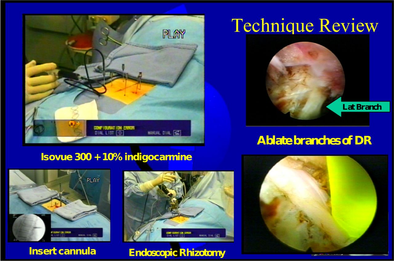
- Fig. 1
Surgical setup for ablation of the medial, intermediate and lateral branches of the dorsal ramus.
- Fig. 2
Richard Wolf YESS Rhizotomy Set. The cannulas, endoscope, bitip and surgical bipolar RF probes by Elliquence are configured ergonomically to provide excellent focal length imaging to keep image in focus with the endoscope scope resting on cannula. The bitip probe cuts tissue, and the RF probe thermally ablates tissue efficiently.
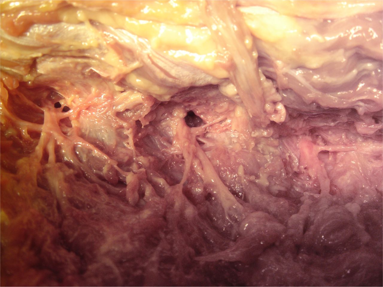
- Fig. 3
Cadaver dissection of the dorsal ramus and its branches out- lining the areas where branches of the dorsal ramus may be visualized and ablated before it reaches the facet joint.
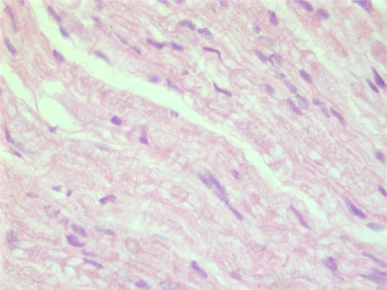
- Fig. 4
This H and E slide of the biopsied specimen is consistent with a peripheral nerve fiber.
- Fig. 5
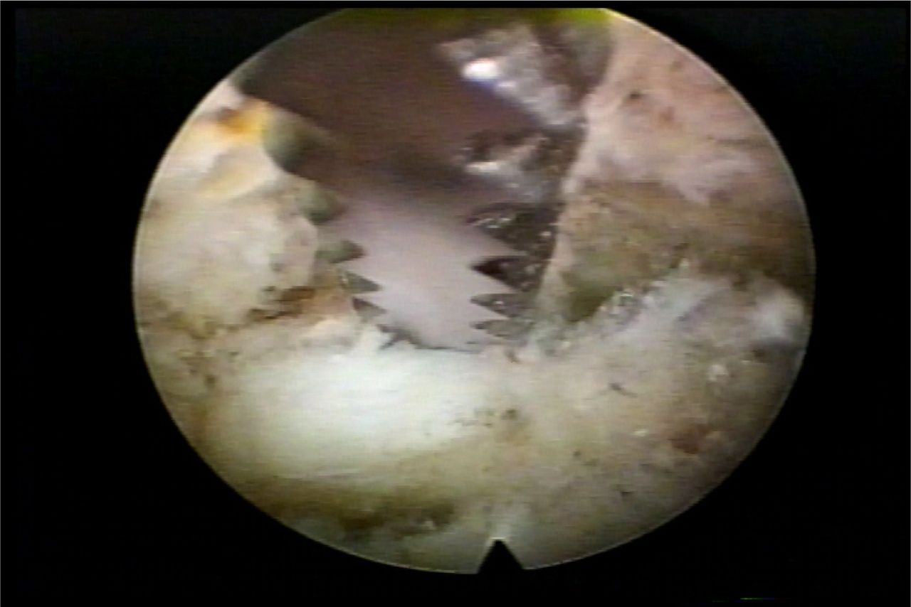
This foraminal view of a branch of the dorsal ramus is in the foramen at the level of the SAP. The nerve runs along the ventral lateral aspect of the superior facet to the tip, and can also run in the vicinity of the foraminal ligament. Endoscopic rasps, trephines, kerrisons, and burrs can be used for foraminoplasty. The nerve should be preserved, if possible, but transection of a branch of the dorsal ramus contributes to axial back pain relief. Branches of the dorsal ramus originates in the foramen before exiting to traverse the transverse process. These nerves are difficult to differentiate from furcal nerves arising from the spinal nerves. Palpating the nerve using local anesthesia can sometimes demonstrate a pain response, but not always, depending on the level of sedation and anesthetic use.
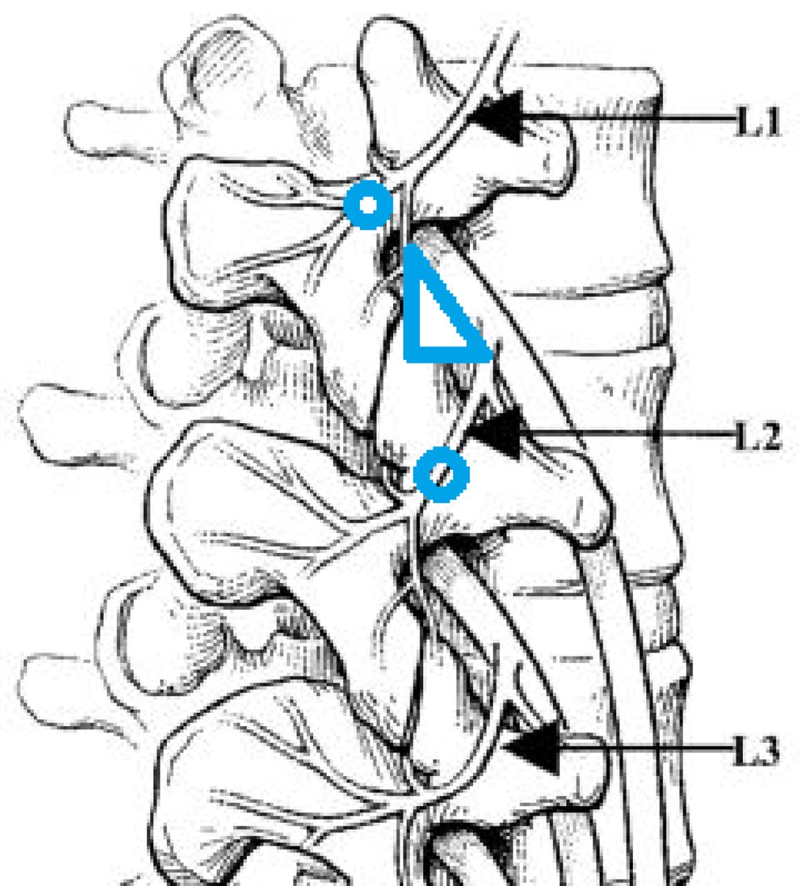
- Fig. 6
Schematic drawing of Kambin's triangle demonstrating where the medial branch can be visualized at the level of the tip of the SAP and at the transverse process. The medial branch can be visualized endoscopically and transected in the foramen, or more consistently, at the interosseous tunnel crossing the transverse.


In this issue


Jump to section
Related Articles
Cited By...
- Insights From the ISASS Webinar Series on Current and Emerging Techniques in Endoscopic Spine Surgery | Part 1: Polytomous Rasch Analysis of Surgeon Endorsement of Endoscopic Discectomy/Foraminotomy, Interbody Fusion, and Importance of Patient Feedback During Surgery
- Radiofrequency treatments for lumbar facet joint syndrome: a systematic review and network meta-analysis
- Transforaminal Endoscopic Decompression of the Lumbar Spine for Stable Isthmic Spondylolisthesis as the Least Invasive Surgical Treatment Using the YESS Surgery Technique