


Article Figures & Data
Figures
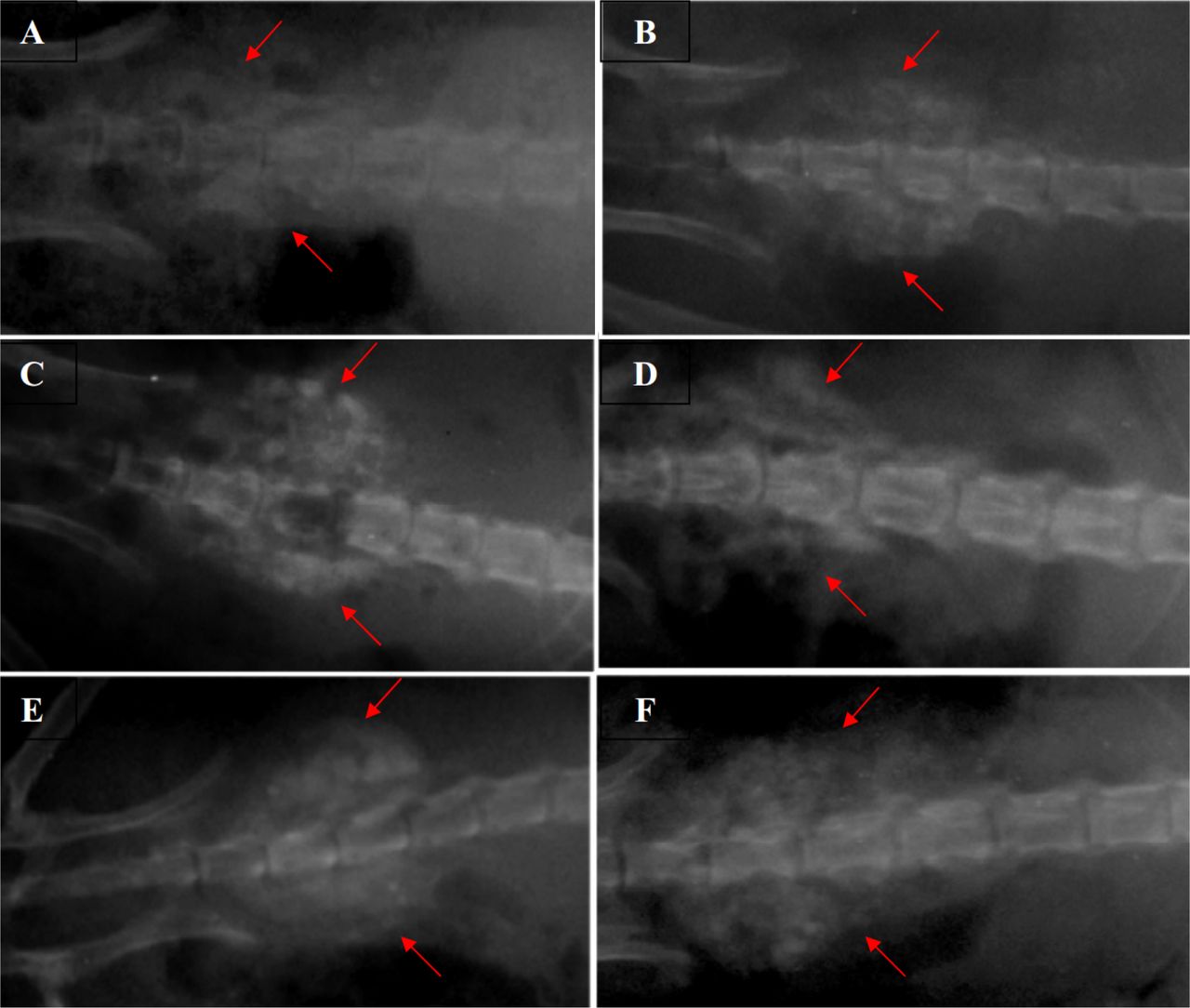
- Fig. 1
Radiographic evaluation on the 14th (A and B), 28th (C and D) and 42nd (E and F) postoperative days. Samples of DCFGP treated spine with fusion (A, C and E) and commercial treated sample (B, D, and F). The red arrows identify the radiopaque tissue masses on both sides of spine at the L4 and L5 segments.
- Fig. 2
Histology of 6-week samples of fusion by DCFGP (A and B) and commercial DBM (C and D). Immature and woven bone in DCFGP (A, 10X H & E Staining). Trabecular bone (white arrow) and marrow formation (black arrow) are seen in the fusion area of the same figure in a high magnification view (B, 40X H&E Staining). More mature bone (black right angle) with osteocyte cells are seen in the lesion of the commercial DBM group (C, 10X H & E Staining). Higher power view of the same picture shows bony tissue (white arrow) and marrow formation (black arrow) (D, 40X H&E Staining)


Tables
Postoperative days DCFGF (n = 8; Median(Minimum- Maximum)) Commercial DBM (n = 8; Median(Minimum- Maximum)) P (pairwise Mann-Whitney U Test) 14 3(2-4) 3(2-4) 0.6 28 4(2-4) 3(2-4) 0.4 42 4(3-4) 4(2-4) 0.1 Bone type evaluation DCFGP (n = 8; Median (Minimum-Maximum)) Commercial DBM (n = 8; Median (Minimum-Maximum)) P (Mann-Whitney U test) Macroscopic union* 2(2-3) 2(2-3) 0.6 Microscopic evaluation† 5 (5-7) 4 (4-6) 0. 1 ↵* Complete union (+3 score), presence of cartilage (+2 score), soft tissue or cracks within the defect indicating a possible unstable union (+ 1 score), complete instability at the defect site indicating nonunion (0 score)
↵† Empty (0 score), fibrous tissue only (1 score), more fibrous tissue than fibrocartilage (2 score), more fibrocartilage than fibrous tissue (3 score), fibrocartilage only (4 score), more fibrocartilage than bone (5 score), more bone than fibrocartilage (6 score) and bone only (7 score)
In this issue


Jump to section
Related Articles
Cited By...
- No citing articles found.