


Article Figures & Data
Figures
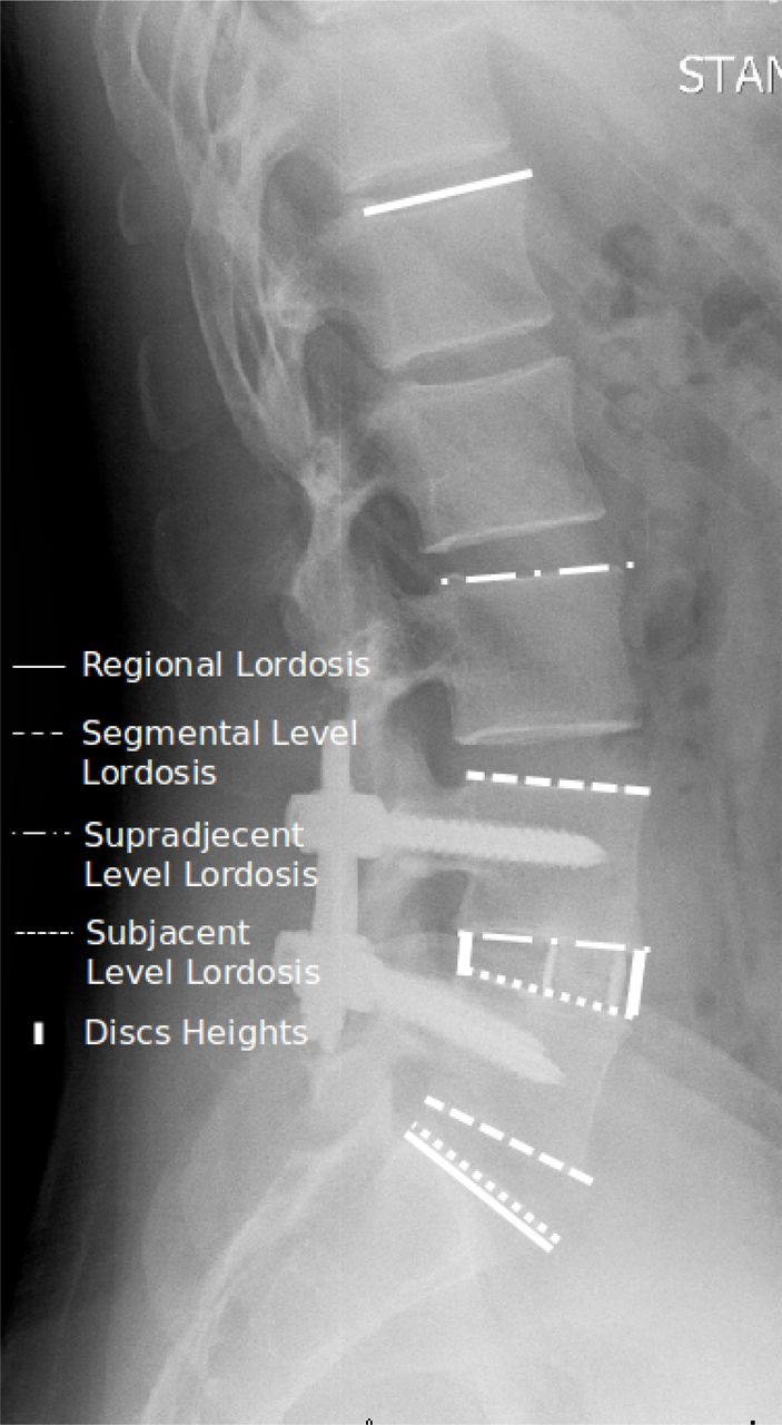
- Fig. 1
Lateral standing radiograph of the lumbosacral spine showing measurements of lordosis evaluated in the study. Regional lordosis was measured using the superior endplate of L1 and S1. Segmental lordosis measurements were measured using the superior endplate of upper vertebra and the inferior endplate of lower vertebra, except at L5-S1 where the superior endplate of S1 was used.
Tables
LLIF ALIF TLIF PSF DDD 17 DDD 17 DDD 16 DDD 5 ASD 9 ASD 4 ASD 3 ASD 2 Degen Spondylolisthesis 5 Degen Spondylolisthesis 2 Degen Spondylolisthesis 15 Degen Spondylolisthesis 12 Isthmic Spondylolisthesis 1 Isthmic Spondylolisthesis 10 Isthmic Spondylolisthesis 2 Spondylosis 2 Spondylosis 2 Spondylosis 4 Degen Scoliosis 1 Degen Scoliosis 2 Degen Scoliosis 3 Degen Scoliosis 1 Spondylolysis 1 HNP (recurrent/far lateral) 9 Pars fracture 2 Total 35 Total 36 Total 50 Total 26 Operative Level Regional (L1-S1) Pre-oplordosis (mean ± SD) Post-oplordosis (mean ± SD) p value Pre-oplordosis (mean ± SD) Post-oplordosis (mean ± SD) p value LLIF 12.1 ± 7.9° 15.3 ± 8.5° p<0.01 51.5 ± 11.3° 54.0 ± 10.0° p<0.01 ALIF 15.8 ± 11.9° 19.6 ± 11.7° p<0.01 51.6 ± 13.0° 55.8 ± 12.6° p<0.01 TLIF 13.0 ± 10.3° 14.9 ± 9.7° p<0.01 47.2 ± 14.2° 49.3 ± 14.4° p=0.02 PSF 14.8 ± 9.6° 15.5 ± 9.7° p=0.13 49.5 ± 15.0° 48.9 ± 16.8° p=0.66 - Table 3
Comparison of Preoperative and Postoperative Anterior Disk Height (ADH) and (Posterior Disk Height (PDH) within groups.
Pre-op ADH [mean (mm) ± SD] Post-op ADH [mean (mm) ± SD] p value Pre-op PDH [mean (mm) ± SD] Post-op PDH [mean (mm) ± SD] p value LLIF 7.9 ± 4.3 13.8 ± 3.8 p<0.01 3.7 ± 2.5 6.5 ± 3.1 p<0.01 ALIF 9.7 ± 5.6 18.4 ± 5.1 p<0.01 3.8 ± 2.0 7.1 ± 2.6 p<0.01 TLIF 9.8 ± 5.1 12.6 ± 3.8 p<0.01 4.5 ± 3.0 5.9 ± 2.9 p<0.01 PSF 9.9 ± 4.2 10.2 ± 3.8 p=0.25 4.3 ± 2.1 3.9 ± 2.0 p=0.04 Pre-op sup lordosis (mean ± SD) Post-op sup lordosis (mean ± SD) p value Pre-op inf lordosis (mean ± SD) Post-op inf lordosis (mean ± SD) p value LLIF 9.1 ± 8.1° 8.7 ± 7.9° p=0.27 22.2 ± 7.3° 22.2 ± 7.7° p=0.99 ALIF 17.3 ± 10.1° 14.8 ± 9.5° p<0.01 21.8 ± 8.5° 18.3 ± 8.2° p=0.25 TLIF 12.6 ± 10.2° 12.1 ± 10.4° p=0.07 15.1 ± 6.3° 15.1 ± 6.0° p=0.99 PSF 11.7 ± 8.8° 12 ± 9.1° p=0.54 18.7 ± 8.1° 18.0 ± 8.1° p=0.20 Pre-op lordosis (mean ± SD) Post-op lordosis (mean ± SD) Mean Change p value LLIF (n=18) 54.6 ± 9.2° 56.4 ± 8.0° 1.8 ± 3.7° p=0.05 ALIF (n=23) 54.8 ± 11.3° 57.2 ± 12.7° 2.4 ± 5.1° p=0.03 TLIF (n=38) 49.3 ± 13.2° 50.1 ± 13.6° 1.4 ± 4.5° p=0.06 PSF (n=18) 50.8 ± 16.1° 51.1 ± 17.6° 0.3 ± 6.9° p=0.86 Pre-op lordosis (mean ± SD) Post-op lordosis (mean ± SD) Mean Change p value LLIF (n=17) 48.0 ± 12.7° 51.2 ± 11.5° 3.3 ± 4.5° p=0.01 ALIF (n=13) 46.0 ± 14.3° 53.3 ± 12.6° 7.7 ± 5.9° p<0.01 TLIF (n=12) 40.6 ± 15.9° 44.9 ± 16.7° 4.3 ± 9.3° p=0.13 PSF (n=8) 46.5 ± 12.5° 44.1 ± 14.7° -2.4 ± 3.9° p=0.12 Studies Segmental Lordosis Regional Lordosis N (levels) Pre-op Post-op Change P value N (patients) Pre-op Post-op Change P value Sharma, et al. 201115 87 5.4° 8.5° 3.1° p=0.001 43 47.8±15.1 48.3±12.0 0.5 p=0.86 Acosta, et al. 201114 66 5.3° 8.2° 2.9° p<0.0001 36 42.1 46.2° 4.1 p >0.05 Watkins et al. 201418 86 8.2 ° 10.4° 2.2° p<0.001 - - - - - Tohmeh et al. 201420 223 10.7° 13.7° 3° p<0.001 - - - - - Present Study 54 12.1° 15.3° 3.2° p<0.0.1 18 51.5 54.0 2.5 P<0.01 - Table 8
Facetectomies used in TLIF Pre-op lordosis (mean ± SD) Post-op lordosis (mean ± SD) Mean Change p value Unilateral 16.1 ± 10.4° 17.1 ± 9.5° 1.0 ± 2.9° p=0.07 Bilateral 10.0 ± 9.5° 12.9 ± 9.6° 2.9 ± 4.5° p<0.01
In this issue


