


Article Figures & Data
Figures
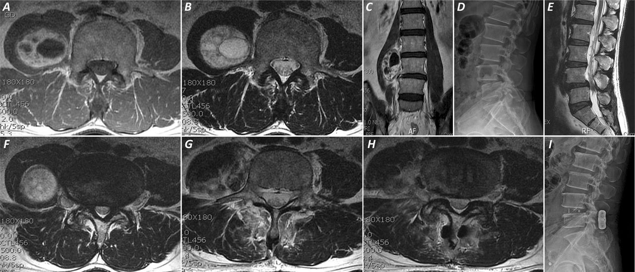
- Fig. 1
T1W (A), T2W (B) and Contrast enhanced (C) axial MRI sections showing an extradural tumour on left side of L4 body with large foraminal component. Axial CT scan section (D) at the level of tumour showing scalloping of L4 body due to the tumour with a widened foramina. T2W axial section (E) at the level of L4-5 disc showing diffuse disc bulge with lateral recess stenosis. Post-operative MRI (F) showing gross total tumour resection and right medial facetectomy and spinal decompression. Grade 1 signal changes in the paraspinal muscle due to the far lateral track on the left side (double arrowheads) and paramedian track on the right side (single arrowhead) can be noted (22).
- Fig. 2
T1W (A) and T2W (B) axial sections showing a large paraspinal tumour with a small extension into the left L1-2 foramina (single arrowheads) without any foraminal widening. The close relation to renal artery (double arrows) can be noted. Saggital T1W (C), T2W (D), contrast enhanced saggital (E) and coronal (F) images showing tumour extension across two segmental levels in the paraspinal area medial to the left kidney. Post-operative T2W (G) and contrast enhanced (H) axial sections showing gross total tumour resection. The paramedian entry point on the skin can be noted (G, double arrowheads) with grade 1 signal changes in the underlying paraspinal muscles (22).
- Fig. 3
Axial T1W (A), T2W (B) and contrast enhanced coronal (C) images showing tumour extension in case 3. The tumour imbedding in the psoas muscle and its relative anterior location as compared to the tumour in case 2 can be noted. Lateral neutral radiograph (D) and T2W saggital MRI showing grade 1 spondylolisthesis at L4-5. T2W axial MRI section (F) at the level of L4-5 disc showing lateral recess stenosis and the lateral location of the tumour. Post-operative MRI (G,H) showing gross total tumour resection with a faint outline of interbody cage. Post-operative lateral radiograph (I) showing the interbody cage and interspinous SPIRE in situ. The relative increase in posterior disc height and foraminal height (indirect decompression) in comparison to pre-op radiograph can be noted.
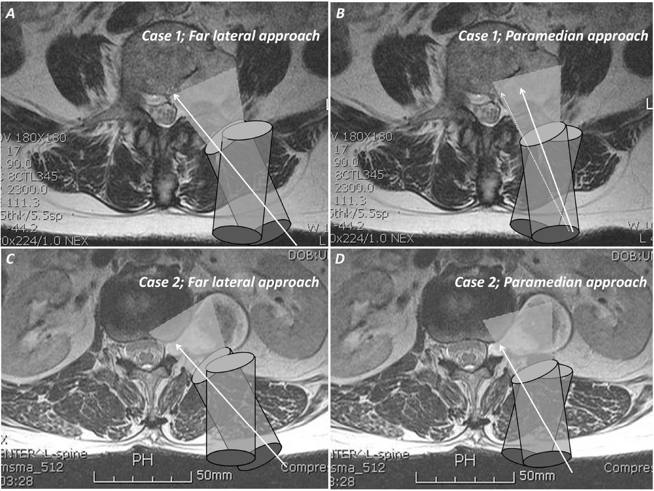
- Fig. 4
Diagrammatic illustration of the size of foraminal component influencing choice of approach and field of vision through a tubular retractor in case 1 (A & B) and case 2 (C & D). The white arrows indicate the line of sight. The large foraminal portion of case 1 could be easily visualised with minimal lateral facet resection from a far lateral angle (A), while a paramedian approach in this case would have led to complete facetectomy to visualise the foraminal component (B; Solid arrow indicates line of sight with safe facet resection. Dotted arrow shows degree of facet resection to visualise the medial portion of the tumour). In case 2 (C & D), foraminal portion could be adequately visualised with paramedian approach which also helped in visualising inferolateral portion of the tumour from a superomedial angle.


In this issue


Jump to section
Related Articles
Cited By...
- No citing articles found.