


Article Figures & Data
Figures
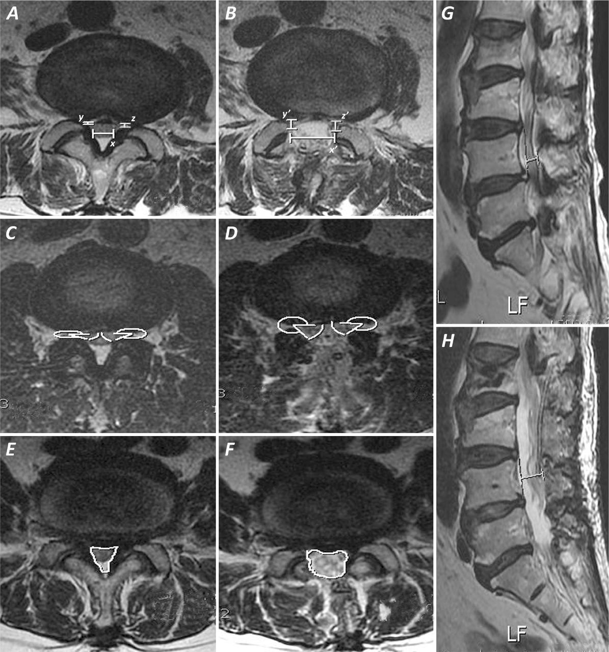
- Fig. 1
Images demonstrating the criteria used for measuring spinal canal dimensions in the present study. A & B - Interfacet distance (A-x – pre-op; B-x’ – post-op) and lateral recess depth (A-y,z – pre-op; B-y’,z’ – post-op). C & D – Lateral recess angle (C – pre-op; D – post-op). E & F – Cross-sectional spinal canal area (E – pre-op; F – post-op). G & H – Effective AP canal diameter (G – pre-op; H – post-op)
- Fig. 2
MR axial T2W images showing severe preoperativecentral and lateral recess stenosis (A) and postoperativeresult after decompression by SPSL technique (B). The preserved spinous process and minimal signal changes in teh paraspinal muscles can be made out. Follow-up CT scan of same case at 3 months showing fusion of the split spinous process (C).
- Fig. 3
Radiographs demonstrating safety of SPSL in patients with pre-existing degenerative coronal and saggital plane deformities. A & B – Preoperative(A) and 14 months follow-up (B) standing, lateral flexion radiographs in an illustrative case with two level degenerative listhesis showing no progression at either levels at follow-up. C & D – Preoperative (C) and 12 months follow-up (D) standing AP radiographs in an illustrative case with degenerative scoliosis showing no progression at follow-up.


Tables
- Table 1
Parameter Imaging Modality Sequence Level where measured Description Interfacet distance MRI Axial T2W Distance on a line connecting the Lower intermedial joint space of facet joints vertebral (post-op) (Fig. 1 B-x') or inner disc flaval surfaces along the same line (pre-op)18 (Fig. 1 A-x) Effective AP distance MRI Sag T2W Mid-body Distance between a vertical line connecting the annulus of adjacent discs and the upper spino-laminar junction (pre-op) (Fig. 1 G) or the dorsal dural margin (post-op) (Fig. 1 H) Lateral recess depth MRI Axial T2W Lower intervertebral disc Distance between the posterior surface of disc and anteromedial portion of superior articular process (post-op) (Fig. 1 B - y' & z') or its attached ligamentum flavum (pre-op) (Fig. 1 A - y & z)19 Lateral recess angle MRI Axial T2W Lower intervertebral disc Angle between the floor (posterior disc margin) and roof (anterome-dial edge of superior articular process [post-op] (Fig. 1 D) or its attached ligamentum flavum [pre-op] (Fig. 1 C)) of the lateral recess20* Cross-sectional area of the spinal canal MRI Axial T2W Lower intervertebral disc (Fig. 1 E & F) Cobb's angle AP standing radiograph Angle between the superior end-plates of the uppermost and lowermost split laminar levels ↵* modified to be used in MRI to include the effect of posterior annular bulge and hypertrophied ligamentum flavum on lateral recess angle. MRI – Magnetic resonance Imaging; AP – Antero-posterior; T1W – T1-weighted sequence; T2W – T2 weighted sequence.
VAS (Back pain) Pre-op 7.8 ± 1.8 (4 - 10) Post-op 3.7 ± 1.3 (2 - 7) JOA score Pre-op 6.3 ± 2.4 (-1 - 11) Post-op 11.2 ± 2.6 (1 - 14) Recovery rate (%) 57.3 ± 26.4 (-40% to 90%) McNab's grade of improvement Excellent 7 cases (18%) Good 23 cases (59%) Fair 7 cases (18%) Poor 2 cases (5%) Values are read as “Mean ± Standard deviation (Minimum value – Maximum value).” VAS – Visual analog scale; JOA – Japanese Orthopaedic Association.
- Table 3
Preoperativeand postoperativecomparative mean values for spinal canal dimensions at the split laminar levels.
n=118 Pre-op Post-op Difference Ratio increase (%) Interfacet distance (mm) 9.2 ± 3.4 (2.8 - 20.8) 17.6 ± 2.0 (11.2 -22.1) 8.4 ± 3.1 (0.7 - 15.8) 116.6 ± 83.4 (4.5 -564.2) AP canal diameter (mm) 9.3 ± 1.6 (5.8 - 13.8) 15.2 ± 2.0 (11.1 -21.0) 5.8 ± 2.3 (1.0 - 12.3) 67.6 ± 34.4 (8.7 -161.8) Lateral recess depth (mm) Right 1.6 ± 0.6 (1.0 -4.1) 3.9 ± 1.0 (1.3 - 6.3) 2.3 ± 1.0 (0 -5.0) 165.1 ± 101.3 (0 - 500.0) Left 1.7 ± 0.6 (1.0 - 4.9) 3.9 ± 0.9 (1.7 - 6.0) 2.1 ± 0.9 (-0.7 -4.6) 149.3 ± 92.1 (-20.0 -460.0) Lateral recess angle (in °) Right 17.6 ± 5.6 (6.7 -38.4) 39.1 ± 9.5 (12.4 - 56.5) 21.5 ± 10.6 (0.7 -45.4) 145.5 ± 103.3 (3.4 -422.0) Left 17.9 ± 5.8 (7.2 -32.0) 37.8 ± 10.4 (16.6 - 58.2) 19.9 ± 11.1 (0.9 -44.9) 133.6 ± 106.5 (3.5 ± 449.0) Cross-sectional area (mm 2 ) 80.9 ± 25.3 (30.6 - 169.0) 194.7 ± 30.2 (114.5 -264.2) 113.8 ± 33.8 (18.1 -189.1) 163.8 ± 90.2 (15.4 -412.8) Values are read as 'Mean ± Standard deviation (Minimum value – Maximum value).'
- Table 4
Preoperativeand postoperativecomparison of various spinal canal dimensions among different levels.
L2-3 (n=19) L3-4 (n=36) L4-5 (n=36) L5-S1 (n=25) Interfacet distance (mm) Pre-op 8.0 7.5 9.0 12.9 Post-op 16.0 16.9 18.4 18.9 Difference 8.0 9.3 9.4 6.0 Ratio increase (%) 115.8 137.0 133.6 61.6 AP canal diameter (mm) Pre-op 9.8 9.1 8.9 9.8 Post-op 14.8 15.0 15.4 15.5 Difference 5.0 5.8 6.5 5.7 Ratio increase (%) 55.3 67.7 78.0 63.6 Right lateral recess depth (mm) Pre-op 1.6 1.6 1.5 1.7 Post-op 3.9 4.0 3.9 3.9 Difference 2.3 2.3 2.3 2.1 Ratio increase (%) 172.2 165.0 172.8 133.0 Left lateral recess depth (mm) Pre-op 1.7 1.6 1.6 1.9 Post-op 4.2 3.8 3.9 3.5 Difference 2.4 2.2 2.2 1.5 Ratio increase (%) 160.9 157.8 172.5 94.4 Right lateral recess angle (°) Pre-op 18.7 16.4 16.8 19.7 Post-op 39.9 41.9 39.0 34.4 Difference 21.1 25.5 22.1 14.7 Ratio increase (%) 123.3 183.2 163.8 80.5 Left lateral recess angle (°) Pre-op 18.9 17.4 15.3 20.8 Post-op 37.5 39.7 36.6 36.5 Difference 18.5 22.2 21.3 15.6 Ratio increase (%) 112.6 152.8 159.7 87.8 Cross-sectional canal area (mm2) Pre-op 77.4 72.13 78.1 101.1 Post-op 193.2 196.3 189.5 200.7 Difference 115.8 124.2 111.4 99.6 Ratio increase (%) 165.6 190.6 168.5 115.5 Values represented are mean values.
Postoperative grade of foraminal stenosis Grade 0 Grade 1 Grade 2 Grade 3 Preoperative grade of Foraminal stenosis Grade 1 (n=15) 6 9 (60%) - - Grade 2 (n=21) 2 8 11 (52.4%) - Grade 3 (n=8) 0 0 3 5 (62.5%)
In this issue


