


Article Figures & Data
Figures
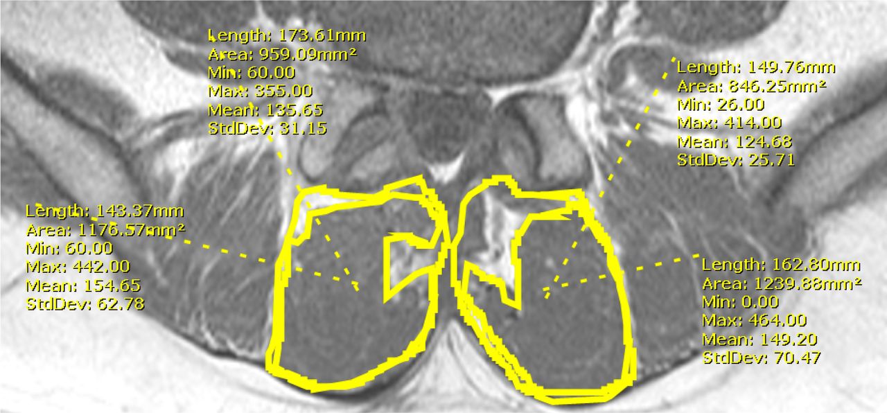
- Fig. 1
41 year-old male presents with a herniated nucleus pulposus at L4-5. Preoperative axial T1-weighted MRI image demonstrates lean cross-sectional area measurements at the index level (L4-5).
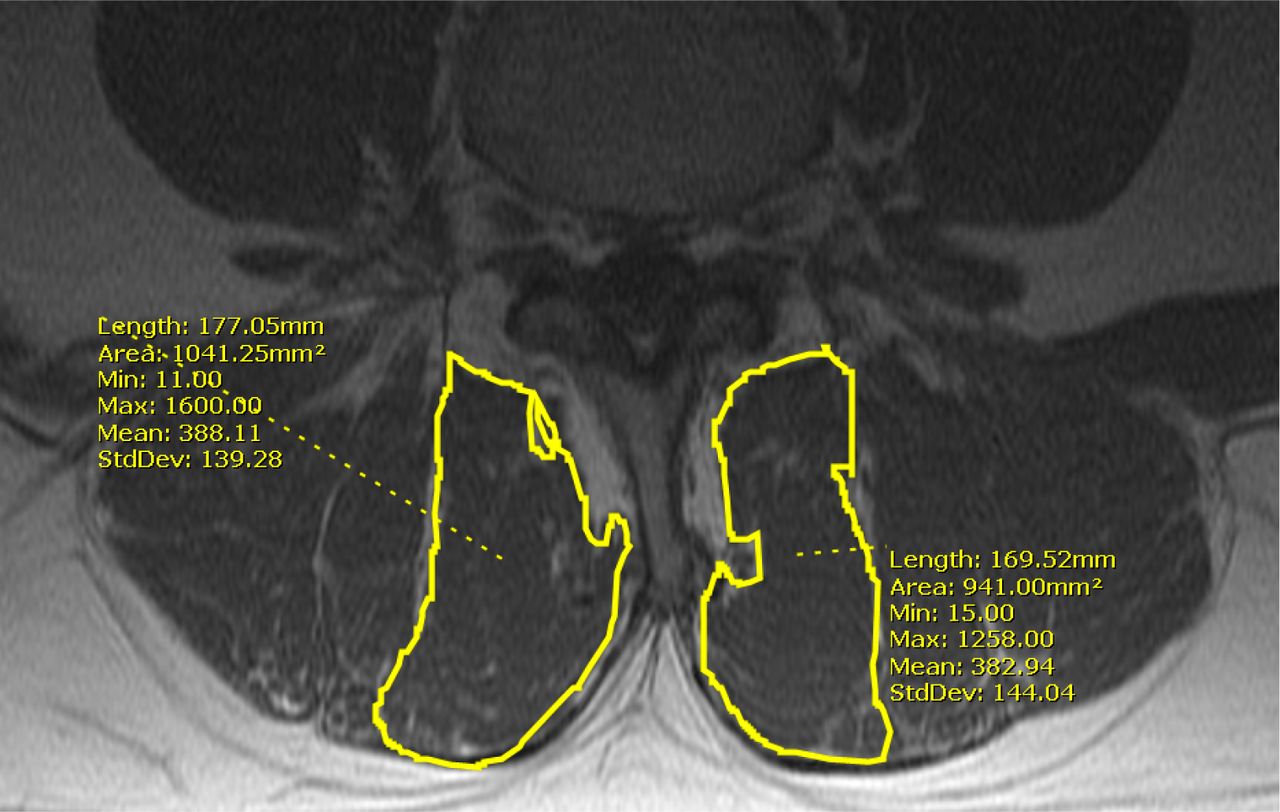
- Fig. 2
Postoperative axial T1-weighted MRI image demonstrates total and lean cross-sectional measurements at the index level (L4-5).

Tables
- Table 1
Patient Characteristics, Operative Levels, and Timing for Primary 1-level MIS Lumbar Decompression.
MIS (n = 24) Age (Years, SD) 47.8 (14.2) Female (n) 58.3% (14) Operative Levels (%, n) L2L3 4.2% (1) L3L4 12.5% (3) L4L5 58.3% (14) L5S1 25.0% (6) Time between Surgery and MRI Preoperative MRI (Days, SD) 61.1 (55.4) Postoperative MRI (Days, SD) 182.5 (194.4) MIS: Minimally Invasive; SD: Standard Deviation; MRI: Magnetic Resonance Imaging.
- Table 2
MRI-Documented Changes in Multifidus Muscle Following Primary 1-Level MIS Lumbar Decompression.
Ipsilateral Multifidus Total Cross Sectional Area (mm2) Multifidus Lean Cross Sectional Area (mm2) Level Preoperative Postoperative % Change pvalue Preoperative Postoperative % Change pvalue Supra Disc 794.0±233.0 811.6±259.0 +2.0% 0.38 582.0±203.0 569.3±232.7 − 3.1% 0.52 Supra Pedicle 953.4±290.0 957.4±341.5 − 1.3% 0.91 688.1±283.2 660.5±270.0 − 2.9% 0.37 Index 1047.4±246.2 1000.3±238.7 − 4.9% <0.01 726.2±234.4 680.4±221.6 − 6.2% <0.05 Infra Pedicle 1148.3±288.9 1088.6±278.0 − 5.3% 0.08 738.0±234.1 641.7±213.2 − 13.0% <0.01 Infra Disc † 1135.5±245.8 1117.3±269.1 − 1.6% 0.66 743.0±230.8 600.3±209.5 − 18.6% <0.01 Contralateral Multifidus Total Cross Sectional Area (mm2) Multifidus Lean Cross Sectional Area (mm2) Level Preoperative Postoperative % Change pvalue Preoperative Postoperative % Change pvalue Supra Disc 787.9±224.5 778.3±212.7 − 0.5% 0.65 582.2±207.3 574.3±205.7 − 1.4% 0.57 Supra Pedicle 961.3±315.0 936.5±323.5 − 0.8% 0.52 714.4±283.2 673.8±267.0 − 4.4% 0.17 Index 1029.9±239.6 1021.5±213.9 + 0.4% 0.71 707.1±206.1 671.6±193.3 − 4.2% 0.052 Infra Pedicle 1121.7±264.4 1138.7±282.3 + 1.7% 0.52 721.7±216.1 706.2±204.5 − 0.1% 0.55 Infra Disc † 1117.4±215.4 1131.9±243.3 + 1.1% 0.59 734.6±206.3 698.5±212.3 − 4.4% 0.15 Ipsilateral Multifidus Psoas T1 Signal Intensity Ratio Lean CSA to Total CSA Ratio Level Preoperative Postoperative % Change pvalue Preoperative Postoperative % Change pvalue Supra Disc 1.81±0.63 1.98±0.72 + 26.0% 0.43 0.73±0.10 0.69±0.11 − 5.2% <0.05 Supra Pedicle 1.91±0.69 2.06±0.80 + 21.7% 0.53 0.71±0.14 0.69±0.10 − 1.5% 0.33 Index 2.23±0.91 2.27±0.89 + 21.8% 0.90 0.69±0.11 0.67±0.12 − 1.2% 0.47 Infra Pedicle 2.35±0.93 2.68±1.20 + 35.0% 0.32 0.65±0.12 0.59±0.11 − 8.4% <0.01 Infra Disc † 2.76±1.24 3.42±1.82 + 51.7% 0.25 0.66±0.14 0.55±0.16 − 17.2% <0.001 Contralateral Multifidus Psoas T1 Signal Intensity Ratio Lean CSA to Total CSA Ratio Level Preoperative Postoperative % Change pvalue Preoperative Postoperative % Change pvalue Supra Disc 1.83±0.65 1.98±0.69 + 22.6% 0.49 0.73±0.12 0.73±0.11 − 0.3% 0.59 Supra Pedicle 1.90±0.69 1.94±0.64 + 15.2% 0.81 0.75±0.19 0.72±0.10 0% 0.41 Index 2.20±0.93 2.41±0.80 + 33.9% 0.47 0.69±0.11 0.65±0.10 − 3.8% 0.09 Infra Pedicle 2.48±1.04 2.65±1.03 + 27.9% 0.61 0.64±0.11 0.63±0.12 − 1.9% 0.37 Infra Disc † 2.80±1.31 3.24±1.75 + 45.7% 0.44 0.66±0.13 0.62±0.14 − 5.3% <0.05 ↵† Patients whose operative levels included S1 were excluded from measurement at the inferior disc level. MIS: Minimally Invasive; MRI: Magnetic Resonance Imaging; Supra: Superior; Infra: Inferior; CSA: Cross Sectional Area.
In this issue


Jump to section
Related Articles
Cited By...
- Qualitative Evaluation of Paraspinal Musculature After Minimally Invasive Lumbar Decompression: A Prospective Study
- Clinical utility and reproducibility of surface electromyography in individuals with chronic low back pain: a protocol for a systematic review and meta-analysis
- Magnetic Resonance Imaging Documentation of Approach Trauma With Lumbar Endoscopic Interlaminar, Translaminar, Compared to Open Microsurgical Discectomy
- Minimal Clinically Important Difference in Patient-Reported Outcome Measures with the Transforaminal Endoscopic Decompression for Lateral Recess and Foraminal Stenosis