


Article Figures & Data
Figures
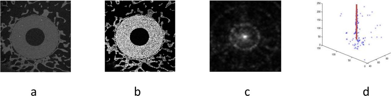
- Fig. 1
Detection of the cylindrical dowel with original image (a), dowel probability map (b), Hough transformed image (c), and line fitting through the maxima of the Hough images (d).
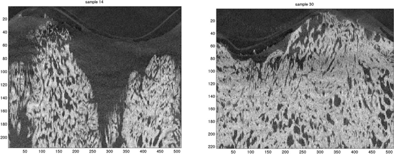
- Fig. 2
Images sampled at 30 voxels from the implant surface and stratified for visualization.
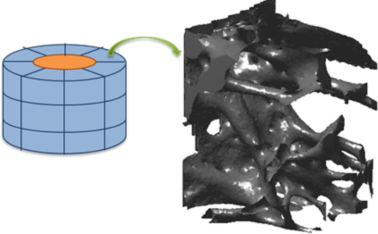
- Fig. 3
Volumes of interest (blue) around the dowel (orange). In this figure, 24 VOIs are drawn (3 times 8). In our analysis we use 60 VOIs (5 times 12).
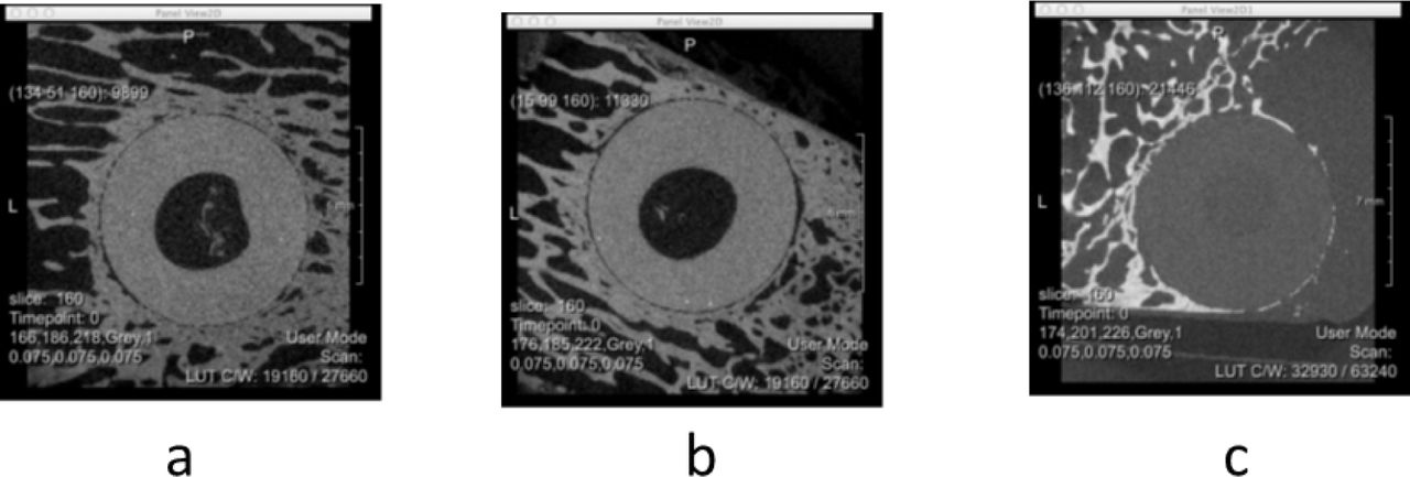
- Fig. 4
Representative slices for titanium, (A), CaP, (B) and control (C).
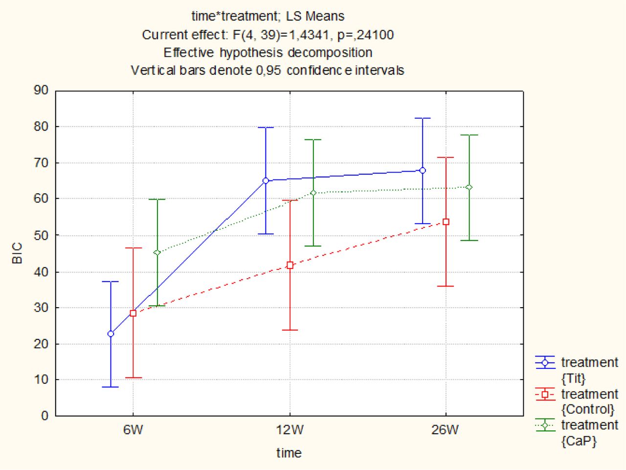
- Fig. 5
Time * Treatment statistics for BIC in the 6 to 26 weeks period. No significant differences were observed (p = 0.24100).
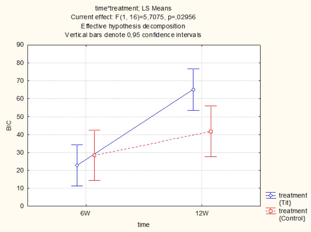
- Fig. 6
Time * Treatment statistics for BIC in the 6 to 12 weeks period. BIC was significantly higher in the titanim group compared to the control.
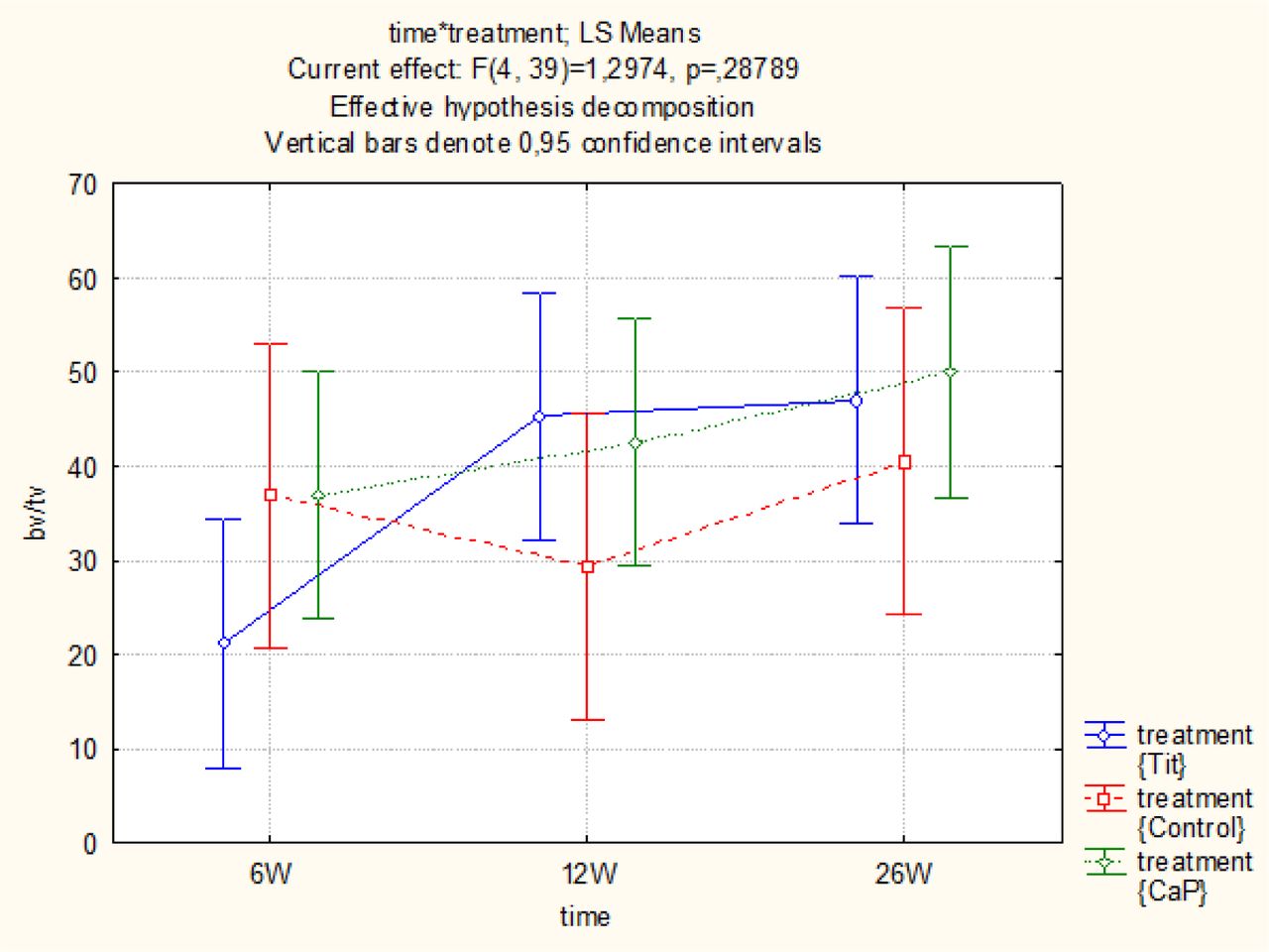
- Fig. 7
Time * Treatment statistics for BV/TV in the 6 to 26 weeks period. No significant differences were observed (p = 0.28789).
- Fig. 8
Time * Treatment statistics for BIC in the 6 to 12 weeks period. BIC was significantly higher in the titanium group compared to the control.
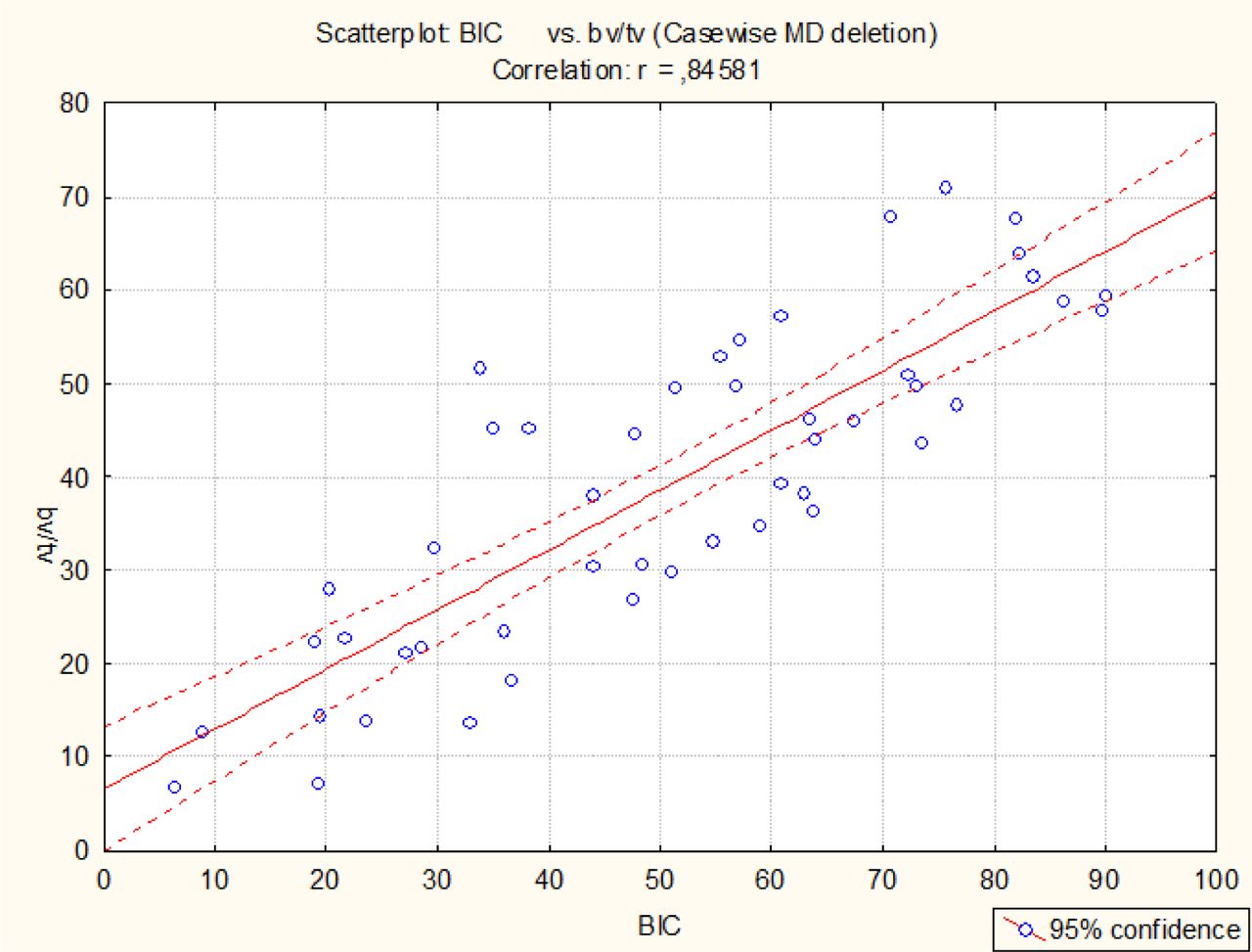
- Fig. 9
Correlation between BIC and BV/TV.
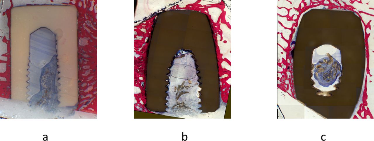
- Fig. 10
Histological sections stained with Stevenel's blue and Von Gieson's picrofuchsin of (a) titanium with direct bone implant contact, (b) CaP with direct bone implant contact (c) Control presence of cartilage tissue and fibrotic layers between the dowel and the surrounding bone tissue.


Tables
Study Design (Animals n = 6) Follow-up 6 weeks 12 weeks 26 weeks Animals n = 2 n = 2 n = 2 Implants n = 16 n = 16 n = 16 Location Tibia n = 8 Femur n = 8 Tibia n = 8 Femur n = 8 Tibia n = 8 Femur n = 8 Coating Control n =4
Titanium n = 6
CaP n = 6Control n =4
Titanium n = 6
CaP n = 6Control n =4
Titanium n = 6
CaP n = 6BIC 6 weeks 12 weeks 26 weeks Titanium N 6 6 6 Mean 22.783 65.040 67,.902 Std.Err. 7.228 7.228 7.228 CaP N 6 6 6 Mean 45.170 61.663 63.184 Std.Err. 7.228 7.228 7.228 Control N 4 4 4 Mean 28.437 41.670 53.793 Std.Err. 8.852 8.852 8.852 BV/TV 6 weeks 12 weeks 26 weeks Titanium N 6 6 6 Mean 21.217 45.236 47.036 Std.Err. 6.538 6.538 6.538 CaP N 6 6 6 Mean 37.027 42.533 49.995 Std.Err. 6.538 6.538 6.538 Control N 4 4 4 Mean 36.889 29.315 40.498 Std.Err. 8.007 8.007 8.007
In this issue


