
Main menu

  • Home
  • Content
    • Current Issue
    • Advance Online Publication
    • Archive
  • About Us
    • About ISASS
    • About the Journal
    • Author Instructions
    • Editorial Board
    • Reviewer Guidelines & Publication Criteria
  • More
    • Advertise
    • Subscribe
    • Alerts
    • Feedback
  • Join Us
  • Reprints & Permissions
  • Sponsored Content
  • Other Publications
    • ijss

User menu

  • My alerts

Search

  • Advanced search
International Journal of Spine Surgery
  • My alerts
International Journal of Spine Surgery

Advanced Search

  • Home
  • Content
    • Current Issue
    • Advance Online Publication
    • Archive
  • About Us
    • About ISASS
    • About the Journal
    • Author Instructions
    • Editorial Board
    • Reviewer Guidelines & Publication Criteria
  • More
    • Advertise
    • Subscribe
    • Alerts
    • Feedback
  • Join Us
  • Reprints & Permissions
  • Sponsored Content
  • Follow ijss on Twitter
  • Visit ijss on Facebook
Research ArticleOther & Special Categories

Hemivertebra Resection and Spinal Arthrodesis by Single-Stage Posterior Approach in Congenital Scoliosis and Kyphoscoliosis: Results at 9.6 Years Mean Follow-up

Marco Crostelli, Osvaldo Mazza, Massimo Mariani, Dario Mascello, Federico Tundo and Carlo Iorio
International Journal of Spine Surgery February 2022, 8188; DOI: https://doi.org/10.14444/8188
Marco Crostelli
Spine Surgery Unit - Bambino Gesù Children’s Hospital, Rome, Italy
  • Find this author on Google Scholar
  • Find this author on PubMed
  • Search for this author on this site
Osvaldo Mazza
Spine Surgery Unit - Bambino Gesù Children’s Hospital, Rome, Italy
  • Find this author on Google Scholar
  • Find this author on PubMed
  • Search for this author on this site
Massimo Mariani
Spine Surgery Unit - Bambino Gesù Children’s Hospital, Rome, Italy
  • Find this author on Google Scholar
  • Find this author on PubMed
  • Search for this author on this site
Dario Mascello
Spine Surgery Unit - Bambino Gesù Children’s Hospital, Rome, Italy
  • Find this author on Google Scholar
  • Find this author on PubMed
  • Search for this author on this site
Federico Tundo
Spine Surgery Unit - Bambino Gesù Children’s Hospital, Rome, Italy
  • Find this author on Google Scholar
  • Find this author on PubMed
  • Search for this author on this site
Carlo Iorio
Spine Surgery Unit - Bambino Gesù Children’s Hospital, Rome, Italy
  • Find this author on Google Scholar
  • Find this author on PubMed
  • Search for this author on this site
  • Article
  • Figures & Data
  • Info & Metrics
  • PDF
Loading

Article Figures & Data

Figures

  • Figure 1
    • Download figure
    • Open in new tab
    • Download powerpoint
    Figure 1

    Preoperative x-ray images (a, b), magnetic resonance images (c), computed tomographic (CT) images (d, e), three-dimensional CT images (f, g), and clinical presentation (h–k) of the patient. Postoperative x-ray images (l, m).
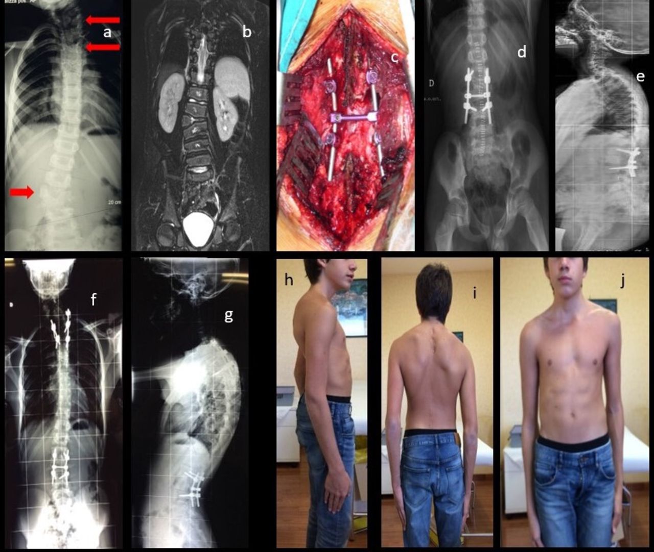
  • Figure 2
    • Download figure
    • Open in new tab
    • Download powerpoint
    Figure 2

    Preoperative x-ray image (a) showing 3 hemivertebras (arrows); preoperative magnetic resonance image (b); intraoperative picture showing lumbar instrumentation (c); postoperative x-ray images (d, e); postoperative x-ray images after the second procedure (f, g); and clinical pictures at latest follow-up 8.8 years after lumbar procedure and 4.7 years after thoracic hemivertebra resection (h-j).
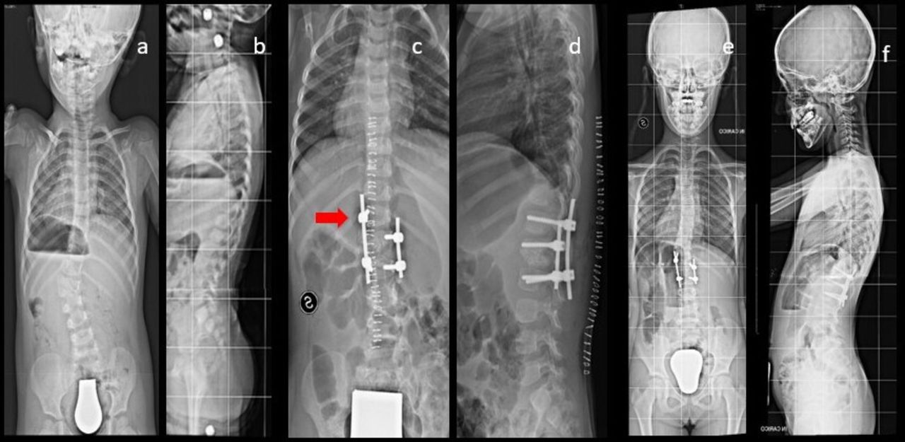
  • Figure 3
    • Download figure
    • Open in new tab
    • Download powerpoint
    Figure 3

    Preoperative x-ray images of a 25-month-old patient (a, b); postoperative x-ray images showing T12 left pedicle screws (arrow) after rupture of L1 left pedicle (c, d); and x-ray images at 7.3 years follow-up (e, f) showing no loss of correction.

  • Figure 4
    • Download figure
    • Open in new tab
    • Download powerpoint
    Figure 4

    Clinical presentation (a–c) and preoperative x-ray images (d, e); postoperative x-ray images with cast (f, g); and 6 months follow-up x-ray images showing proximal junctional kyphosis (h, i).
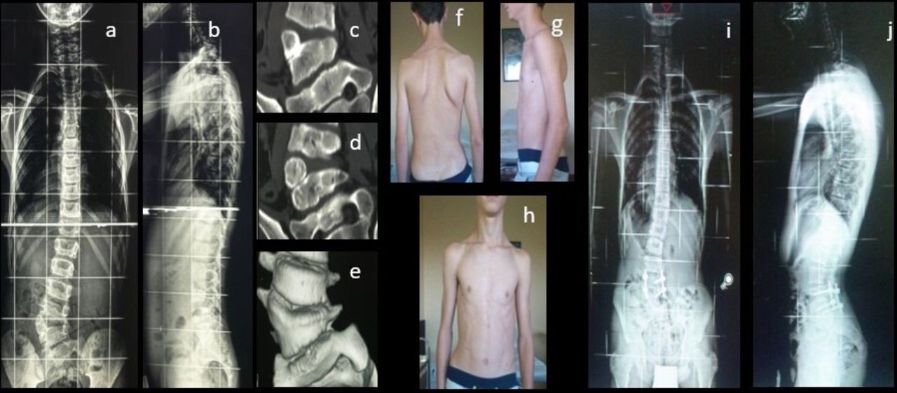
  • Figure 5
    • Download figure
    • Open in new tab
    • Download powerpoint
    Figure 5

    Preoperative x-ray images (a, b), computed tomographic (CT) images (c, d), and three-dimensional CT image (e); and clinical (f–h) and radiologic (i, j) findings at 8 months follow-up, showing small upper scoliotic curve development and slight cosmetic impairment.

Next
Back to top

In this issue

International Journal of Spine Surgery: 19 (S2)
International Journal of Spine Surgery
Vol. 19, Issue S2
1 Apr 2025
  • Table of Contents
  • Index by author

Print
Download PDF
Article Alerts
Sign In to Email Alerts with your Email Address
Email Article

Thank you for your interest in spreading the word on International Journal of Spine Surgery.

NOTE: We only request your email address so that the person you are recommending the page to knows that you wanted them to see it, and that it is not junk mail. We do not capture any email address.

Enter multiple addresses on separate lines or separate them with commas.
Hemivertebra Resection and Spinal Arthrodesis by Single-Stage Posterior Approach in Congenital Scoliosis and Kyphoscoliosis: Results at 9.6 Years Mean Follow-up
(Your Name) has sent you a message from International Journal of Spine Surgery
(Your Name) thought you would like to see the International Journal of Spine Surgery web site.
CAPTCHA
This question is for testing whether or not you are a human visitor and to prevent automated spam submissions.
Citation Tools
Hemivertebra Resection and Spinal Arthrodesis by Single-Stage Posterior Approach in Congenital Scoliosis and Kyphoscoliosis: Results at 9.6 Years Mean Follow-up
Marco Crostelli, Osvaldo Mazza, Massimo Mariani, Dario Mascello, Federico Tundo, Carlo Iorio
International Journal of Spine Surgery Feb 2022, 8188; DOI: 10.14444/8188

Citation Manager Formats

  • BibTeX
  • Bookends
  • EasyBib
  • EndNote (tagged)
  • EndNote 8 (xml)
  • Medlars
  • Mendeley
  • Papers
  • RefWorks Tagged
  • Ref Manager
  • RIS
  • Zotero
Share
Hemivertebra Resection and Spinal Arthrodesis by Single-Stage Posterior Approach in Congenital Scoliosis and Kyphoscoliosis: Results at 9.6 Years Mean Follow-up
Marco Crostelli, Osvaldo Mazza, Massimo Mariani, Dario Mascello, Federico Tundo, Carlo Iorio
International Journal of Spine Surgery Feb 2022, 8188; DOI: 10.14444/8188
Twitter logo Facebook logo Mendeley logo
  • Tweet Widget
  • Facebook Like
  • Google Plus One

Jump to section

  • Article
    • Abstract
    • INTRODUCTION
    • MATERIALS AND METHODS
    • RESULTS
    • DISCUSSION
    • CONCLUSIONS
    • Footnotes
    • References
  • Figures & Data
  • Info & Metrics
  • PDF

Related Articles

  • No related articles found.
  • PubMed
  • Google Scholar

Cited By...

  • No citing articles found.
  • Google Scholar

More in this TOC Section

  • Prevalence and Significance of Vitamin D Deficiency in Patients Undergoing Corrective Surgery for Adolescent Idiopathic Scoliosis
  • Adult Congenital Spine Deformity: Clinical Features and Motivations for Surgical Treatment
Show more Other & Special Categories

Similar Articles

Keywords

  • congenital scoliosis
  • hemivertebra resection
  • posterior vertebral arthrodesis

Content

  • Current Issue
  • Latest Content
  • Archive

More Information

  • About IJSS
  • About ISASS
  • Privacy Policy

More

  • Subscribe
  • Alerts
  • Feedback

Other Services

  • Author Instructions
  • Join ISASS
  • Reprints & Permissions

© 2025 International Journal of Spine Surgery

International Journal of Spine Surgery Online ISSN: 2211-4599

Powered by HighWire