


Article Figures & Data
Figures
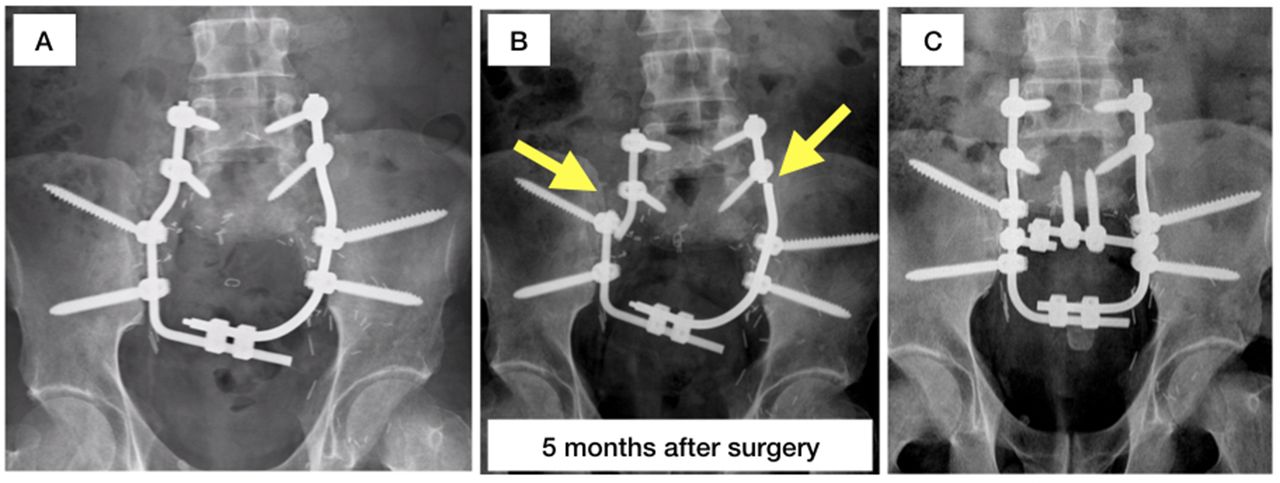
- Figure 1
Clinical case. During the initial surgery, reconstruction was performed according to model 2 (A). After 5 mo, the rods broke at the fusion site of the L5 pedicle screws and the proximal iliacs (B). A second reconstruction was subsequently performed on the basis of model 4 (C).
- Figure 2
Sacral prosthesis substitution.
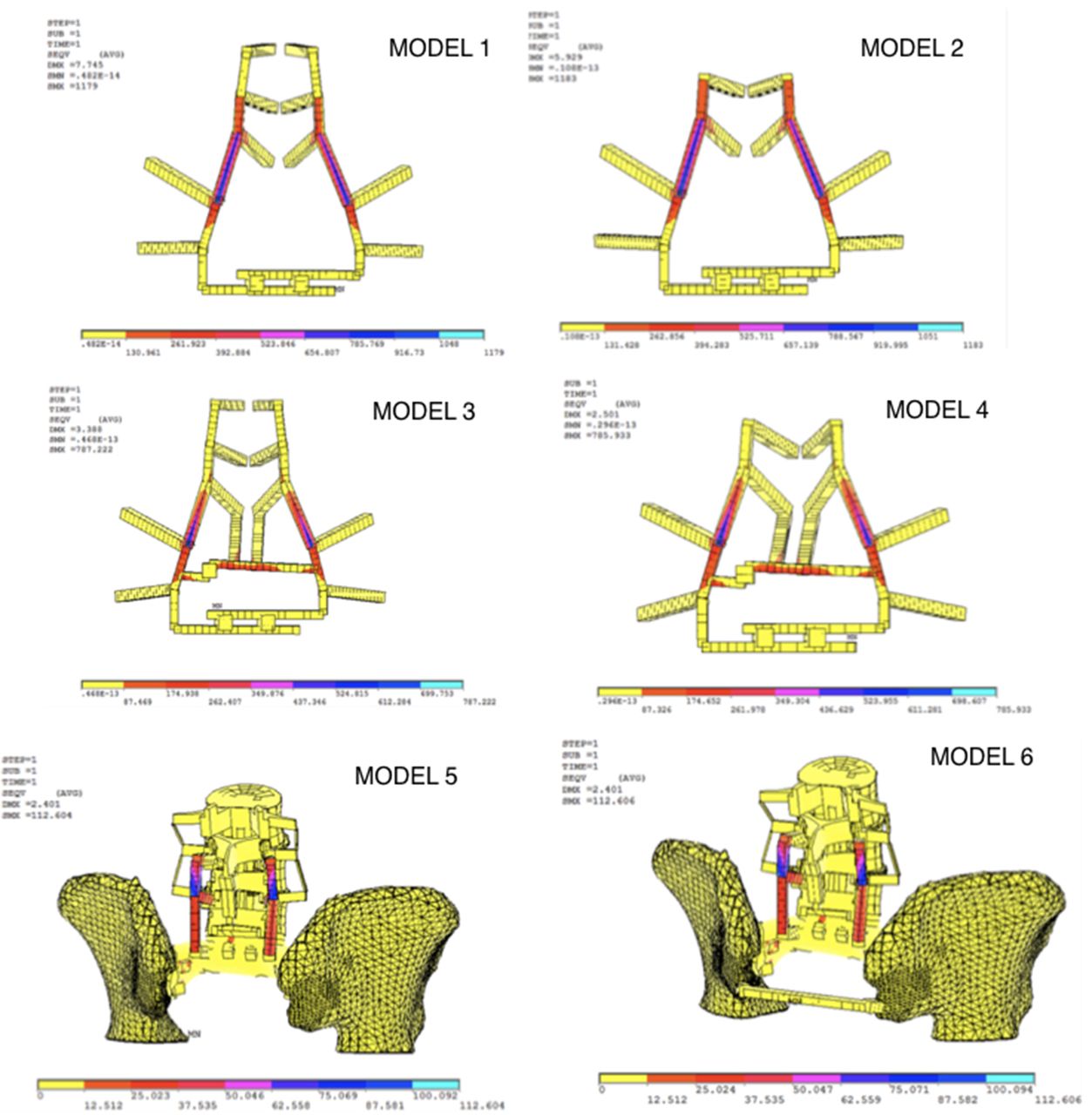
- Figure 3
Finite element models.
- Figure 4
Stress distributions resulting from application of loads in the finite element models.


Tables
Elements Diameter, mm Material Rods 5.5 Cobalt-chrome (Co28Cr6Mo) Pedicle screws 7 Titanium (Ti6Al4V) Iliac screws 7.5 Titanium (Ti6Al4V) Connectors - Titanium (Ti6Al4V) - Table 2
Properties of the materials cobalt-chrome, titanium, and porous titanium, as determined by the Biomechanics Institute of Valencia.
Material Young’s Modulus, MPa Elastic Limit, MPa Breaking Stress, MPa Poisson Coefficient Cobalt-chrome (Co28Cr6Mo) 220 925 1200 0.45 Titanium (Ti6Al4V) 115 1000 1050 0.3 Porous titanium 3 180 230 0.33 Posterior ligamentous complex 20 20 0.3 Note: Properties of complex o.
Vertical Displacement, mm Models 1 2 3 4 5 6 L3 −5.45 −5 −2.3 −1.73 −0.03 −0.03 L4 −5.45 −5.6 −2.15 −2.14 −0.5 −0.5 L5 −5.08 −5.15 −1.69 −1.7 −0.13 −0.13 Models 1 2 3 4 5 6 Maximum von Mises stress, MPa 1179 1182 787 786 112 112 Models 1 2 3 4 5 6 Rigidity, Nm/mm 232.1 292.7 465.7 462.4 861.5 861.5 Angle Preoperative(Measured Using CT) First Reconstruction(Measured Using CT) Second Reconstruction(Measured Using CT) Reconstruction With Sacral Replacement Piece (Theoretical Value) Modified pelvic incidence angle 44.0° 24.8° 25.1° 44.0° CT, computed tomography.
- Table 7
Literature review of finite element model studies of spinopelvic reconstructions following total sacrectomies and a comparison with the results of the present study.
Vertical Displacement of L5, mm Maximum von Mises Stress Values,MPa Rigidity,Nm/mm Area of Maximum Tension Modified Galveston reconstruction 1042 (Kawahara)71042 (Murakami) 8 Murakami 8: Area of the spinal rod spanning the L5 and iliac screws Triangular frame reconstruction 222 (Kawahara)7229 (Murakami stainless steel)8222 (Murakami titanium alloy)8 Kawahara7 and Murakami 8: Point at which the sacral rod inserts into L5, between L5 and the iliac bone Sacral rod reconstruction 2.5 (Zhu)9 309 (Zhu)9400 (Kawahara)7 123.6 (Zhu)9 Zhu9: In the longitudinal or sacral rod proximal to the connection between screw and rod Four-rod reconstruction 7.2 (Zhu)9 324 (Zhu)9 45 (Zhu)9 Zhu9: Middle part of the rods between the short and long iliac screws Bilateral fibular flap reconstruction 1.3 (Zhu)9 221 (Zhu)9 179 (Zhu)9 Zhu9: In the longitudinal or sacral rod near the connection between the screws and the rods Improved compound reconstruction 0.70 (Zhu)9 108 (Zhu)9222 (Kawahara)7 154.3 (Zhu)9 Zhu9: In the longitudinal or sacral bar near the connection between the screws and rods Model 1 5.08 1179 232.1 In the rods that join L5 with the screws anchored in the pelvis Model 2 5.15 1182 292.7 In the rods that join L5 with the screws anchored in the pelvis Model 3 1.69 787 465.7 In the rods joining L5 with the screws anchored in the pelvis; tension values are also high in the cross-connecting elements Model 4 1.7 786 462.7 In the rods joining L5 with the screws anchored in the pelvis; tension values are also high in the cross-connecting elements Model 5 0.13 112 861.5 In the rods joining L5 with the screws anchored to the sacral prosthesis Model 6 0.13 112 861.5 In the rods that joining L5 with the screws anchored to the sacral prosthesis; values of up to 112 MPa
In this issue


