
Main menu

  • Home
  • Content
    • Current Issue
    • Advance Online Publication
    • Archive
  • About Us
    • About ISASS
    • About the Journal
    • Author Instructions
    • Editorial Board
    • Reviewer Guidelines & Publication Criteria
  • More
    • Advertise
    • Subscribe
    • Alerts
    • Feedback
  • Join Us
  • Reprints & Permissions
  • Sponsored Content
  • Other Publications
    • ijss

User menu

  • My alerts

Search

  • Advanced search
International Journal of Spine Surgery
  • My alerts
International Journal of Spine Surgery

Advanced Search

  • Home
  • Content
    • Current Issue
    • Advance Online Publication
    • Archive
  • About Us
    • About ISASS
    • About the Journal
    • Author Instructions
    • Editorial Board
    • Reviewer Guidelines & Publication Criteria
  • More
    • Advertise
    • Subscribe
    • Alerts
    • Feedback
  • Join Us
  • Reprints & Permissions
  • Sponsored Content
  • Follow ijss on Twitter
  • Visit ijss on Facebook
Research ArticleNew Technology

Robotic-Assisted Revision Spine Surgery

Alexander M. Satin, Stanley Kisinde and Isador H. Lieberman
International Journal of Spine Surgery June 2022, 8272; DOI: https://doi.org/10.14444/8272
Alexander M. Satin
1 Texas Back Institute, Plano, Texas, USA
MD
  • Find this author on Google Scholar
  • Find this author on PubMed
  • Search for this author on this site
Stanley Kisinde
1 Texas Back Institute, Plano, Texas, USA
MBChB, MMed
  • Find this author on Google Scholar
  • Find this author on PubMed
  • Search for this author on this site
Isador H. Lieberman
1 Texas Back Institute, Plano, Texas, USA
MD, MBA, FRCSC
  • Find this author on Google Scholar
  • Find this author on PubMed
  • Search for this author on this site
  • Article
  • Figures & Data
  • Info & Metrics
  • PDF
Loading

Article Figures & Data

Figures

  • Figure 1
    • Download figure
    • Open in new tab
    • Download powerpoint
    Figure 1

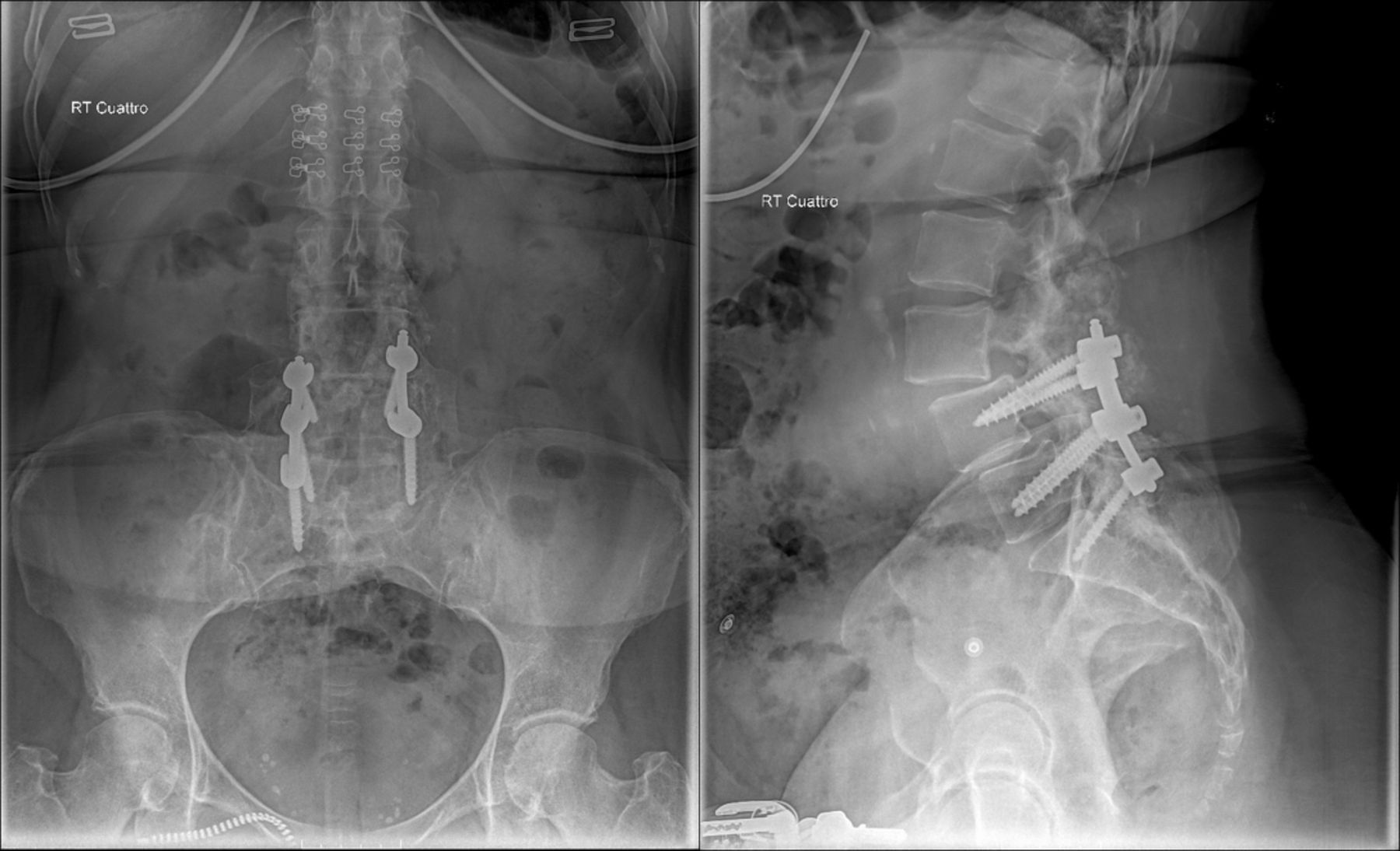
    Case 1: Lumbar spine anteroposterior and lateral standing radiographic images of the lumbosacral spine demonstrated intact hardware from L4 to S1, evidence of an iatrogenic flat back, grade I spondylolisthesis of L4-L5 level, grade I adjacent segment spondylolisthesis at the L3-L4 level, and increased proximal lumbar lordosis to compensate for her iatrogenic flat back.

  • Figure 2
    • Download figure
    • Open in new tab
    • Download powerpoint
    Figure 2

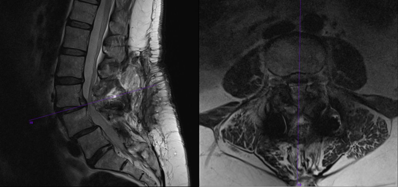
    Case 1: Lumbar spine magnetic resonance images verified adjacent segment deterioration at the L3-L4 level as well as reduction in the degree of spondylolisthesis in comparison to the standing radiographic images. There was also evidence of central and lateral recess stenosis and a disc herniation at this level.

  • Figure 3
    • Download figure
    • Open in new tab
    • Download powerpoint
    Figure 3

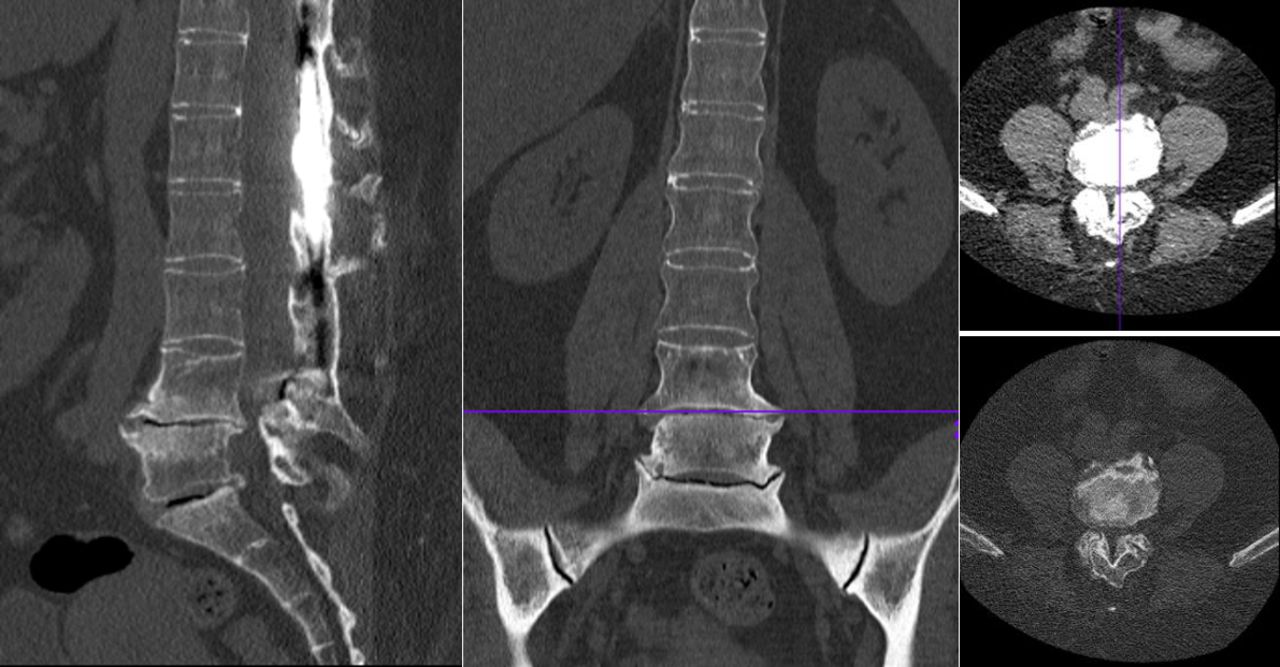
    Case 1: Computed tomography images after stage II confirmed substantial restoration of lordosis and reduction of the spondylolisthesis at both the previously fused L4-L5 level as well as the adjacent L3-L4 level.

  • Figure 4
    • Download figure
    • Open in new tab
    • Download powerpoint
    Figure 4

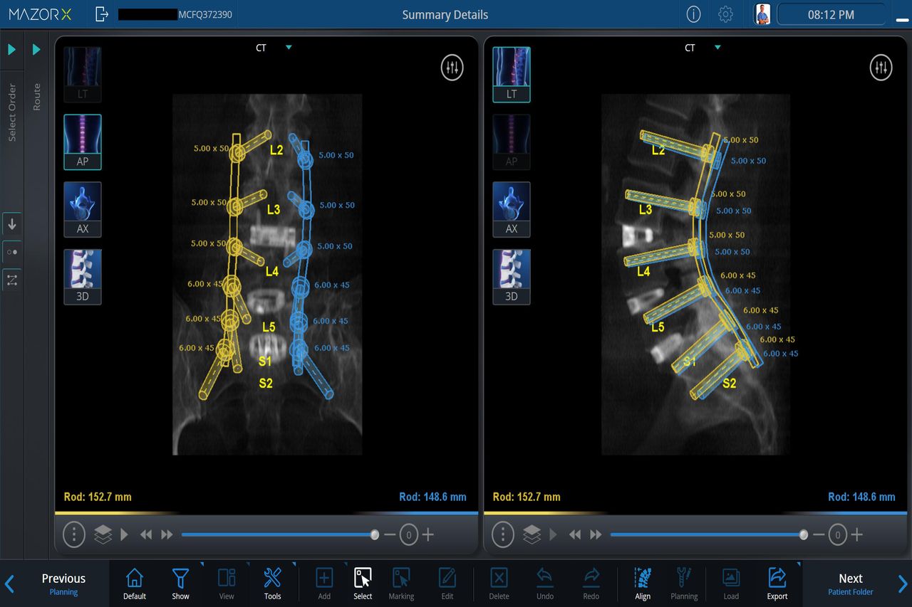
    Case 1: Preoperative planning for robotic-assisted placement of pedicle screws.

  • Figure 5
    • Download figure
    • Open in new tab
    • Download powerpoint
    Figure 5

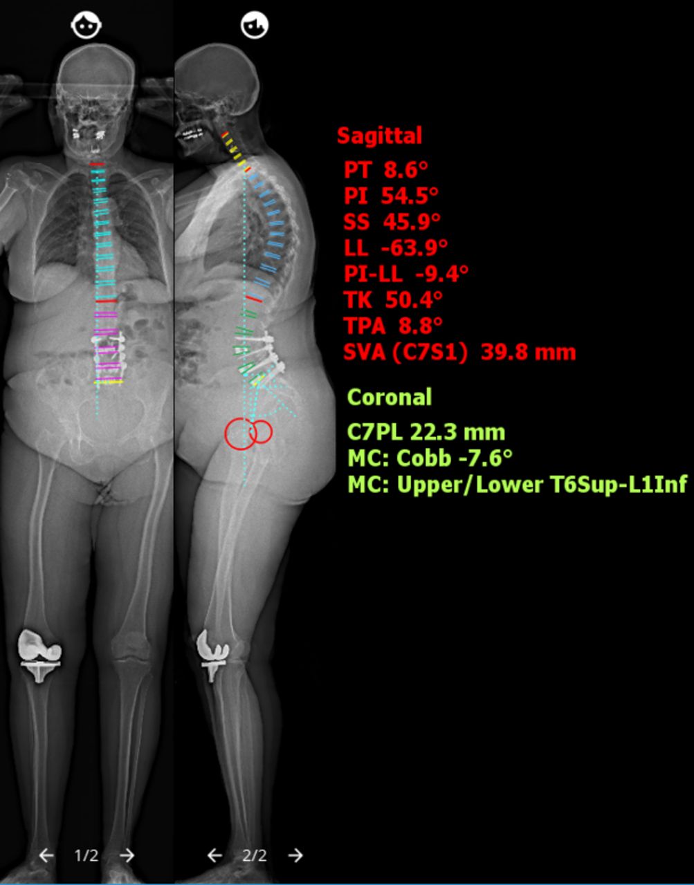
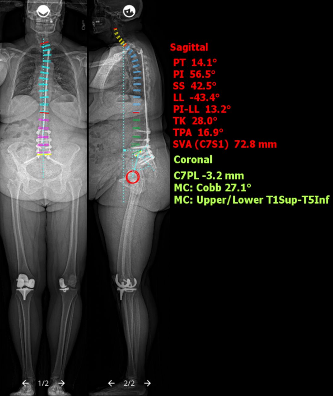
    Case 1: Postoperative EOS ImagingTM (Electro Optical System) full-length spine radiographs demonstrate reduction of the spondylolisthesis and restoration of spinopelvic and sagittal parameters. PT, sagittal pelvic tilt; PI, pelvic incidence; SS, sacral slope; LL, lumbar lordosis; PI-LL, PI-LL mismatch; TK, thoracic kyphosis; TPA, T1 pelvic angle; SVA, sagittal vertical axis; C7PL, C7 plumb line; MC, major Cobb angle.

  • Figure 6
    • Download figure
    • Open in new tab
    • Download powerpoint
    Figure 6

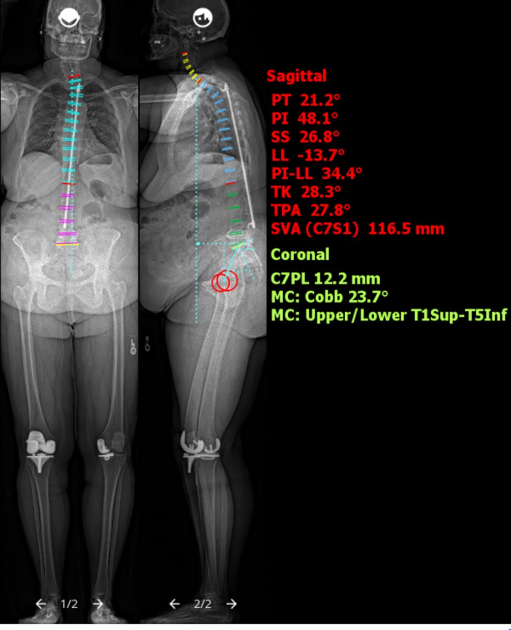
    Case 2: Preoperative EOS ImagingTM (Electro Optical System) full-length spine films revealed Harrington rod fixation across the thoracolumbar spine with degenerative changes most pronounced from L4 to S1. PT, sagittal pelvic tilt; PI, pelvic incidence; SS, sacral slope; LL, lumbar lordosis; PI-LL, PI-LL mismatch; TK, thoracic kyphosis; TPA, T1 pelvic angle; SVA, sagittal vertical axis; C7PL, C7 plumb line; MC, major Cobb angle.

  • Figure 7
    • Download figure
    • Open in new tab
    • Download powerpoint
    Figure 7

    Case 2: Computed tomography images revealed end-stage deterioration at the L4-L5 and L5-S1 levels, with foraminal stenosis at both those levels. The proximal fusion was well healed with a solid posterior fusion mass.

  • Figure 8
    • Download figure
    • Open in new tab
    • Download powerpoint
    Figure 8

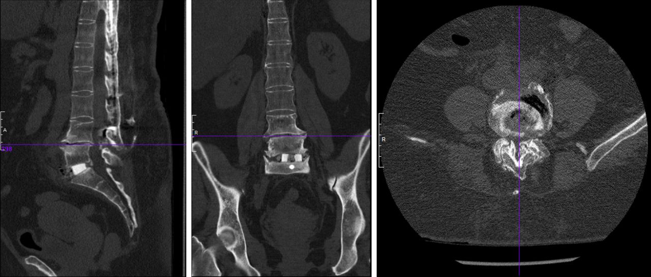
    Case 2: Computed tomography images after stage I anterior fusion.

  • Figure 9
    • Download figure
    • Open in new tab
    • Download powerpoint
    Figure 9

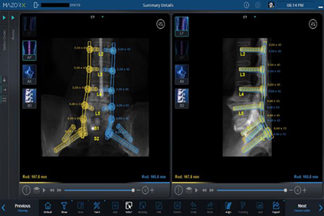
    Case 2: Preoperative planning for robotic-assisted placement of pedicle screws. Note that screws placed through fusion mass where anatomic landmarks are lost.

  • Figure 10
    • Download figure
    • Open in new tab
    • Download powerpoint
    Figure 10

    Case 2: EOS ImagingTM (Electro Optical System) full-length spine radiographic images after stage II posterior reconstruction revealed significant improvement in lumbar lordosis and sagittal parameters. PT, sagittal pelvic tilt; PI, pelvic incidence; SS, sacral slope; LL, lumbar lordosis; PI-LL, PI-LL mismatch; TK, thoracic kyphosis; TPA, T1 pelvic angle; SVA, sagittal vertical axis; C7PL, C7 plumb line; MC, major Cobb angle.

  • Figure 11
    • Download figure
    • Open in new tab
    • Download powerpoint
    Figure 11

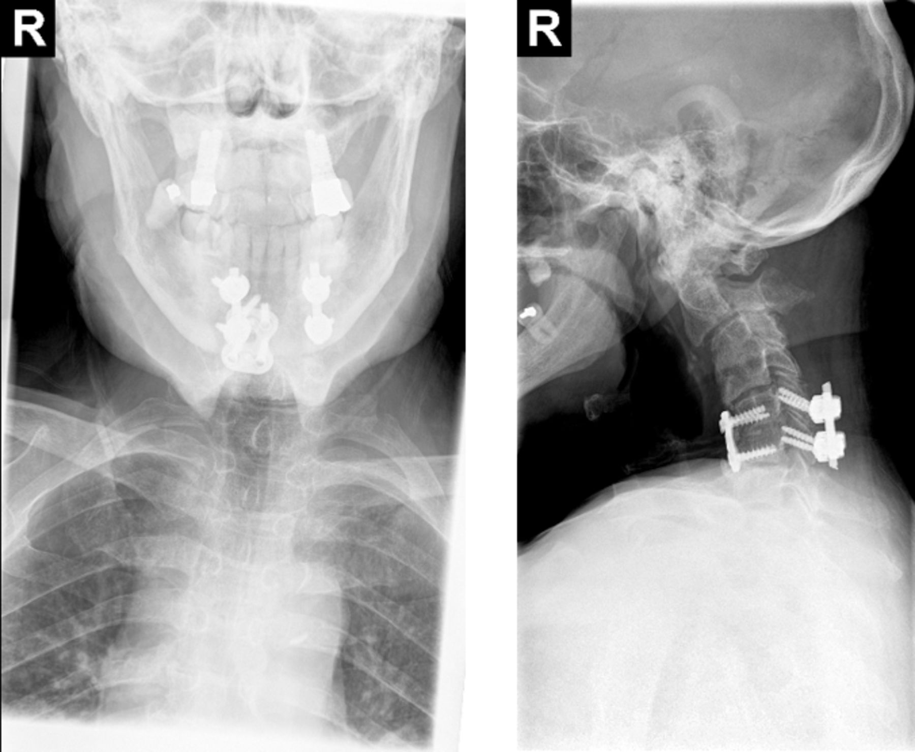
    Case 3: Cervical spine anteroposterior and lateral radiographs demonstrate kyphotic sagittal alignment of the cervical spine with a grade II anterolisthesis of C2 on C3, autofusion of C3 and C4, and retrolisthesis of C4 on C5. Prior instrumented fusion of C5-C6 with anterior plating and posterior lateral mass screw that appeared to be intact.

  • Figure 12
    • Download figure
    • Open in new tab
    • Download powerpoint
    Figure 12

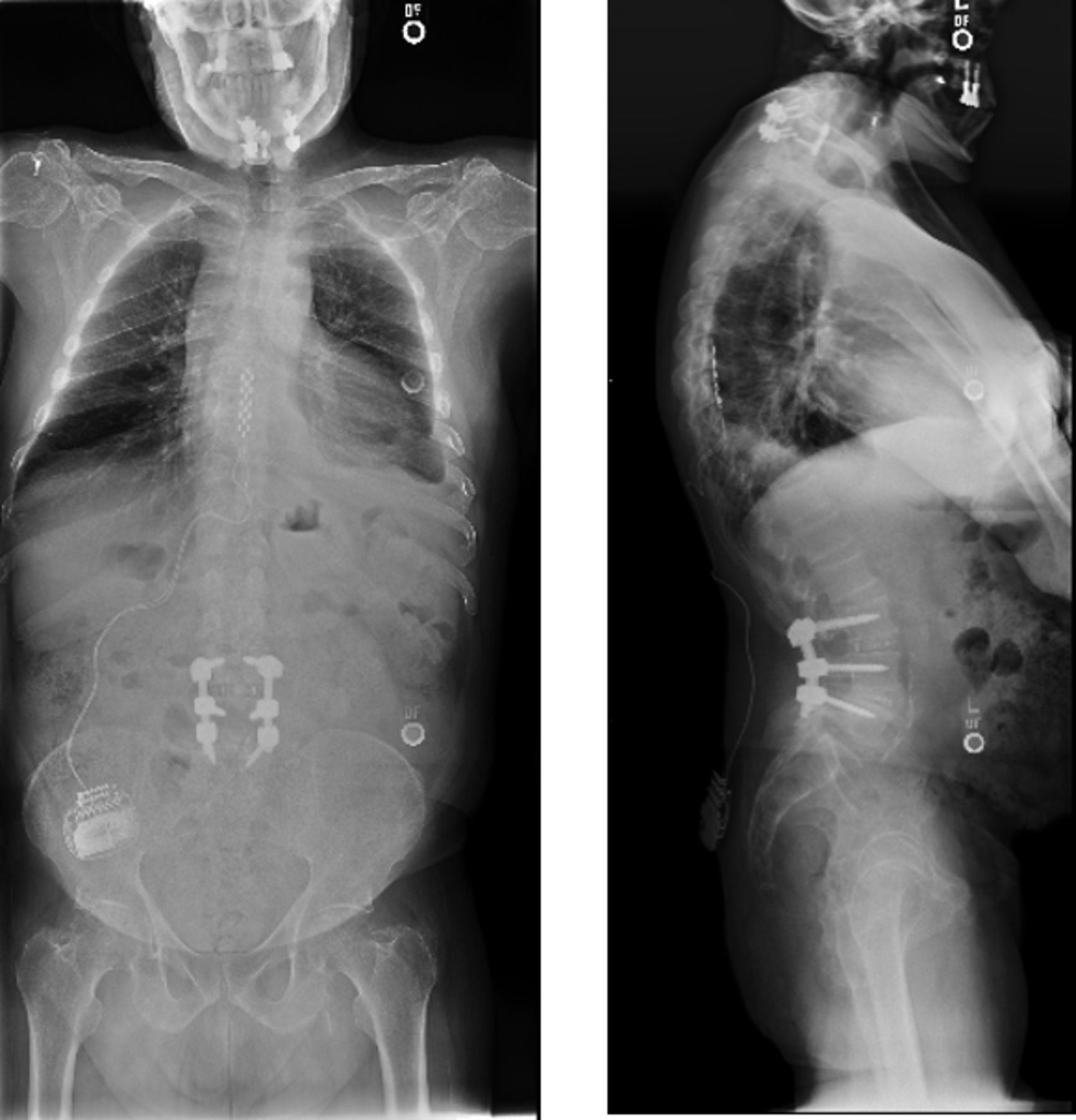
    Case 3: Full-spine radiographs demonstrate the patient’s chin-on-chest deformity.

  • Figure 13
    • Download figure
    • Open in new tab
    • Download powerpoint
    Figure 13

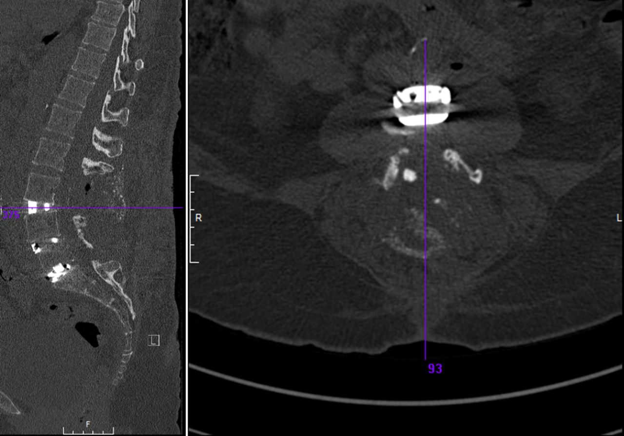
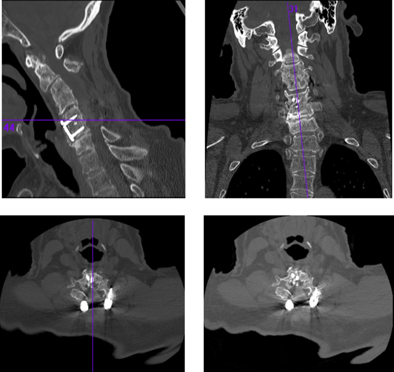
    Case 3: Preoperative cervical spine computed tomography images. The images showed loss of disc space height at C6-C7, evidence of laminectomies from C3 to C6, and evidence of autofusion at the C3-C4 level.

  • Figure 14
    • Download figure
    • Open in new tab
    • Download powerpoint
    Figure 14

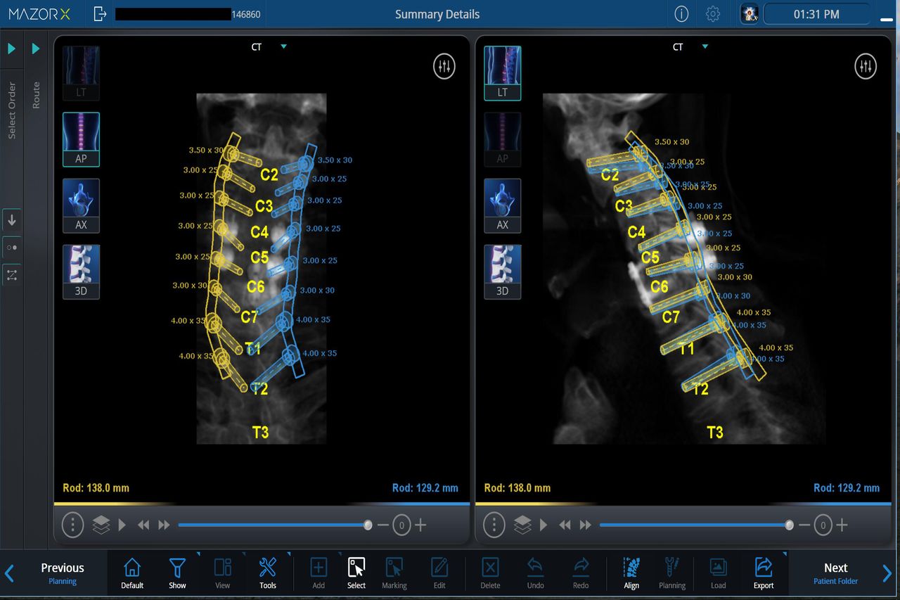
    Case 3: Preoperative planning for robotic-assisted placement of cervical pedicle screws.

  • Figure 15
    • Download figure
    • Open in new tab
    • Download powerpoint
    Figure 15

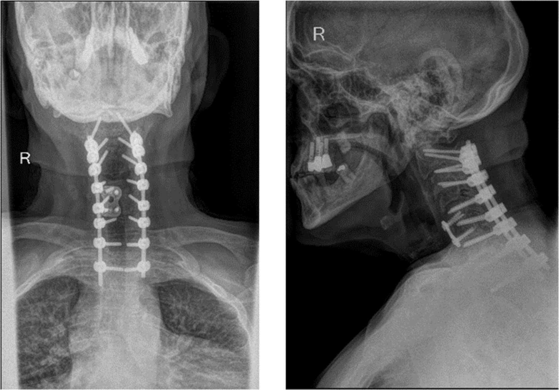
    Case 3: Postoperative radiographic images show the final construct with pedicle screw fixation and deformity correction.

  • Figure 16
    • Download figure
    • Open in new tab
    • Download powerpoint
    Figure 16

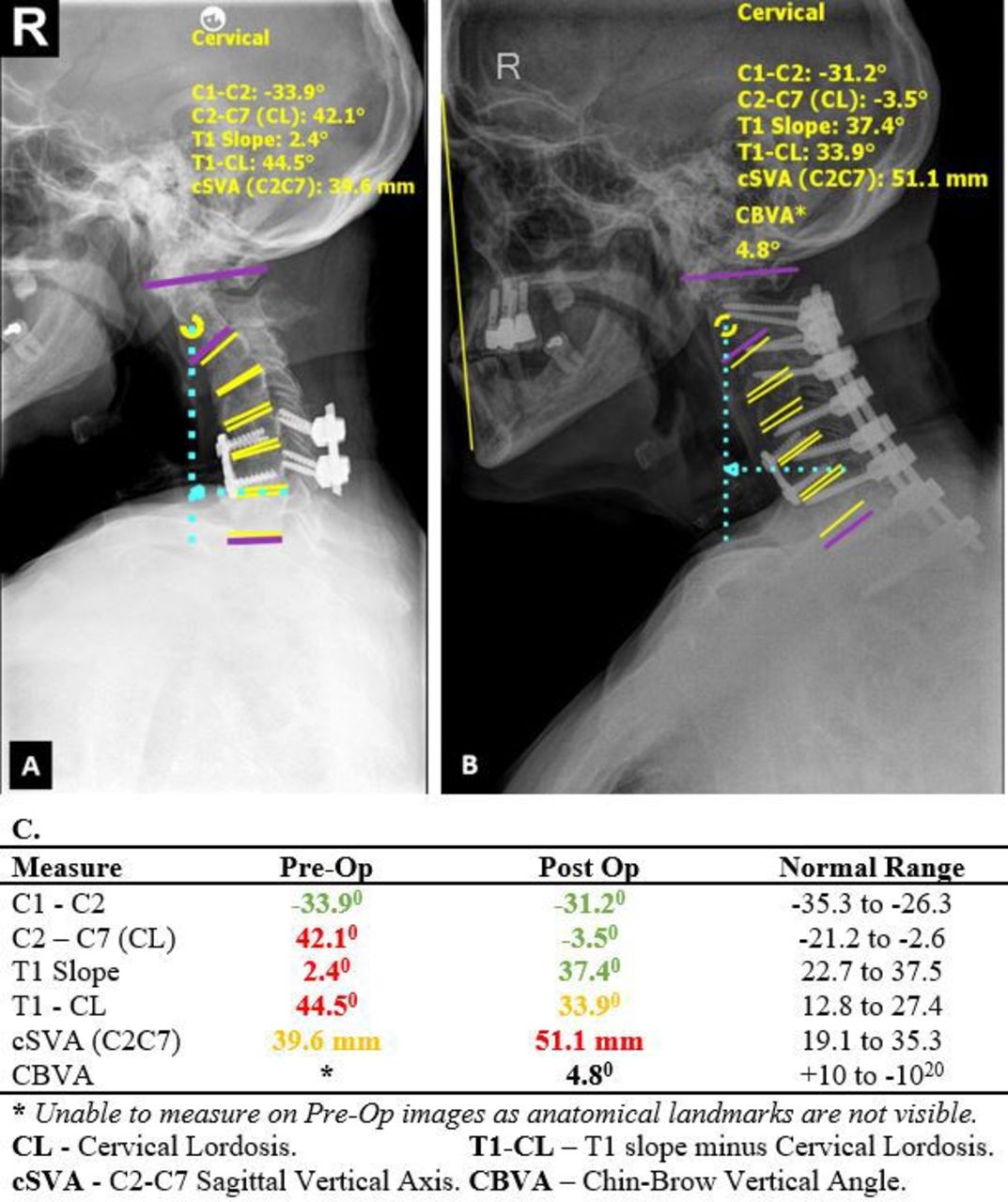
    Case 3: Pre- and postoperative lateral radiographs demonstrate significant improvement in sagittal alignment (normal range for CBVA20.

Next
Back to top

In this issue

International Journal of Spine Surgery: 19 (S2)
International Journal of Spine Surgery
Vol. 19, Issue S2
1 Apr 2025
  • Table of Contents
  • Index by author

Print
Download PDF
Article Alerts
Sign In to Email Alerts with your Email Address
Email Article

Thank you for your interest in spreading the word on International Journal of Spine Surgery.

NOTE: We only request your email address so that the person you are recommending the page to knows that you wanted them to see it, and that it is not junk mail. We do not capture any email address.

Enter multiple addresses on separate lines or separate them with commas.
Robotic-Assisted Revision Spine Surgery
(Your Name) has sent you a message from International Journal of Spine Surgery
(Your Name) thought you would like to see the International Journal of Spine Surgery web site.
CAPTCHA
This question is for testing whether or not you are a human visitor and to prevent automated spam submissions.
Citation Tools
Robotic-Assisted Revision Spine Surgery
Alexander M. Satin, Stanley Kisinde, Isador H. Lieberman
International Journal of Spine Surgery Jun 2022, 8272; DOI: 10.14444/8272

Citation Manager Formats

  • BibTeX
  • Bookends
  • EasyBib
  • EndNote (tagged)
  • EndNote 8 (xml)
  • Medlars
  • Mendeley
  • Papers
  • RefWorks Tagged
  • Ref Manager
  • RIS
  • Zotero
Share
Robotic-Assisted Revision Spine Surgery
Alexander M. Satin, Stanley Kisinde, Isador H. Lieberman
International Journal of Spine Surgery Jun 2022, 8272; DOI: 10.14444/8272
Twitter logo Facebook logo Mendeley logo
  • Tweet Widget
  • Facebook Like
  • Google Plus One

Jump to section

  • Article
    • Abstract
    • INTRODUCTION
    • CASES
    • CONCLUSION
    • Footnotes
    • References
  • Figures & Data
  • Info & Metrics
  • PDF

Related Articles

  • No related articles found.
  • PubMed
  • Google Scholar

Cited By...

  • No citing articles found.
  • Google Scholar

More in this TOC Section

  • High Uptake Detection for Spinal Degenerative Changes: A Comparison Between Bone Scintigraphy and Single Photon Emission Computed Tomography Combined With High-Resolution Computed Tomography
  • Accuracy of Pedicle Screw Placement Using the ExcelsiusGPS Robotic Navigation Platform: An Analysis of 728 Screws
  • A Network Meta-Analysis Comparing the Efficacy and Safety of Pedicle Screw Placement Techniques Using Intraoperative Conventional, Navigation, Robot-Assisted, and Augmented Reality Guiding Systems
Show more New Technology

Similar Articles

Keywords

  • revision spine surgical procedures
  • altered osseous anatomy
  • preoperative planning
  • robotic planning software
  • computer-assisted robotic guidance

Content

  • Current Issue
  • Latest Content
  • Archive

More Information

  • About IJSS
  • About ISASS
  • Privacy Policy

More

  • Subscribe
  • Alerts
  • Feedback

Other Services

  • Author Instructions
  • Join ISASS
  • Reprints & Permissions

© 2025 International Journal of Spine Surgery

International Journal of Spine Surgery Online ISSN: 2211-4599

Powered by HighWire