
Main menu

  • Home
  • Content
    • Current Issue
    • Advance Online Publication
    • Archive
  • About Us
    • About ISASS
    • About the Journal
    • Author Instructions
    • Editorial Board
    • Reviewer Guidelines & Publication Criteria
  • More
    • Advertise
    • Subscribe
    • Alerts
    • Feedback
  • Join Us
  • Reprints & Permissions
  • Sponsored Content
  • Other Publications
    • ijss

User menu

  • My alerts

Search

  • Advanced search
International Journal of Spine Surgery
  • My alerts
International Journal of Spine Surgery

Advanced Search

  • Home
  • Content
    • Current Issue
    • Advance Online Publication
    • Archive
  • About Us
    • About ISASS
    • About the Journal
    • Author Instructions
    • Editorial Board
    • Reviewer Guidelines & Publication Criteria
  • More
    • Advertise
    • Subscribe
    • Alerts
    • Feedback
  • Join Us
  • Reprints & Permissions
  • Sponsored Content
  • Follow ijss on Twitter
  • Visit ijss on Facebook
Research ArticleMinimally Invasive Surgery

Lateral Transpsoas Interbody Fusion

T. Barrett Sullivan, Angel Ordaz and Frank M. Phillips
International Journal of Spine Surgery January 2025, 8711; DOI: https://doi.org/10.14444/8711
T. Barrett Sullivan
1 Department of Orthopedic Surgery, University of California San Diego, La Jolla, CA, USA
MD
  • Find this author on Google Scholar
  • Find this author on PubMed
  • Search for this author on this site
  • For correspondence: tbsullivan@health.ucsd.edu
Angel Ordaz
1 Department of Orthopedic Surgery, University of California San Diego, La Jolla, CA, USA
MD
  • Find this author on Google Scholar
  • Find this author on PubMed
  • Search for this author on this site
Frank M. Phillips
2 Ronald DeWald Endowed Professor of Spinal Deformities, Rush University Medical Center, Chicago, IL, USA
MD
  • Find this author on Google Scholar
  • Find this author on PubMed
  • Search for this author on this site
  • Article
  • Figures & Data
  • Info & Metrics
  • PDF
Loading

Article Figures & Data

Figures

  • Figure 1
    • Download figure
    • Open in new tab
    • Download powerpoint
    Figure 1

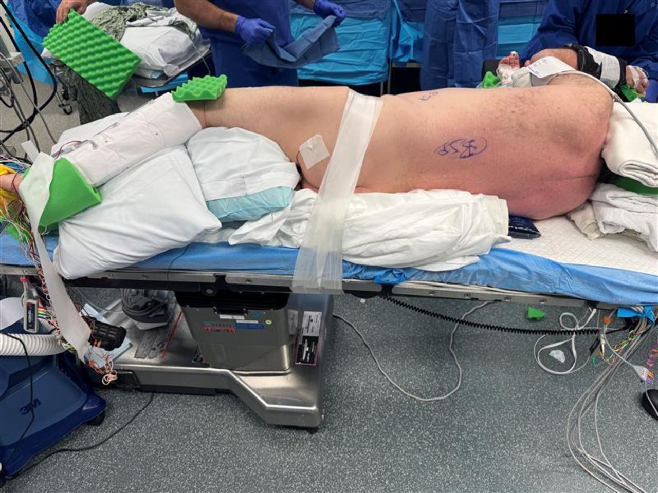
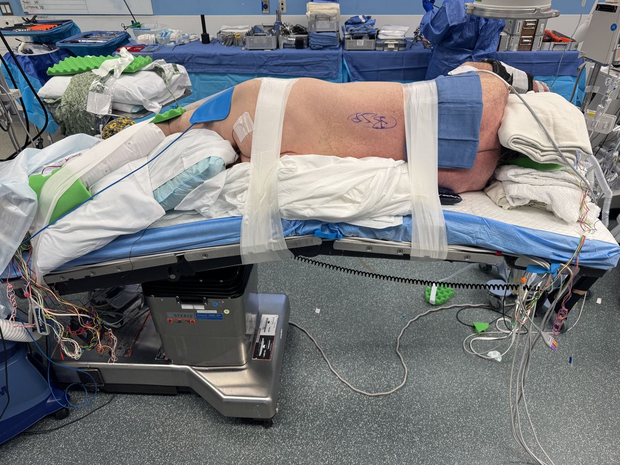
    The pelvis is secured with circumferential tape that is affixed to the bed distal to the bed break. Tape should be proximal to the greater trochanter and distal to the iliac crest to avoid draping out of any planned incisions.

  • Figure 2
    • Download figure
    • Open in new tab
    • Download powerpoint
    Figure 2

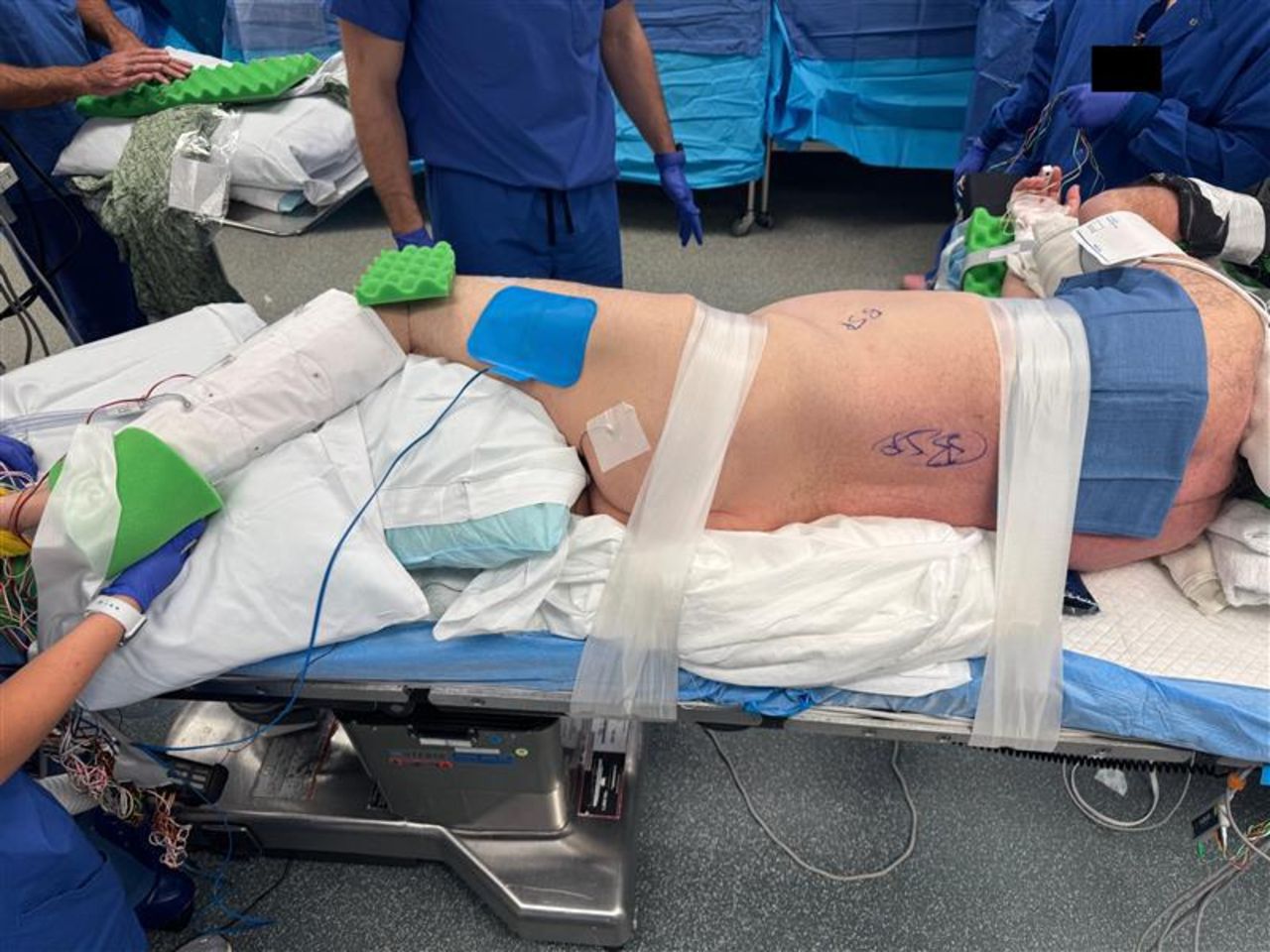
    The chest is secured with circumferential taping.

  • Figure 3
    • Download figure
    • Open in new tab
    • Download powerpoint
    Figure 3

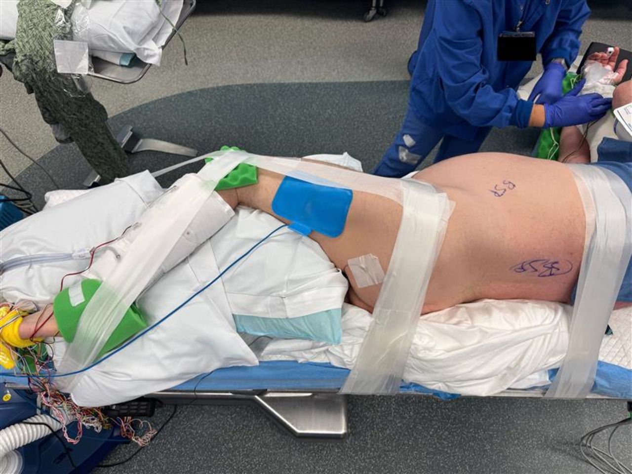
    The pelvis and legs are further secured with circumferential taping.

  • Figure 4
    • Download figure
    • Open in new tab
    • Download powerpoint
    Figure 4

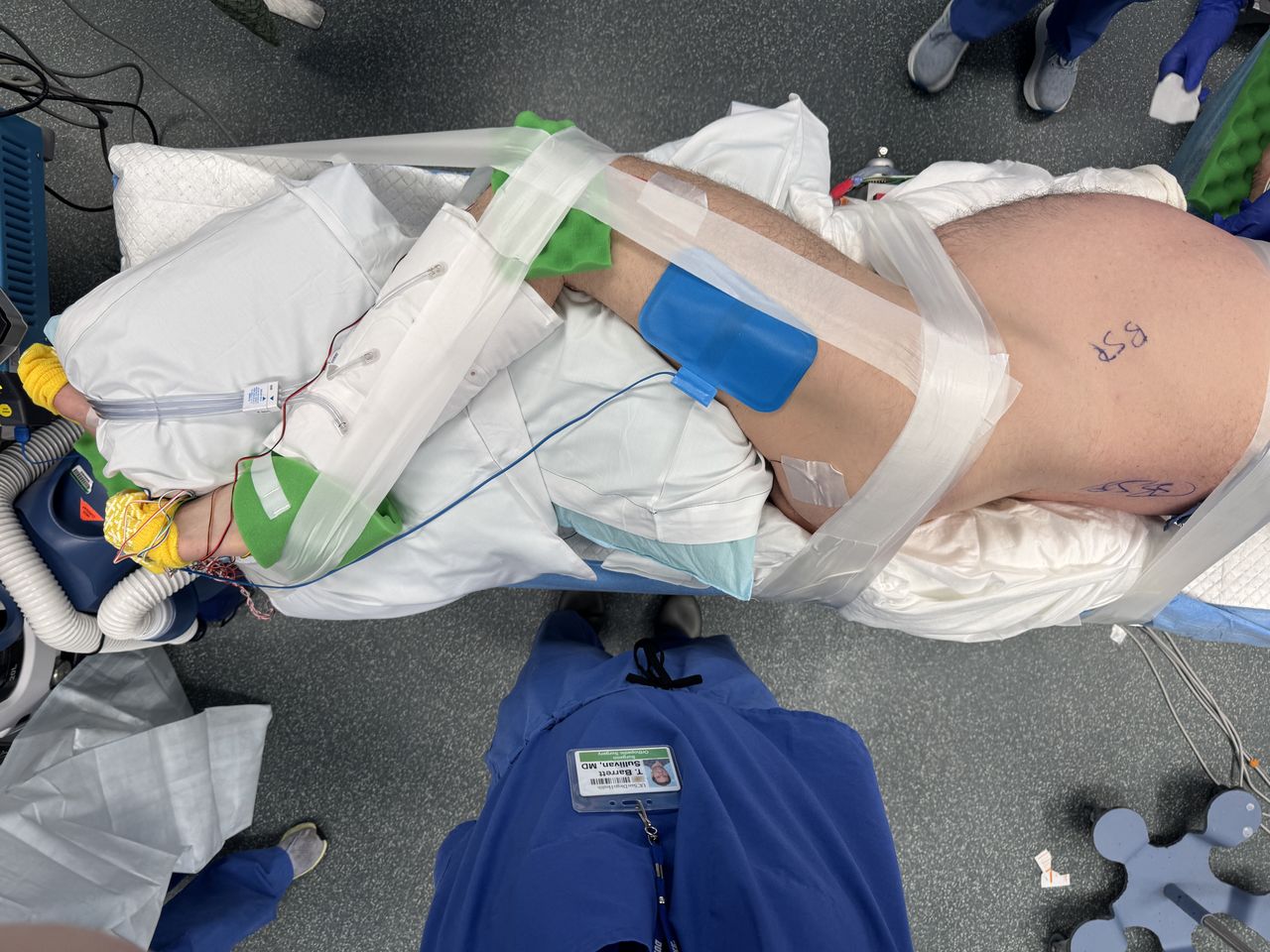
    The pelvis and legs are further secured with circumferential taping (view from above).

  • Figure 5
    • Download figure
    • Open in new tab
    • Download powerpoint
    Figure 5

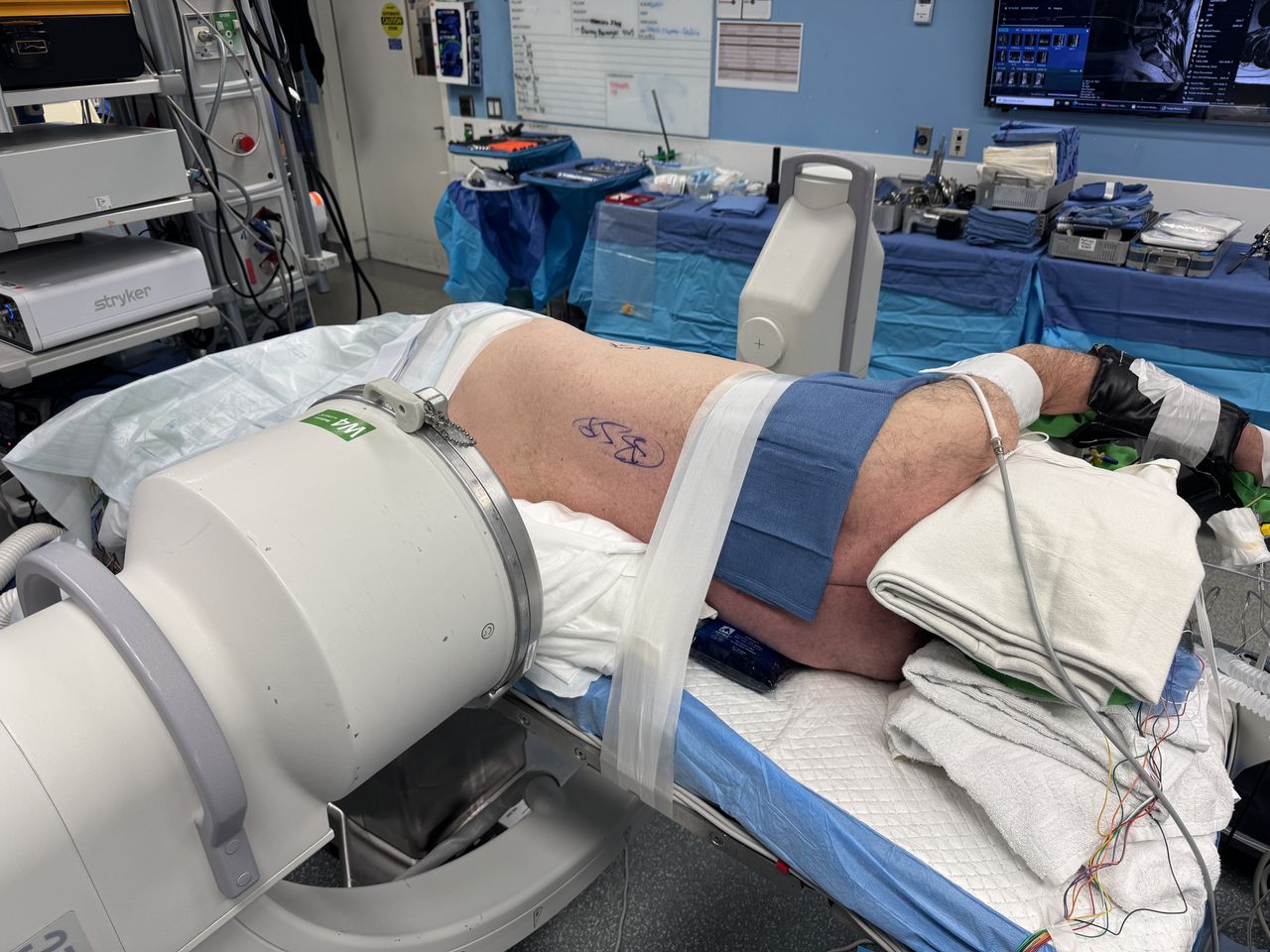
    The table is then “broken” to improve access between the 12th rib and the iliac crest. The amount of table break is greater for levels more cephalad or more caudal due to these structures.

  • Figure 6
    • Download figure
    • Open in new tab
    • Download powerpoint
    Figure 6

    After securing the patient to the bed, the bed is rotated to obtain a perfect anteroposterior fluoroscopic image. C-arm position is impacted by room orientation and size. Having the intensifier on the posterior reduces surgeon radiation but comes at the cost of ergonomic challenges working around the larger portion of the C-arm.

  • Figure 7
    • Download figure
    • Open in new tab
    • Download powerpoint
    Figure 7

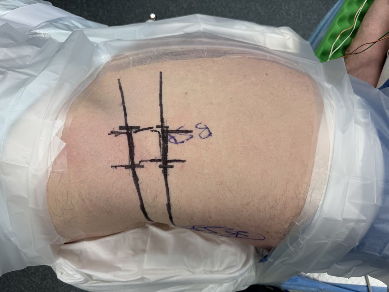
    Example of marking for L3-L4 and L4-L5. Each disc space angle is marked more broadly, in addition to anterior and posterior aspects of the vertebral body and superior and inferior endplates.
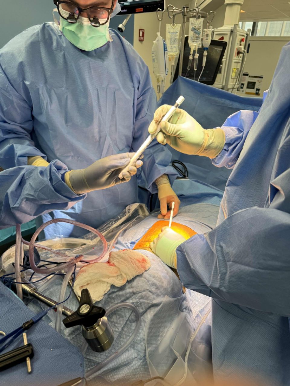
  • Figure 8
    • Download figure
    • Open in new tab
    • Download powerpoint
    Figure 8

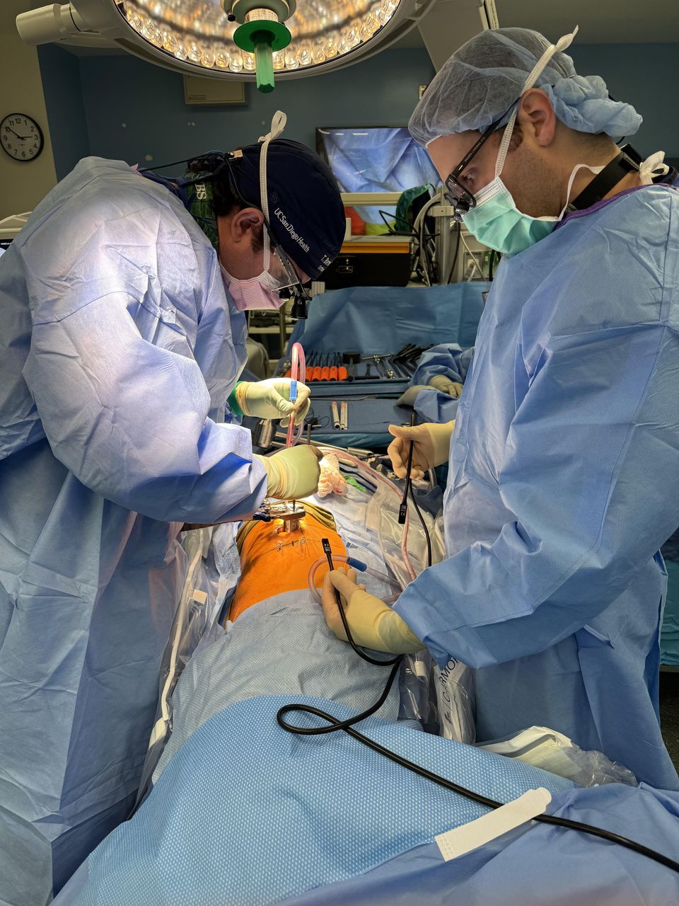
    Example of shallow docking technique. After seating the initial dilator on top of the psoas and dilating up to the retractor, the retractor is then opened superficially to the muscle, allowing the surgeon direct visualization of the muscle prior to piercing the psoas with the initial dilator. An endoscopic Kittner sponge can be used to clear any retroperitoneal fat if there is an obscured view of the psoas.

  • Figure 9
    • Download figure
    • Open in new tab
    • Download powerpoint
    Figure 9

    After the initial dilator is seated in the psoas muscle under electromyography stimulation, the superficially positioned retractor is then removed.
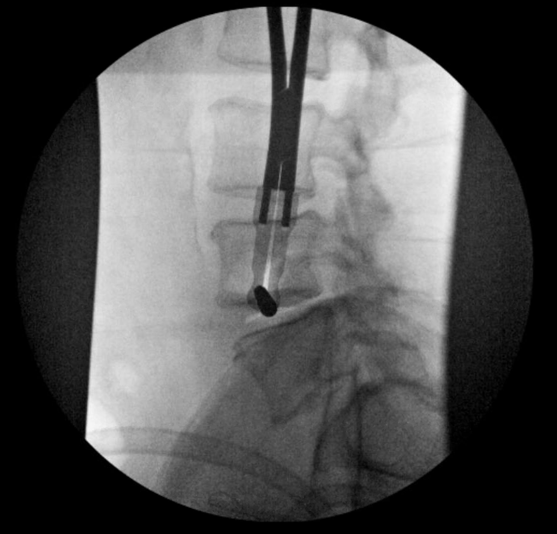
  • Figure 10
    • Download figure
    • Open in new tab
    • Download powerpoint
    Figure 10

    Position of the initial dilator is confirmed at the level of the disc space on lateral fluoroscopy.
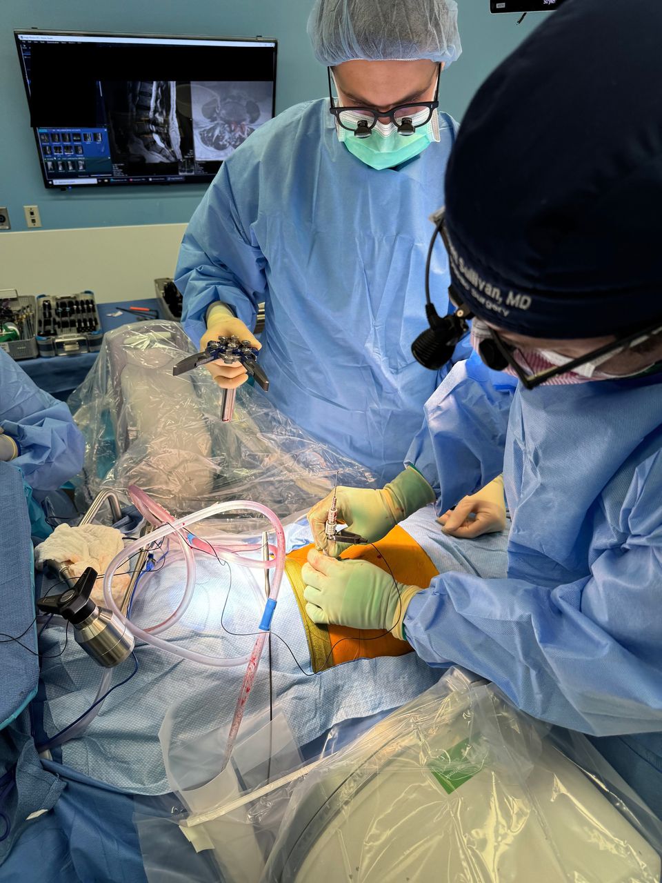
  • Figure 11
    • Download figure
    • Open in new tab
    • Download powerpoint
    Figure 11

    Sequential dilation through the psoas is performed under live, directional electromyography. The position of the plexus should be identified posteriorly.
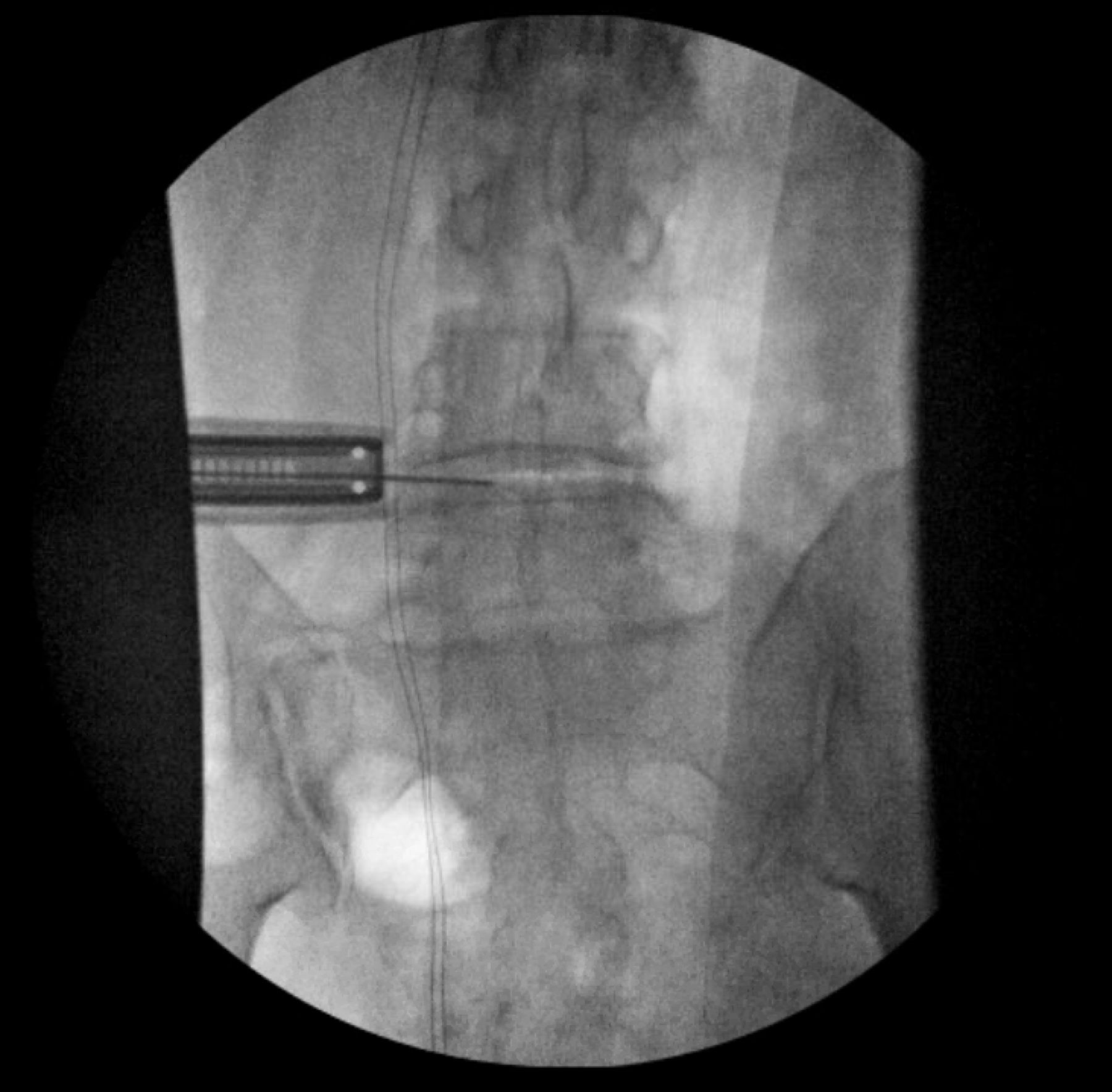
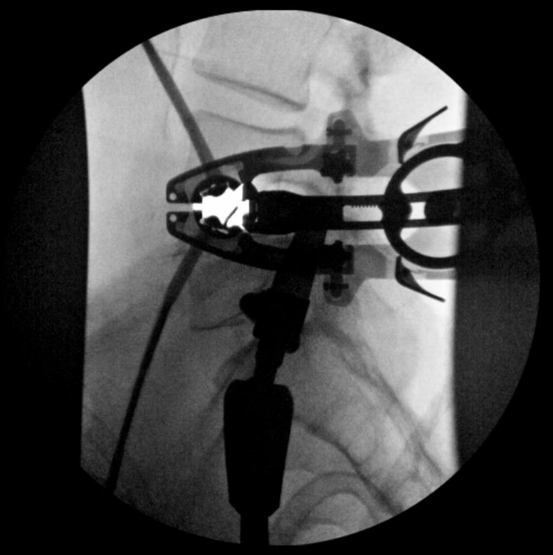
  • Figure 12
    • Download figure
    • Open in new tab
    • Download powerpoint
    Figure 12

    Orientation of the retractor is confirmed relative to the disc space on anteroposterior view.
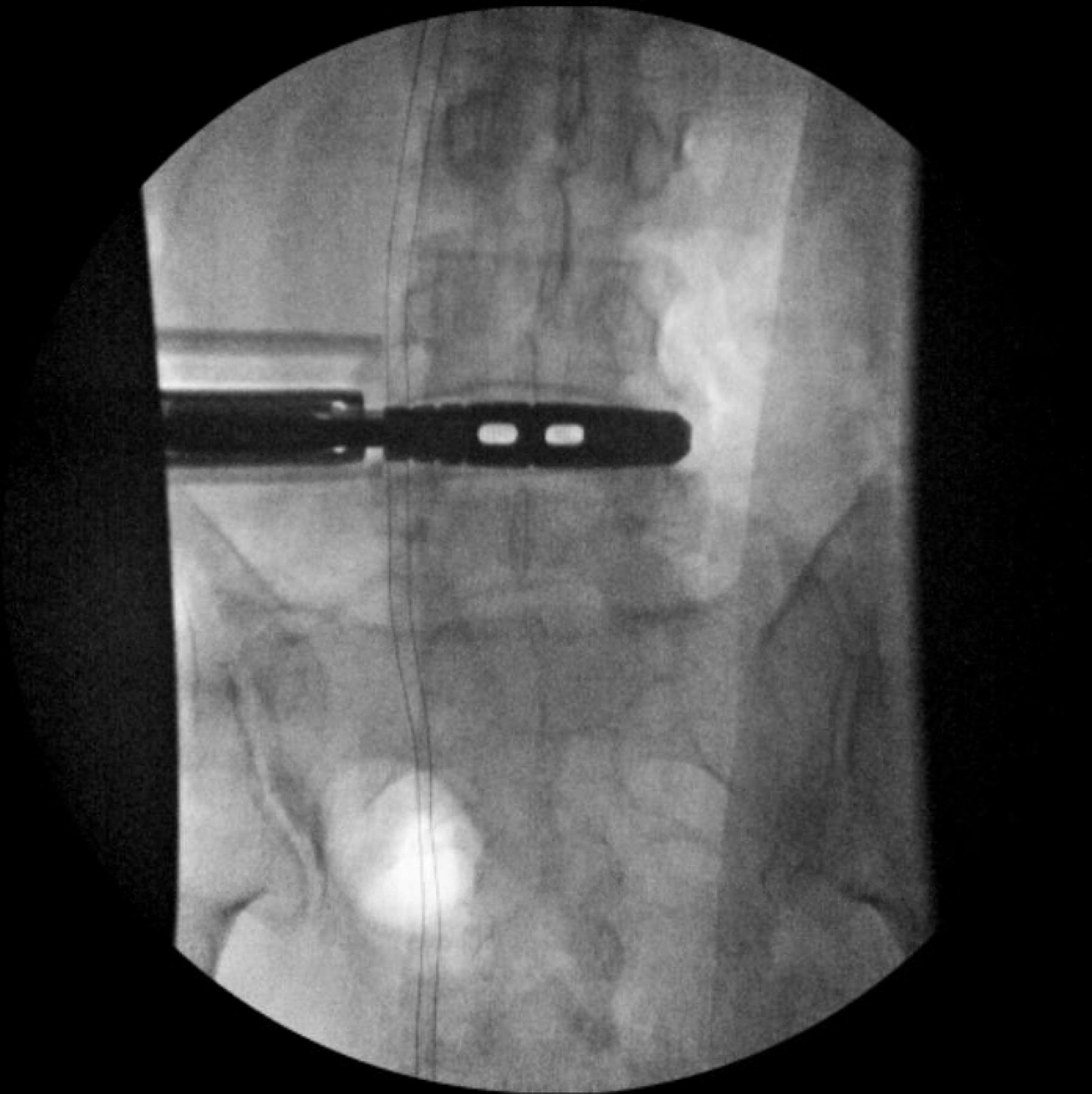
  • Figure 13
    • Download figure
    • Open in new tab
    • Download powerpoint
    Figure 13

    Orientation of the retractor is confirmed relative to the disc space on the lateral view. The retractor is slightly opened to allow electromyography stimulation along the anterior aspect of the posterior blade prior to shim placement.
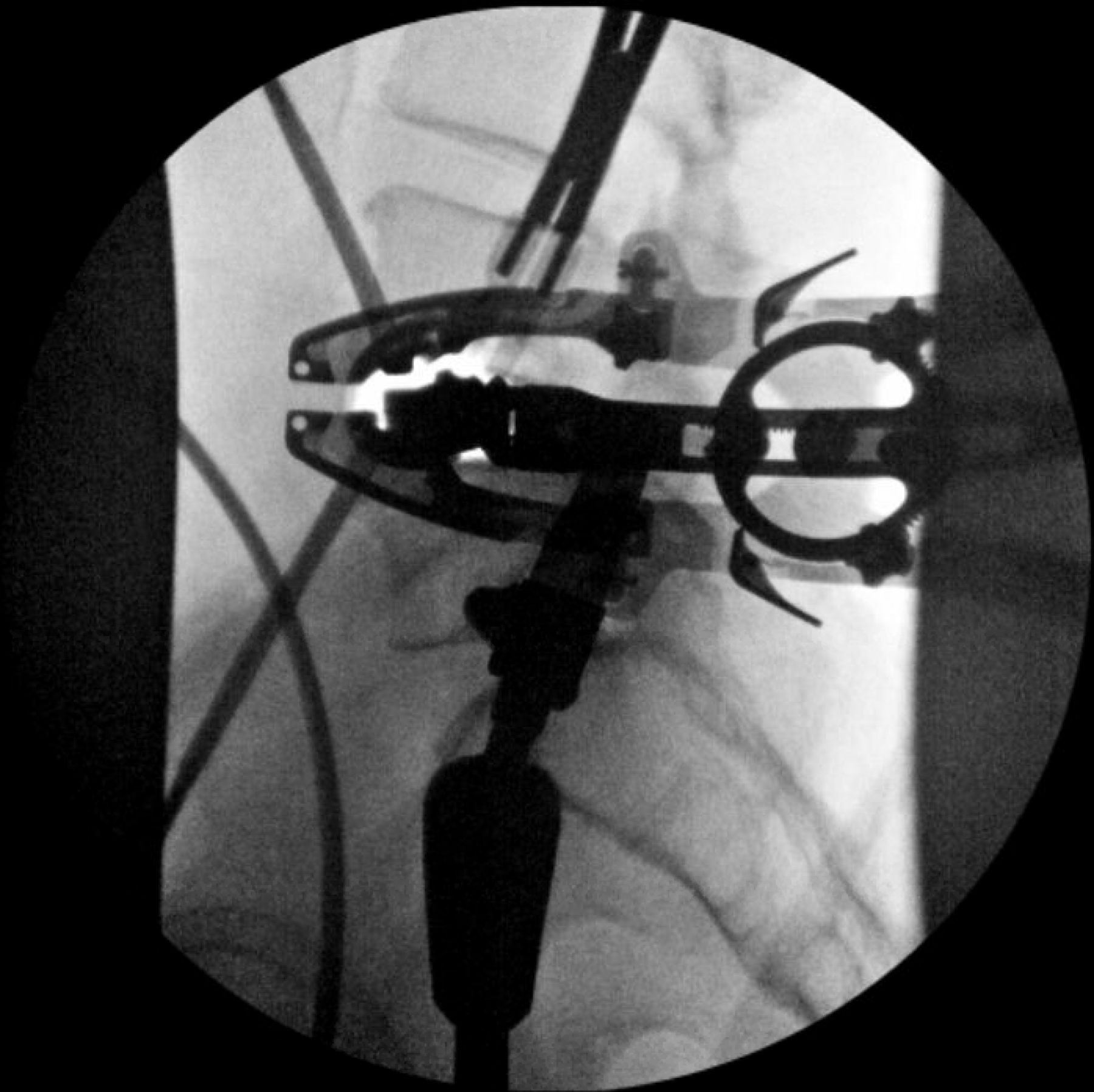
  • Figure 14
    • Download figure
    • Open in new tab
    • Download powerpoint
    Figure 14

    The shim is then placed under anteroposterior fluoroscopy.
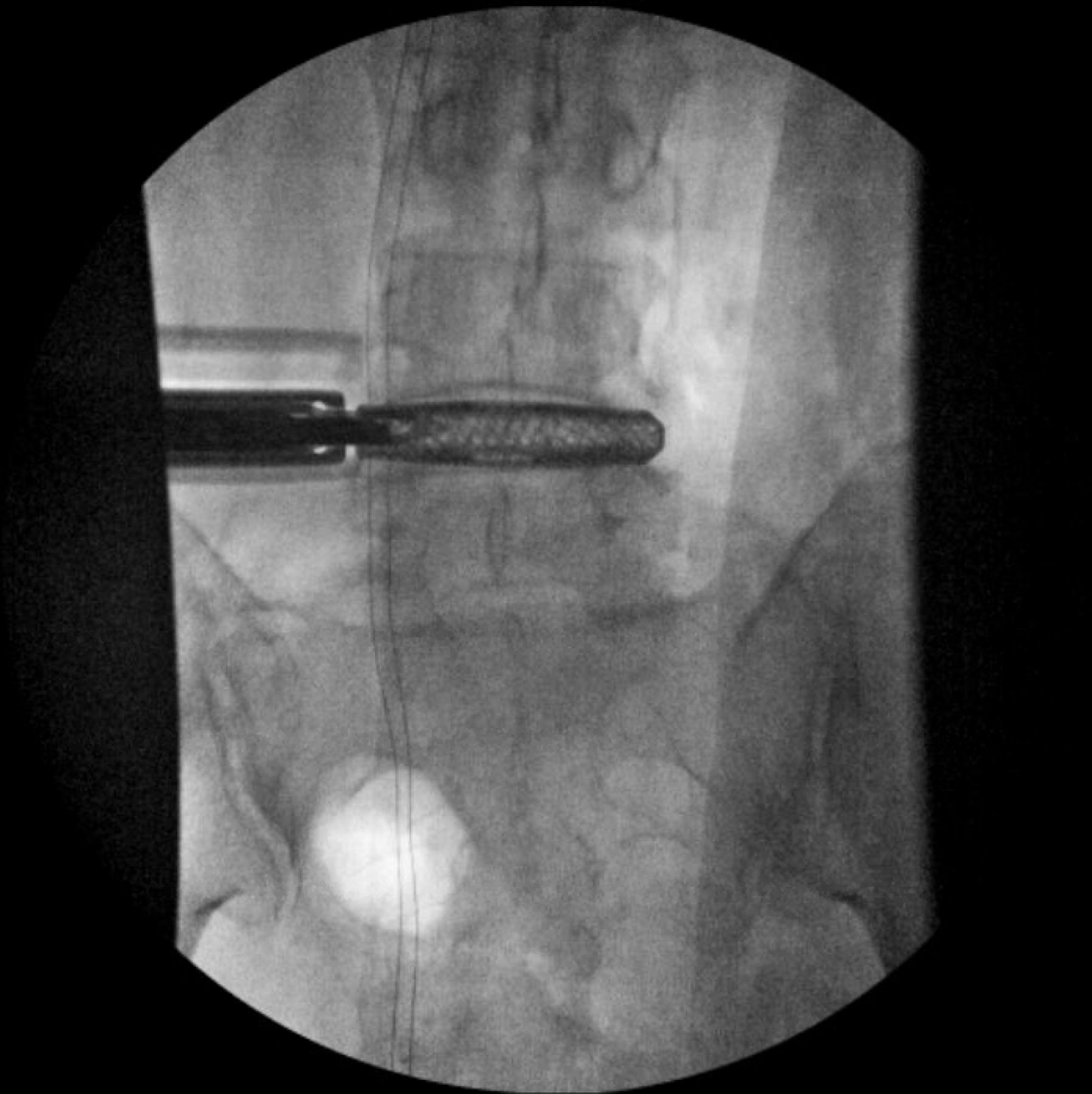
  • Figure 15
    • Download figure
    • Open in new tab
    • Download powerpoint
    Figure 15

    The retractor is then opened. The position of the anterior and posterior blades as well as the retractor orientation/angle should be noted to allow for further visual landmarks of the operative “safe zone.”

  • Figure 16
    • Download figure
    • Open in new tab
    • Download powerpoint
    Figure 16

    Trialing is performed under anteroposterior fluoroscopy. After determining the appropriate implant length and height, position of the trial is confirmed on lateral imaging.

  • Figure 17
    • Download figure
    • Open in new tab
    • Download powerpoint
    Figure 17

    After determining the appropriate implant length and height on the anteroposterior view, position of the trial is confirmed on lateral imaging.
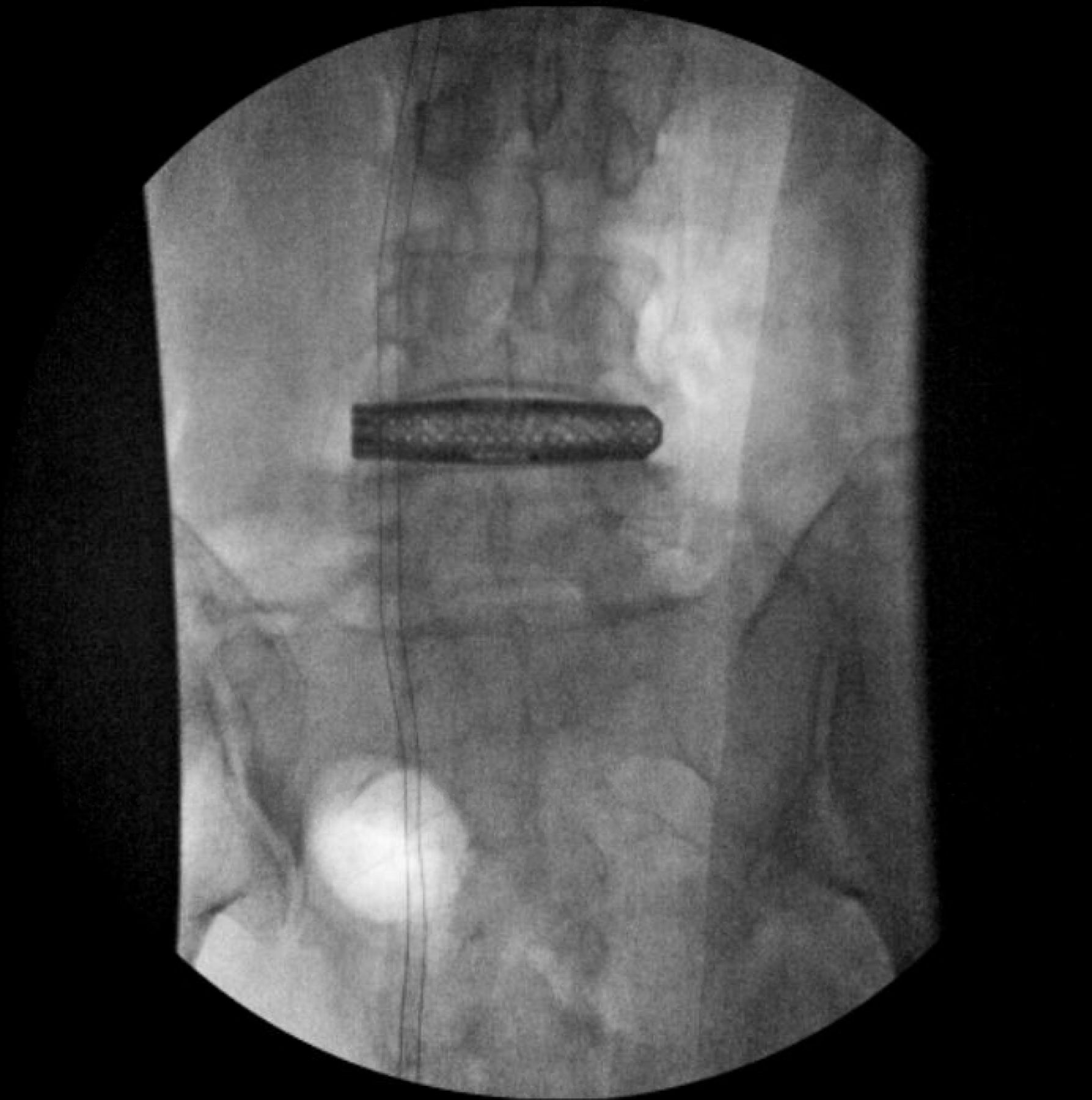
  • Figure 18
    • Download figure
    • Open in new tab
    • Download powerpoint
    Figure 18

    The final implant is then placed on anteroposterior fluoroscopy.

  • Figure 19
    • Download figure
    • Open in new tab
    • Download powerpoint
    Figure 19

    The insertion handle is removed, and final position of the implant is confirmed on the anteroposterior view.
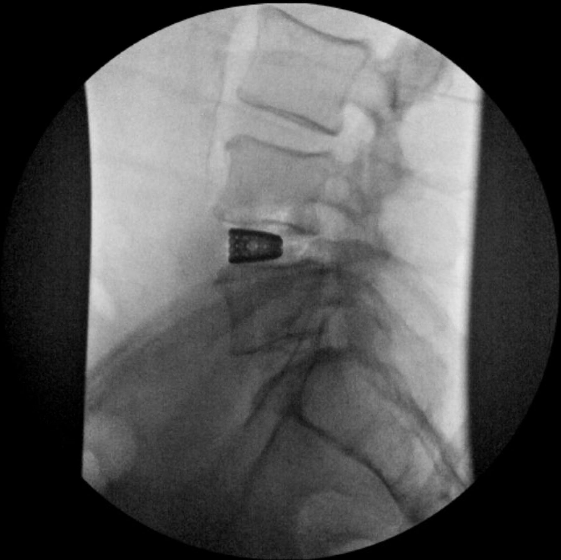
  • Figure 20
    • Download figure
    • Open in new tab
    • Download powerpoint
    Figure 20

    Final position of the implant is confirmed on the lateral view.

PreviousNext
Back to top

In this issue

International Journal of Spine Surgery: 19 (S2)
International Journal of Spine Surgery
Vol. 19, Issue S2
1 Apr 2025
  • Table of Contents
  • Index by author

Print
Download PDF
Article Alerts
Sign In to Email Alerts with your Email Address
Email Article

Thank you for your interest in spreading the word on International Journal of Spine Surgery.

NOTE: We only request your email address so that the person you are recommending the page to knows that you wanted them to see it, and that it is not junk mail. We do not capture any email address.

Enter multiple addresses on separate lines or separate them with commas.
Lateral Transpsoas Interbody Fusion
(Your Name) has sent you a message from International Journal of Spine Surgery
(Your Name) thought you would like to see the International Journal of Spine Surgery web site.
CAPTCHA
This question is for testing whether or not you are a human visitor and to prevent automated spam submissions.
Citation Tools
Lateral Transpsoas Interbody Fusion
T. Barrett Sullivan, Angel Ordaz, Frank M. Phillips
International Journal of Spine Surgery Jan 2025, 8711; DOI: 10.14444/8711

Citation Manager Formats

  • BibTeX
  • Bookends
  • EasyBib
  • EndNote (tagged)
  • EndNote 8 (xml)
  • Medlars
  • Mendeley
  • Papers
  • RefWorks Tagged
  • Ref Manager
  • RIS
  • Zotero
Share
Lateral Transpsoas Interbody Fusion
T. Barrett Sullivan, Angel Ordaz, Frank M. Phillips
International Journal of Spine Surgery Jan 2025, 8711; DOI: 10.14444/8711
Twitter logo Facebook logo Mendeley logo
  • Tweet Widget
  • Facebook Like
  • Google Plus One

Jump to section

  • Article
    • Abstract
    • Introduction
    • Indications and Benefits
    • Risks and Complications
    • Technique
    • Neuromonitoring
    • Outcomes
    • Conclusion
    • Footnotes
    • References
  • Figures & Data
  • Info & Metrics
  • PDF

Related Articles

  • No related articles found.
  • PubMed
  • Google Scholar

Cited By...

  • No citing articles found.
  • Google Scholar

More in this TOC Section

  • Comparative Review of Lateral and Oblique Lumbar Interbody Fusion: Technique, Outcomes, and Complications
  • A Spine Surgeon’s Learning Curve With the Minimally Invasive L5 to S1 Lateral ALIF Surgical Approach: Perioperative Outcomes and Technical Considerations
  • Fully Navigated Single-Position Prone Lateral Lumbar Interbody Fusion: A Detailed Technical Report and Description of 15 Cases
Show more Minimally Invasive Surgery

Similar Articles

Keywords

  • lumbar interbody fusion
  • extreme lateral interbody fusion
  • XLIF
  • minimally invasive spine surgery
  • degenerative disease
  • low back pain

Content

  • Current Issue
  • Latest Content
  • Archive

More Information

  • About IJSS
  • About ISASS
  • Privacy Policy

More

  • Subscribe
  • Alerts
  • Feedback

Other Services

  • Author Instructions
  • Join ISASS
  • Reprints & Permissions

© 2025 International Journal of Spine Surgery

International Journal of Spine Surgery Online ISSN: 2211-4599

Powered by HighWire