
Main menu

  • Home
  • Content
    • Current Issue
    • Advance Online Publication
    • Archive
  • About Us
    • About ISASS
    • About the Journal
    • Author Instructions
    • Editorial Board
    • Reviewer Guidelines & Publication Criteria
  • More
    • Advertise
    • Subscribe
    • Alerts
    • Feedback
  • Join Us
  • Reprints & Permissions
  • Sponsored Content
  • Other Publications
    • ijss

User menu

  • My alerts

Search

  • Advanced search
International Journal of Spine Surgery
  • My alerts
International Journal of Spine Surgery

Advanced Search

  • Home
  • Content
    • Current Issue
    • Advance Online Publication
    • Archive
  • About Us
    • About ISASS
    • About the Journal
    • Author Instructions
    • Editorial Board
    • Reviewer Guidelines & Publication Criteria
  • More
    • Advertise
    • Subscribe
    • Alerts
    • Feedback
  • Join Us
  • Reprints & Permissions
  • Sponsored Content
  • Follow ijss on Twitter
  • Visit ijss on Facebook
Case ReportFull Length Article
Open Access

Instrumented reduction of a fixed C1–2 subluxation using occipital and C2/C3 fixation: A case report

Clifton Meals, Rachel Harrison, Warren Yu and Joseph O'Brien
International Journal of Spine Surgery January 2013, 7 e20-e23; DOI: https://doi.org/10.1016/j.ijsp.2013.01.002
Clifton Meals
Department of Orthopedics, George Washington University Medical Center, Washington, DC
MD
  • Find this author on Google Scholar
  • Find this author on PubMed
  • Search for this author on this site
  • For correspondence: cliftongm@gmail.com
Rachel Harrison
Department of Orthopedics, George Washington University Medical Center, Washington, DC
MD
  • Find this author on Google Scholar
  • Find this author on PubMed
  • Search for this author on this site
Warren Yu
Department of Orthopedics, George Washington University Medical Center, Washington, DC
MD
  • Find this author on Google Scholar
  • Find this author on PubMed
  • Search for this author on this site
Joseph O'Brien
Department of Orthopedics, George Washington University Medical Center, Washington, DC
MD
  • Find this author on Google Scholar
  • Find this author on PubMed
  • Search for this author on this site
  • Article
  • Figures & Data
  • Info & Metrics
  • PDF
Loading

Abstract

Background Different strategies exist for reduction of the cervical spine. Placement of C1 lateral mass screws is a powerful technique but may be impossible in a degenerative or revision setting. We report the open, posterior-only, and instrumented reduction of a fixed C1–2 subluxation using occipital and C2/C3 fixation. The patient had rheumatoid arthritis and had undergone previous surgery of the cervical spine.

Methods We performed a retrospective chart review and focused appraisal of the literature.

Results Satisfactory reduction was achieved with this infrequently reported technique.

Conclusions/Level of Evidence Spine surgeons may consider the described procedure a viable treatment alternative in problematic subluxations of the cervical spine. Level V.

  • Cervical spine
  • Subluxation
  • Reduction
  • Instrumented
  • Rheumatoid arthritis

Instrumented reduction has been used to treat lumbar spondylolisthesis,1–3 thoracic fracture-dislocations,4, 5 cervicothoracic deformity,6 and most recently, craniocervical and atlantoaxial subluxation.7–10

Craniocervical subluxation and atlantoaxial subluxation produce a variety of deformities, for example, cranial settling, basilar invagination, and cervical stenosis. Different instrumented reduction techniques may be particularly suited for each. In cases of cervical stenosis and instability, placement of C1 lateral mass screws is a powerful alternative to the traditional use of wiring and transarticular screw fixation.9 C1 lateral mass screws may be difficult to place, however, in a revision or degenerative setting.

As an alternative to the placement of C1 lateral mass screws, we describe occipital plating and fixation of C2 to affect atlantoaxial reduction and correction of cervical stenosis/instability.

Case report

The subject of this case report provided written informed consent for print and electronic publication of this report. The use of bone morphogenetic protein in this case was off-label.

History

A 34-year-old woman with rheumatoid arthritis presented to our clinic 6 months after undergoing a C1–2 fusion. This procedure had been designed to correct subluxation at the atlantoaxial articulation and was accomplished with iliac crest autograft and cables. The patient had been diagnosed with rheumatoid arthritis at age 20 years, was unable to tolerate Enbrel (Entanercept; Pfizer, New York, New York), and took prednisone only. She had recently been placed in halo fixation to prevent paresthesias with neck range of motion (L'hermitte phenomenon).

Examination

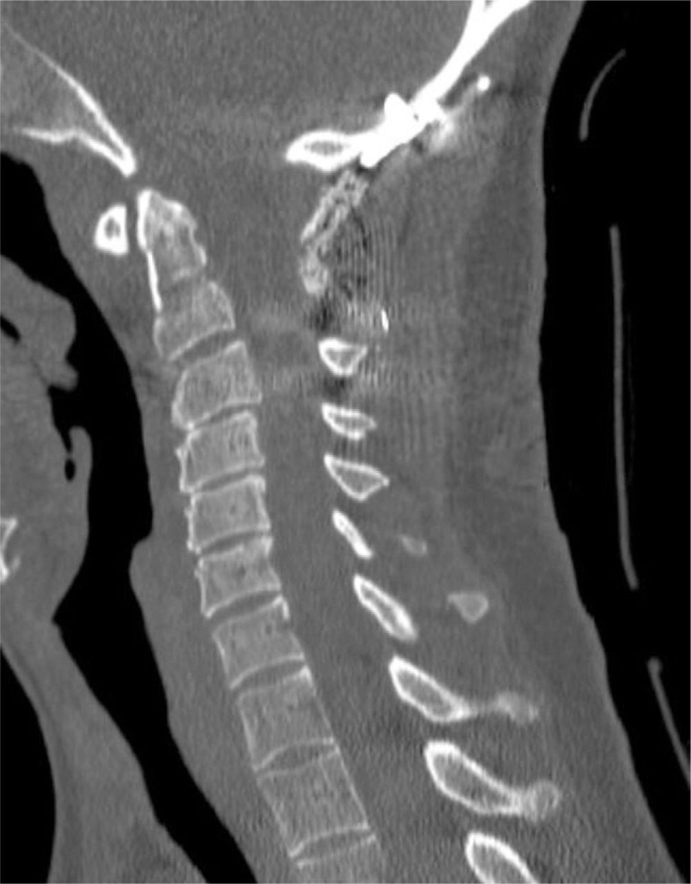
The patient's pin sites were clear, and an examination of her cranial nerves yielded benign findings. Additional examination of the extremities showed her to be neurologically intact and normoreflexic. The patient was noted to have a boutonnière deformity of her left ring finger and a swan-neck deformity of her left middle finger. A cervical computed tomography scan obtained at the time of presentation to our clinic is shown in Fig. 1. The space available for the spinal cord at C1–2 was 6 mm. Revision decompression and fusion of the cervical spine were recommended.

Fig. 1
  • Download figure
  • Open in new tab
  • Download powerpoint
Fig. 1

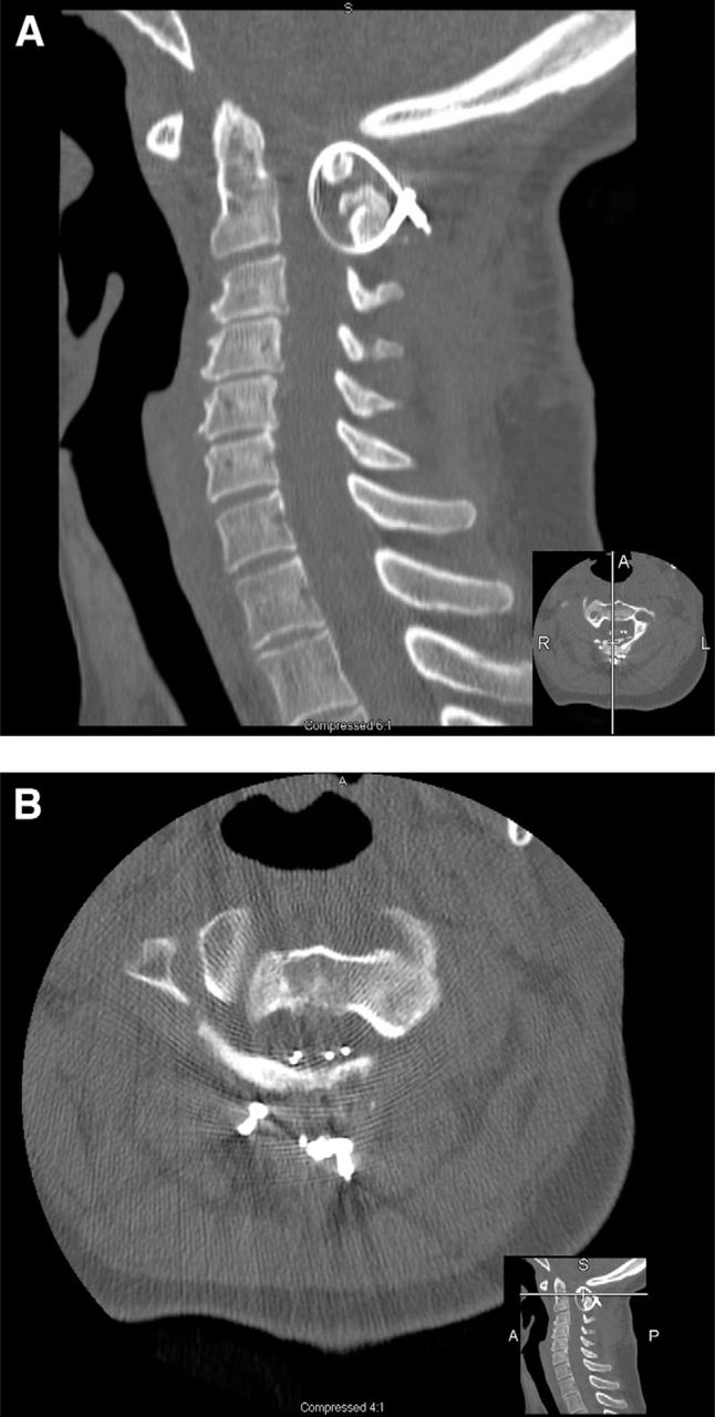
Sagittal (A) and axial (B) computed tomography images of cervical spine obtained at time of presentation to clinic.

Operation

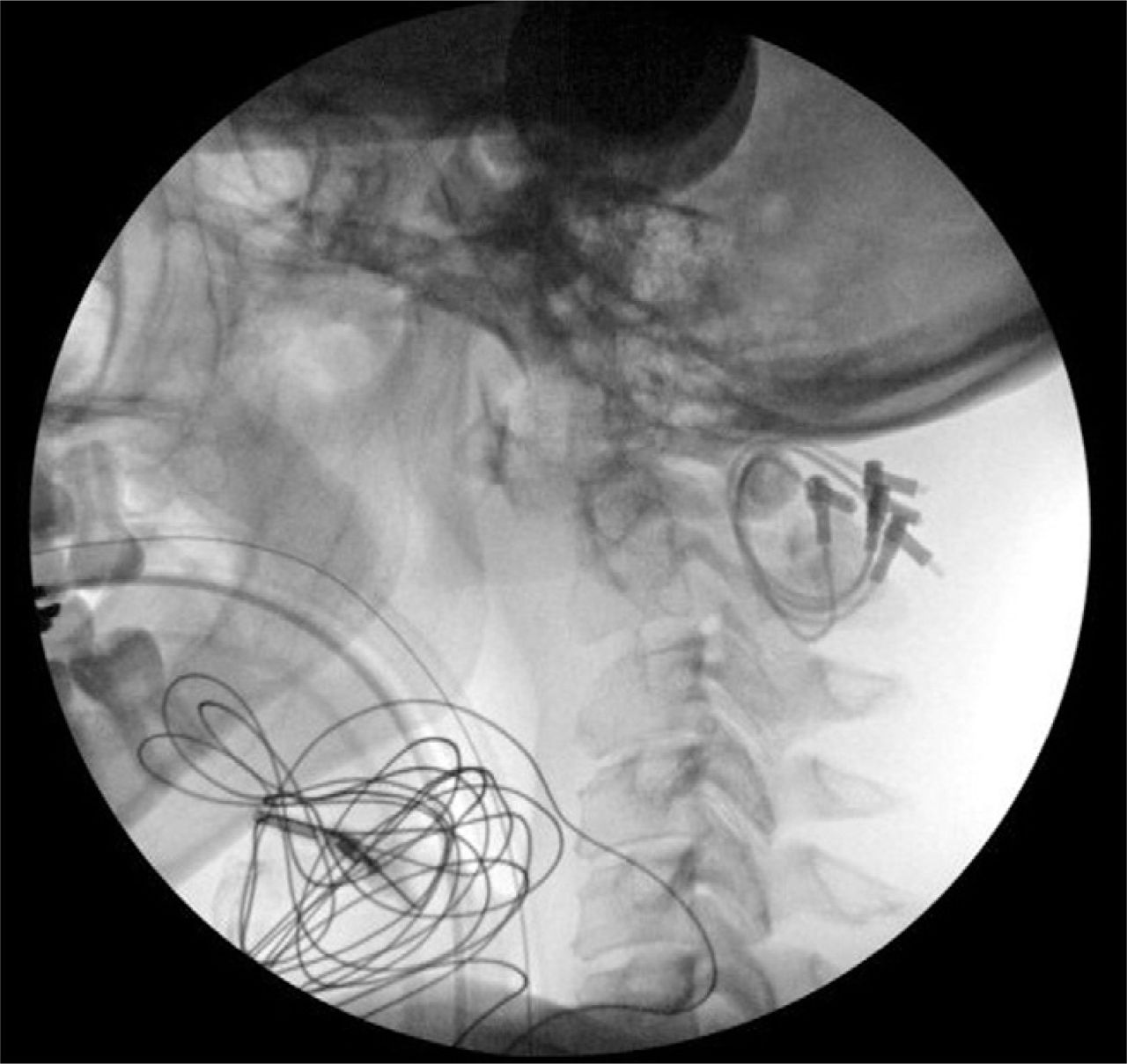
Intravenous antibiotics were administered, fiberoptic intubation was performed with succinylcholine, and both somatosensory evoked potentials (SSEPs) and motor evoked potentials (MEPs) were obtained in the supine position. The halo was removed, and the patient was positioned prone with her head attached to a Jackson frame via a Mayfield clamp. Prone SSEPs and MEPs were stable. Attempted closed reduction of the C1–2 subluxation was unsuccessful (Fig. 2).

Fig. 2
  • Download figure
  • Open in new tab
  • Download powerpoint
Fig. 2

Fluoroscopic intraoperative image showing failed closed reduction.

The patient's neck was then prepared and draped, and a midline subperiosteal approach was performed with sharp dissection and bipolar electrocautery to avoid monopolar contact with the C1–2 cables. With the cervical spine exposed from the occiput to C3, laminectomy of C1 and C2 was achieved with a high-speed bur. The laminae and cables were removed en bloc, and no cerebral spinal leak occurred.

Bone graft from the patient's prior procedure was fused to the undersurface of C1 and obscured the C1 lateral mass. Scarring made exposure of this area additionally difficult. As such, the decision was made to perform an occipitalcervical fusion. A midline occipital plate, C2 pars screws, and C3 lateral mass screws were placed. Reduction was performed as follows: A rod was contoured to connect the occipital plate and distal fixation. The cervical screws were locked, and the end caps in the occipital plate were left loose. A rod holder was placed adjacent to the occipital plate (Fig. 3). Distraction between the rod holder and the occipital plate was used to translate C2 anteriorly, effecting the reduction. The end caps on the occipital plate were then locked. Bone graft derived from the laminectomy and fusion mass was placed. Ceramic and a small strip of bone morphogenetic protein was used to completely fill the gap between the occiput and the cervical spine. The soft tissues were then closed in layers, a cervical collar was placed, and the patient was taken to the recovery room. SSEPs and MEPs were noted to be stable throughout the procedure. An immediate postoperative X-ray showed satisfactory reduction (Fig. 4).

Fig. 3
  • Download figure
  • Open in new tab
  • Download powerpoint
Fig. 3

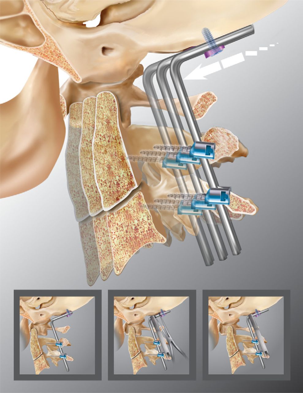
Instrumented reduction of atlantoaxial articulation using occipital plate.

Fig. 4
  • Download figure
  • Open in new tab
  • Download powerpoint
Fig. 4

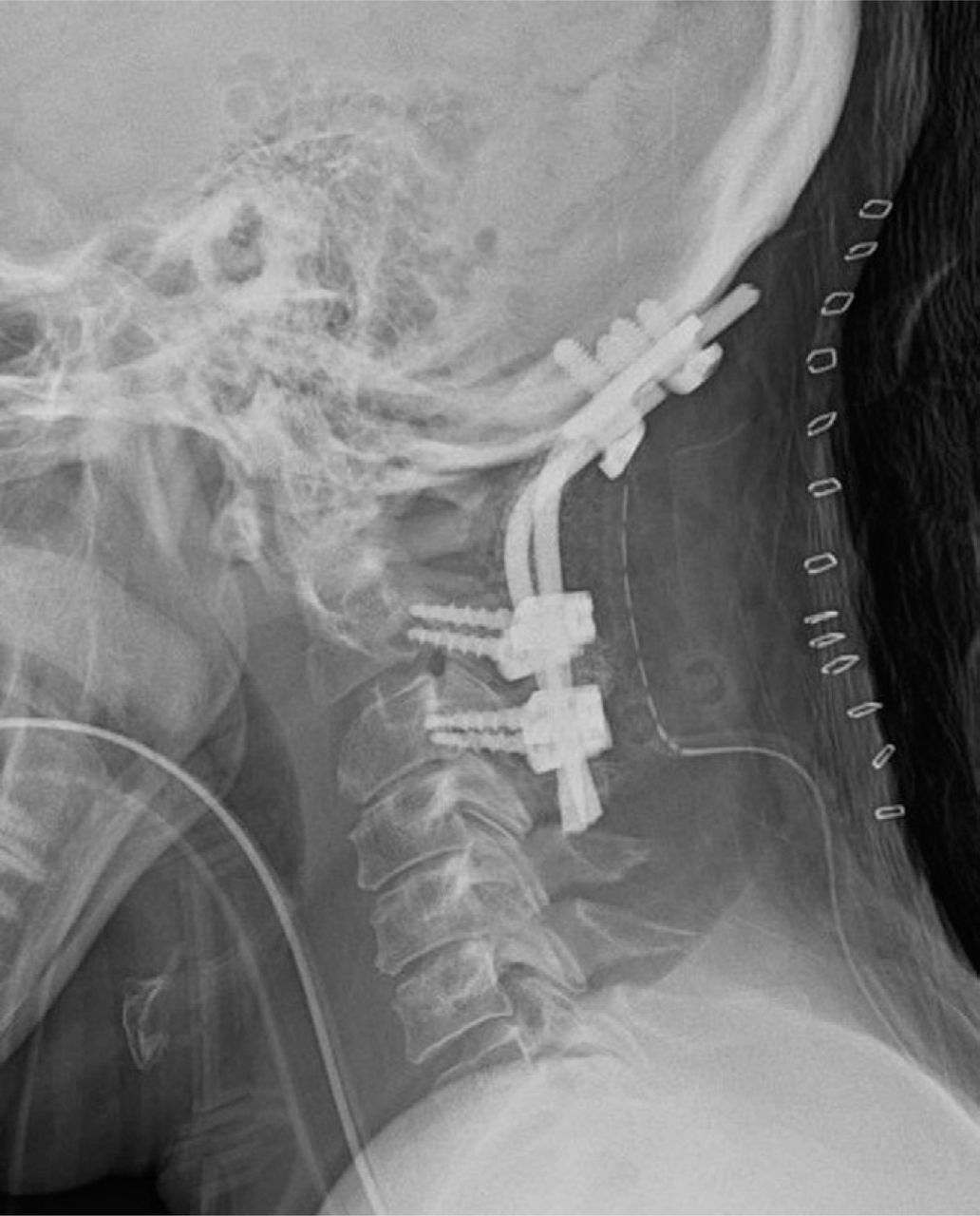
Immediate postoperative X-ray shows satisfactory reduction.

Postoperative course

A postoperative computed tomography scan showed satisfactory decompression and reduction (Fig. 5).

Fig. 5
  • Download figure
  • Open in new tab
  • Download powerpoint
Fig. 5

Postoperative axial computed tomography image showing maintenance of reduction.

The patient's recovery was unremarkable, and she was discharged home on postoperative day 4. In the early postoperative period, a right iliac vein deep venous thrombosis developed where a femoral line had been placed. (The patient's halo had made preoperative placement of an internal jugular or subclavian line impractical.) Nine months of warfarin therapy was begun with an international normalized ratio goal of 2.5 to 3. X-rays taken 3 months after surgery showed maintenance of the patient's cervical reduction.

Discussion

As in the reported case, inflammatory arthritis may both produce cervical instability and present obstacles to its resolution. The incidence of C1–2 instability in patients with rheumatoid arthritis, in particular, is 20% to 80%.11–13 Of note, lower extremity joint arthroplasty may mark an important opportunity for prophylaxis or intervention. Fifty percent of rheumatoid arthritis patients undergoing joint replacement surgery will have some form of cervical instability. Of these patients, 8% will show cervical subluxation requiring surgical treatment.14

Inflammatory or otherwise, craniocervical and atlantoaxial subluxation may produce cranial settling, basilar invagination, cervical stenosis, or a combination of each. Anterior resection of the odontoid with posterior fixation may beused in these scenarios, especially in cases of irreducible deformity.8 This strategy necessitates anterior dissection, however, and does not facilitate reduction. Accordingly, and corresponding to the diversity of subluxation-associated deformity, a variety of posteriorly based reduction techniques have been described. Goel and Laheri7 have treated cranial settling associated with C1–2 instability by release of the C1–2 joints and atlantoaxial or occipito-atlantoaxial instrumentation with plates and screws. Several authors have described treatment of basilar invagination due to atlantoaxial instability with posterior instrumented reduction using an occipital plate.8, 10 Other instrumented reduction techniques rely on C1 lateral mass screws.9 The reported technique draws on aspects of these modern procedures7, 8, 10 and may be used to treat cervical stenosis/instability, especially in cases in which placement of a C1 lateral mass screw is impossible (Fig. 6).

Fig. 6
  • Download figure
  • Open in new tab
  • Download powerpoint
Fig. 6

Example of lateral mass erosion preventing access to C1 lateral mass.

  • © 2013 ISASS - International Society for the Advancement of Spine Surgery. Published by Elsevier Inc. All rights reserved.

This is an Open Access article distributed under the terms of the Creative Commons Attribution-Noncommercial 3.0 Unported License, permitting all non-commercial use, distribution, and reproduction in any medium, provided the original work is properly cited.

References

  1. ↵
    1. Edwards CC,
    2. Bradford DS
    (1994) Instrumented reduction of spondylolisthesis. Spine 19:1538–41.
    1. Floman Y,
    2. Millgram MA,
    3. Ashkenazi E,
    4. Smorgick Y,
    5. Rand N
    (2008) Instrumented slip reduction and fusion for painful unstable isthmic spondylolisthesis in adults. J Spinal Disord Tech 21:477–83.
  2. ↵
    1. Pan J,
    2. Li L,
    3. Qian L,
    4. et al.
    (2011) Spontaneous slip reduction of low-grade isthmic spondylolisthesis following circumferential release via bilateral minimally invasive transforaminal lumbar interbody fusion: technical note and short-term outcome. Spine 36:283–9.
  3. ↵
    1. Korovessis P,
    2. Repantis T,
    3. Petsinis G,
    4. Iliopoulos P,
    5. Hadjipavlou A
    (2008) Direct reduction of thoracolumbar burst fractures by means of balloon kyphoplasty with calcium phosphate and stabilization with pediclescrew instrumentation and fusion. Spine 33:100–8.
  4. ↵
    1. Marco RA,
    2. Meyer BC,
    3. Kushwaha VP
    (2010) Thoracolumbar burst fractures treated with posterior decompression and pedicle screw instrumentation supplemented with balloon-assisted vertebroplasty and calcium phosphate reconstruction. Surgical technique. J Bone Joint Surg Br 92:67–76.
  5. ↵
    1. Mehdian S,
    2. Arun R
    (2011) A safe controlled instrumented reduction technique for cervical osteotomy in ankylosing spondylitis. Spine 36:715–20.
  6. ↵
    1. Goel A,
    2. Laheri V
    (1994) Plate and screw fixation for atlanto-axial subluxation. Acta Neurochir (Wien) 129:47–53.
  7. ↵
    1. Hsu W,
    2. Hasan Z,
    3. Suk I,
    4. Gokaslan Z,
    5. Wolinsky J
    (2010) A new technique for intraoperative reduction of occipitocervical instability operative. Neurosurgery 66:319–23.
  8. ↵
    1. O'Brien J,
    2. Gokaslan Z,
    3. Suk I,
    4. Wolinsky J,
    5. Riley L III.
    (2008) Open reduction of C1-2 subluxation with the use of C1 lateral mass and C2 translaminar screws. Neurosurgery 63:ONS95–8.
  9. ↵
    1. Peng X,
    2. Chen L,
    3. Wan Y,
    4. Zou X
    (2011) Treatment of primary basilar invagination by cervical traction and posterior instrumented reduction together with occipitocervical fusion. Spine (Phila Pa 1976) 36:1528–31.
  10. ↵
    1. Boden S,
    2. Dodge L,
    3. Bohlman H,
    4. Rechtine G
    (1993) Rheumatoid arthritis of the cervical spine. A long-term analysis with predictors of paralysis and recovery. J Bone Joint Surg Br 75:1282–97.
    1. Pellicci P,
    2. Ranawat C,
    3. Tsairis P,
    4. Bryan W,
    5. Röttger J,
    6. Siegel A
    (1981) A prospective study of the progression of rheumatoid arthritis of the cervical spine. J Bone Joint Surg Br 63:342–50.
  11. ↵
    1. Rana NA
    (1989) Natural history of atlanto-axial subluxation in rheumatoid arthritis. Spine (Phila Pa 1976) 14:1054–6.
  12. ↵
    1. Grauer JN,
    2. Tingstad EM,
    3. Rand N,
    4. Christie MJ,
    5. Hilibrand AS
    (2004) Predictors of paralysis in the rheumatoid cervical spine in patients undergoing total joint arthroplasty. J Bone Joint Surg Am 86:1420–4.
PreviousNext
Back to top

In this issue

International Journal of Spine Surgery
Vol. 7
1 Jan 2013
  • Table of Contents
  • Index by author

Print
Download PDF
Article Alerts
Sign In to Email Alerts with your Email Address
Email Article

Thank you for your interest in spreading the word on International Journal of Spine Surgery.

NOTE: We only request your email address so that the person you are recommending the page to knows that you wanted them to see it, and that it is not junk mail. We do not capture any email address.

Enter multiple addresses on separate lines or separate them with commas.
Instrumented reduction of a fixed C1–2 subluxation using occipital and C2/C3 fixation: A case report
(Your Name) has sent you a message from International Journal of Spine Surgery
(Your Name) thought you would like to see the International Journal of Spine Surgery web site.
CAPTCHA
This question is for testing whether or not you are a human visitor and to prevent automated spam submissions.
Citation Tools
Instrumented reduction of a fixed C1–2 subluxation using occipital and C2/C3 fixation: A case report
Clifton Meals, Rachel Harrison, Warren Yu, Joseph O'Brien
International Journal of Spine Surgery Jan 2013, 7 e20-e23; DOI: 10.1016/j.ijsp.2013.01.002

Citation Manager Formats

  • BibTeX
  • Bookends
  • EasyBib
  • EndNote (tagged)
  • EndNote 8 (xml)
  • Medlars
  • Mendeley
  • Papers
  • RefWorks Tagged
  • Ref Manager
  • RIS
  • Zotero
Share
Instrumented reduction of a fixed C1–2 subluxation using occipital and C2/C3 fixation: A case report
Clifton Meals, Rachel Harrison, Warren Yu, Joseph O'Brien
International Journal of Spine Surgery Jan 2013, 7 e20-e23; DOI: 10.1016/j.ijsp.2013.01.002
Twitter logo Facebook logo Mendeley logo
  • Tweet Widget
  • Facebook Like
  • Google Plus One

Jump to section

  • Article
    • Abstract
    • Case report
    • Discussion
    • References
  • Figures & Data
  • Info & Metrics
  • PDF

Related Articles

  • No related articles found.
  • Google Scholar

Cited By...

  • No citing articles found.
  • Google Scholar

More in this TOC Section

  • Kyphoplasty versus vertebroplasty for painful osteoporotic vertebral compression fractures—which one is better? A systematic review and meta-analysis
  • Overtightening of halo pins resulting in intracranial penetration, pneumocephalus, and epileptic seizure
  • Retrospective cost analysis of cervical laminectomy and fusion versus cervical laminoplasty in the treatment of cervical spondylotic myelopathy
Show more Full Length Article

Similar Articles

Keywords

  • cervical spine
  • Subluxation
  • reduction
  • Instrumented
  • rheumatoid arthritis

Content

  • Current Issue
  • Latest Content
  • Archive

More Information

  • About IJSS
  • About ISASS
  • Privacy Policy

More

  • Subscribe
  • Alerts
  • Feedback

Other Services

  • Author Instructions
  • Join ISASS
  • Reprints & Permissions

© 2025 International Journal of Spine Surgery

International Journal of Spine Surgery Online ISSN: 2211-4599

Powered by HighWire