
Main menu

  • Home
  • Content
    • Current Issue
    • Advance Online Publication
    • Archive
  • About Us
    • About ISASS
    • About the Journal
    • Author Instructions
    • Editorial Board
    • Reviewer Guidelines & Publication Criteria
  • More
    • Advertise
    • Subscribe
    • Alerts
    • Feedback
  • Join Us
  • Reprints & Permissions
  • Sponsored Content
  • Other Publications
    • ijss

User menu

  • My alerts

Search

  • Advanced search
International Journal of Spine Surgery
  • My alerts
International Journal of Spine Surgery

Advanced Search

  • Home
  • Content
    • Current Issue
    • Advance Online Publication
    • Archive
  • About Us
    • About ISASS
    • About the Journal
    • Author Instructions
    • Editorial Board
    • Reviewer Guidelines & Publication Criteria
  • More
    • Advertise
    • Subscribe
    • Alerts
    • Feedback
  • Join Us
  • Reprints & Permissions
  • Sponsored Content
  • Follow ijss on Twitter
  • Visit ijss on Facebook
Research ArticleMinimally Invasive Surgery

Feasibility of Endoscopic Inspection of Pedicle Wall Integrity in a Live Surgery Model

KRISTEN RADCLIFF, HARVEY SMITH, BOBBY KALANTAR, ROBERT ISAACS, BARRETT WOODS, ALEXANDER R. VACCARO and JAMES BRANNON
International Journal of Spine Surgery April 2018, 12 (2) 241-249; DOI: https://doi.org/10.14444/5030
KRISTEN RADCLIFF
1Department of Orthopedic Surgery, Thomas Jefferson University, Rothman Institute, Egg Harbor, New Jersey
MD
  • Find this author on Google Scholar
  • Find this author on PubMed
  • Search for this author on this site
HARVEY SMITH
2Department of Orthopedic Surgery, University of Pennsylvania, Philadelphia, Pennsylvania
MD
  • Find this author on Google Scholar
  • Find this author on PubMed
  • Search for this author on this site
BOBBY KALANTAR
3Department of Orthopedic Surgery, Georgetown University, Washington, DC
MD
  • Find this author on Google Scholar
  • Find this author on PubMed
  • Search for this author on this site
ROBERT ISAACS
4Department of Neurological Surgery, Duke University, Durham, North Carolina
MD
  • Find this author on Google Scholar
  • Find this author on PubMed
  • Search for this author on this site
BARRETT WOODS
1Department of Orthopedic Surgery, Thomas Jefferson University, Rothman Institute, Egg Harbor, New Jersey
MD
  • Find this author on Google Scholar
  • Find this author on PubMed
  • Search for this author on this site
ALEXANDER R. VACCARO
1Department of Orthopedic Surgery, Thomas Jefferson University, Rothman Institute, Egg Harbor, New Jersey
MD, PHD, MBA
  • Find this author on Google Scholar
  • Find this author on PubMed
  • Search for this author on this site
JAMES BRANNON
5Orthopedic Sciences, Inc, Seal Beach, California, Joint Preservation Institute of Kansas, Overland Park, Kansas
MD
  • Find this author on Google Scholar
  • Find this author on PubMed
  • Search for this author on this site
  • Article
  • Figures & Data
  • Info & Metrics
  • PDF
Loading

Article Figures & Data

Figures

  • Figure 1
    • Download figure
    • Open in new tab
    • Download powerpoint
    Figure 1

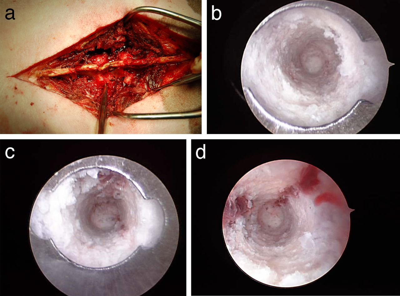
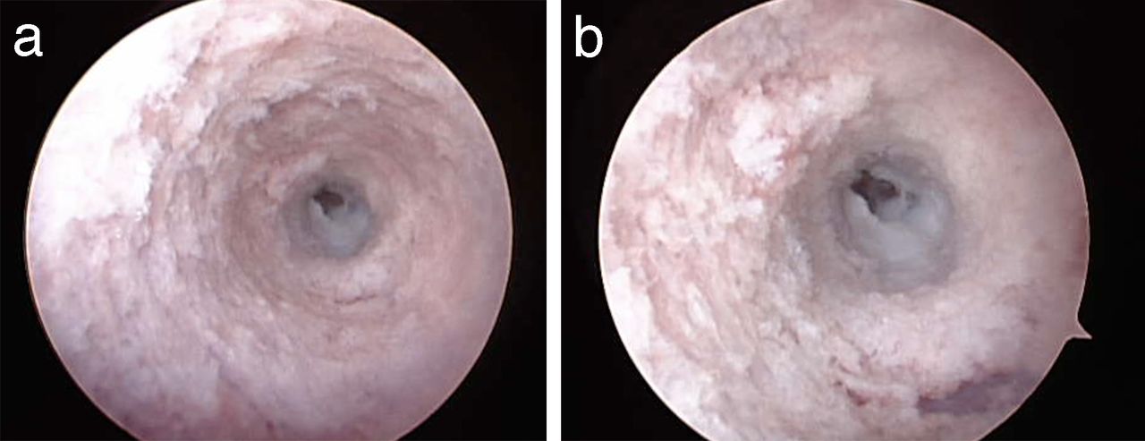
    (a) Photograph of the instrument in the wound while visualization was being performed. The instrument is visible adjacent to the lumbar facet joints. Cranial is to the left and caudal is to the right. There was only trace fluid extravasation of irrigation from into the wound. The endoscopic instrument is designed to achieve a seal with the endosteal pedicle surface. (b) Endoscopic view of an intact pedicle. The distal tip of the cannula with batwing indentation to determine orientation is visible. A complete tube of ivory bone is visible through the cannula. Medial is to the right side of the screen. (c) Endoscopic view of an intact pedicle. A complete tube of white bone is visible through the cannula. At the 12 o'clock position, red, cancellous bone is visible. (d) Endoscopic view of an intact pedicle. A complete tube of white bone is visible through the cannula. At the 1 o'clock and 9 o'clock positions, red, cancellous bone and marrow are visible.

  • Figure 2
    • Download figure
    • Open in new tab
    • Download powerpoint
    Figure 2

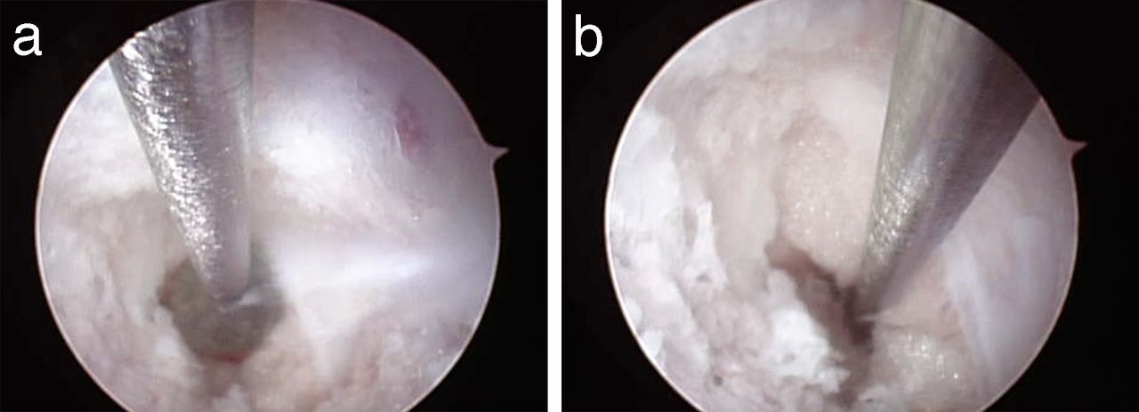
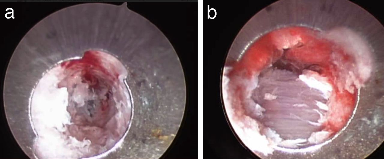
    (a) Endoscopic view of a medial pedicle wall breach into the spinal canal. The triangular breach was deliberately created with a gearshift. Within the breach linear vertical fragments of ligamentum flavum are visible. (b) Close-up view of the medial pedicle breach. At approximately 4 o'clock yellow epidural fat is visible in the defect.

  • Figure 3
    • Download figure
    • Open in new tab
    • Download powerpoint
    Figure 3

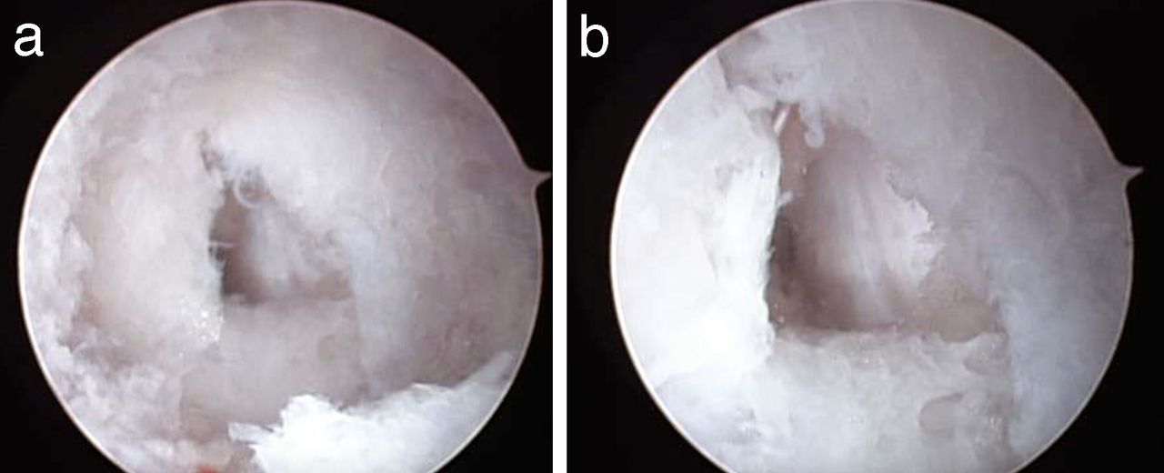
    (a) Endoscopic view of anterior pedicle breach. The pedicle side walls display ivory cortical bone. The white anterior longitudinal ligament is visible in the center of the screen. It is lighter colored than the adjacent, ivory colored bone. There is a square-shared defect in the anterior longitudinal ligament. (b) Close-up view of the anterior pedicle breach. The white anterior longitudinal ligament and anterior defect are visible. The surrounding bone walls are intact.

  • Figure 4
    • Download figure
    • Open in new tab
    • Download powerpoint
    Figure 4

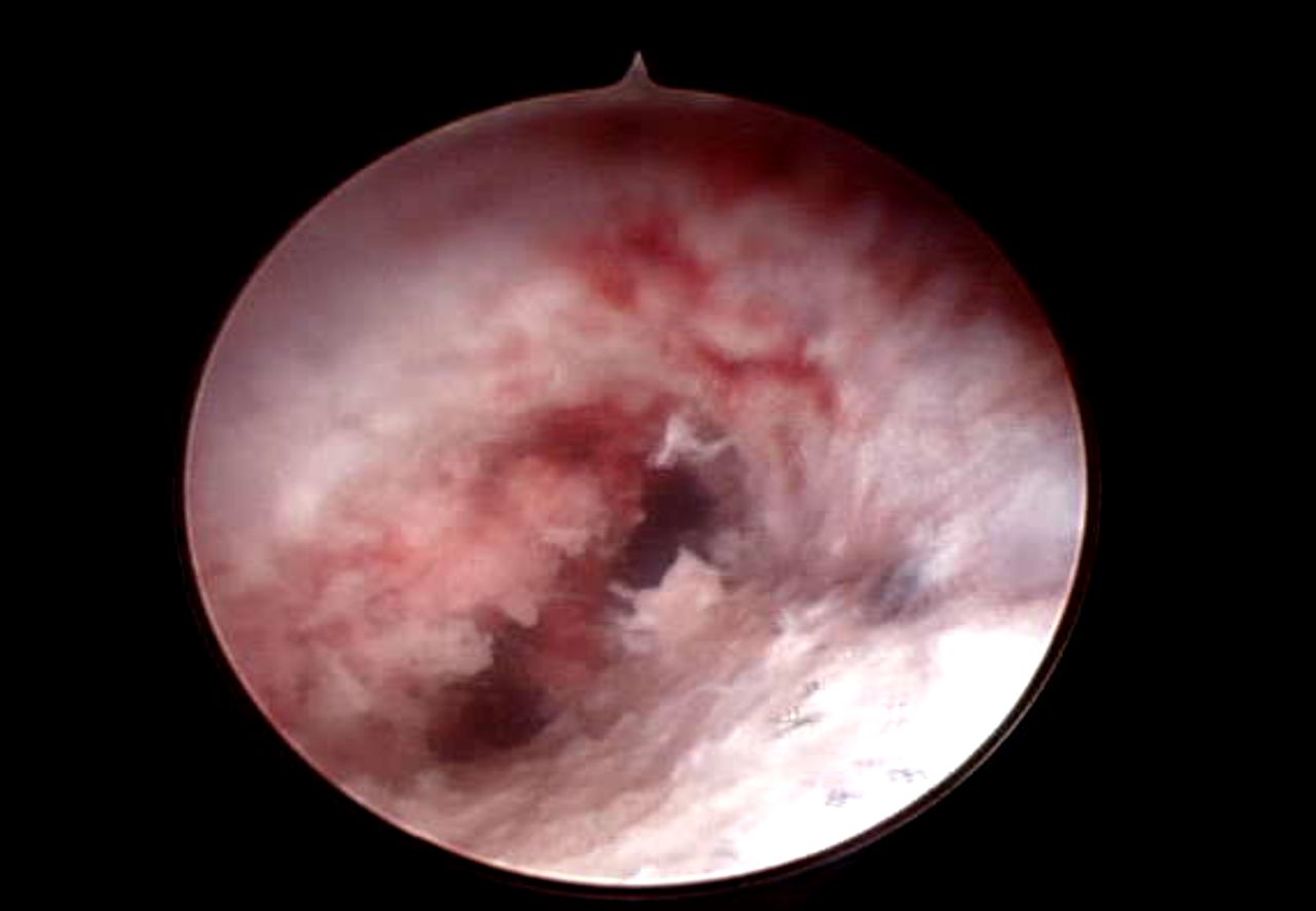
    (a) Endoscopic view of lateral pedicle wall breach. In the center-left of the field there are purple horizontal fibers of paraspinous muscles. (b) Close-up view of lateral pedicle wall breach. The horizontal muscle fibers are visible through the defect.

  • Figure 5
    • Download figure
    • Open in new tab
    • Download powerpoint
    Figure 5

    (a) Endoscopic view with ball-tip probe inside of the pedicle. The endoscopic instrument is alongside the ball-tip probe. The ball is adjacent to a defect in the medial pedicle wall. Epidural fat is distal to the tip of the ball. Ligamentum flavum is to the right side of the ball. (b) Probing the spinal canal defect. The ball-tip probe has been advanced under endoscopic guidance into the spinal canal outside of the pedicle. Epidural fat is adjacent to the shaft of the probe. The ligamentum flavum has been reflected laterally.

  • Figure 6
    • Download figure
    • Open in new tab
    • Download powerpoint
    Figure 6

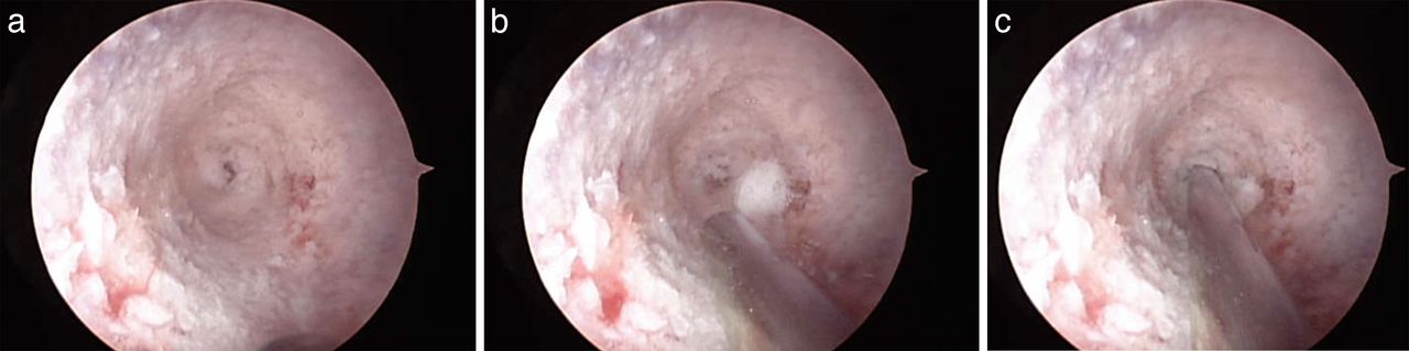
    (a) Endoscopic view with anterior defect in the center of the field. The remainder of the pedicle walls are intact. (b) Endoscopic view with anterior defect in the center of the field. A guidewire is being advanced alongside the endoscopic instrument at the 6 o'clock position. (c) Guidewire placed into the anterior vertebral body defect under endoscopic guidance.

  • Figure 7
    • Download figure
    • Open in new tab
    • Download powerpoint
    Figure 7

    Endoscopic view of a pedicle with 2 divergent paths. One path is at approximately 2 o'clock and one path is at 7 o'clock.

  • Figure 8
    • Download figure
    • Open in new tab
    • Download powerpoint
    Figure 8

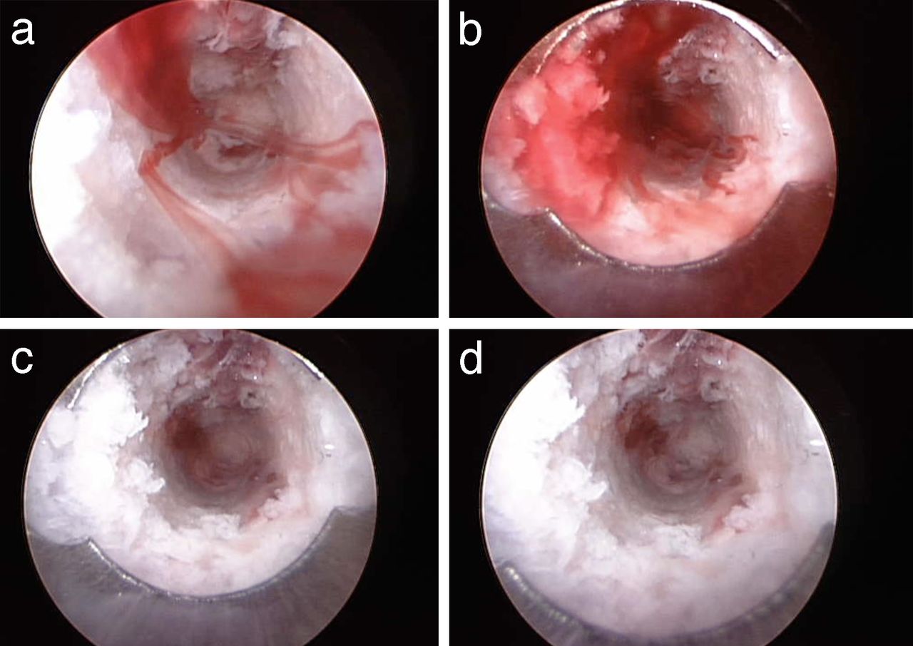
    (a) Endoscopic view of a pedicle with intact walls (Figure 1b) immediately after irrigation fluid was stopped. Bony bleeding resumed immediately. The bleeding appears to have formed linear, ribbon-shaped lamellae. (b) Endoscopic view of a pedicle with intact walls with bleeding allowed to continue further. Blood filled the entire inner aspect of the pedicle. (c) Endoscopic view of pedicle with irrigation reestablished. The blood began to clear. (d) Endoscopic view of pedicle with irrigation reestablished. The blood cleared easily.

PreviousNext
Back to top

In this issue

International Journal of Spine Surgery
Vol. 12, Issue 2
1 Apr 2018
  • Table of Contents
  • Index by author

Print
Download PDF
Article Alerts
Sign In to Email Alerts with your Email Address
Email Article

Thank you for your interest in spreading the word on International Journal of Spine Surgery.

NOTE: We only request your email address so that the person you are recommending the page to knows that you wanted them to see it, and that it is not junk mail. We do not capture any email address.

Enter multiple addresses on separate lines or separate them with commas.
Feasibility of Endoscopic Inspection of Pedicle Wall Integrity in a Live Surgery Model
(Your Name) has sent you a message from International Journal of Spine Surgery
(Your Name) thought you would like to see the International Journal of Spine Surgery web site.
CAPTCHA
This question is for testing whether or not you are a human visitor and to prevent automated spam submissions.
Citation Tools
Feasibility of Endoscopic Inspection of Pedicle Wall Integrity in a Live Surgery Model
KRISTEN RADCLIFF, HARVEY SMITH, BOBBY KALANTAR, ROBERT ISAACS, BARRETT WOODS, ALEXANDER R. VACCARO, JAMES BRANNON
International Journal of Spine Surgery Apr 2018, 12 (2) 241-249; DOI: 10.14444/5030

Citation Manager Formats

  • BibTeX
  • Bookends
  • EasyBib
  • EndNote (tagged)
  • EndNote 8 (xml)
  • Medlars
  • Mendeley
  • Papers
  • RefWorks Tagged
  • Ref Manager
  • RIS
  • Zotero
Share
Feasibility of Endoscopic Inspection of Pedicle Wall Integrity in a Live Surgery Model
KRISTEN RADCLIFF, HARVEY SMITH, BOBBY KALANTAR, ROBERT ISAACS, BARRETT WOODS, ALEXANDER R. VACCARO, JAMES BRANNON
International Journal of Spine Surgery Apr 2018, 12 (2) 241-249; DOI: 10.14444/5030
Twitter logo Facebook logo Mendeley logo
  • Tweet Widget
  • Facebook Like
  • Google Plus One

Jump to section

  • Article
    • ABSTRACT
    • INTRODUCTION
    • METHODS
    • RESULTS
    • DISCUSSION
    • APPENDIX
    • Footnotes
    • REFERENCES
  • Figures & Data
  • Info & Metrics
  • PDF

Related Articles

  • No related articles found.
  • PubMed
  • Google Scholar

Cited By...

  • No citing articles found.
  • Google Scholar

More in this TOC Section

  • Comparative Review of Lateral and Oblique Lumbar Interbody Fusion: Technique, Outcomes, and Complications
  • Key Considerations in Surgical Decision-Making on the Side of Approach for Lumbar Lateral Transpsoas Interbody Fusion Techniques
  • A Spine Surgeon’s Learning Curve With the Minimally Invasive L5 to S1 Lateral ALIF Surgical Approach: Perioperative Outcomes and Technical Considerations
Show more Minimally Invasive Surgery

Similar Articles

Keywords

  • endoscopic spine surgery
  • endoscopy
  • intraoosseous endoscopy
  • spine fusion
  • spine instrumentation
  • pedicle screws
  • pedicle cannulation
  • minimally invasive spine surgery

Content

  • Current Issue
  • Latest Content
  • Archive

More Information

  • About IJSS
  • About ISASS
  • Privacy Policy

More

  • Subscribe
  • Alerts
  • Feedback

Other Services

  • Author Instructions
  • Join ISASS
  • Reprints & Permissions

© 2025 International Journal of Spine Surgery

International Journal of Spine Surgery Online ISSN: 2211-4599

Powered by HighWire