
Main menu

  • Home
  • Content
    • Current Issue
    • Advance Online Publication
    • Archive
  • About Us
    • About ISASS
    • About the Journal
    • Author Instructions
    • Editorial Board
    • Reviewer Guidelines & Publication Criteria
  • More
    • Advertise
    • Subscribe
    • Alerts
    • Feedback
  • Join Us
  • Reprints & Permissions
  • Sponsored Content
  • Other Publications
    • ijss

User menu

  • My alerts

Search

  • Advanced search
International Journal of Spine Surgery
  • My alerts
International Journal of Spine Surgery

Advanced Search

  • Home
  • Content
    • Current Issue
    • Advance Online Publication
    • Archive
  • About Us
    • About ISASS
    • About the Journal
    • Author Instructions
    • Editorial Board
    • Reviewer Guidelines & Publication Criteria
  • More
    • Advertise
    • Subscribe
    • Alerts
    • Feedback
  • Join Us
  • Reprints & Permissions
  • Sponsored Content
  • Follow ijss on Twitter
  • Visit ijss on Facebook
Research ArticleNew Technology

Clinical Application of a High Definition Three-Dimensional Exoscope in Anterior Lumbar Interbody Fusion: Technical Note

MANUELA D'ERCOLE, ELENA SERCHI, MATTEO ZANELLO, TOMMASO TUFO and CARMELO STURIALE
International Journal of Spine Surgery December 2020, 14 (6) 1003-1008; DOI: https://doi.org/10.14444/7150
MANUELA D'ERCOLE
1Department of Neurosurgery, Fondazione Policlinico Universitario A. Gemelli Roma, Italy
MD, PHD
  • Find this author on Google Scholar
  • Find this author on PubMed
  • Search for this author on this site
ELENA SERCHI
2Department of Neurosurgery, IRCCS Istituto delle Scienze Neurologiche Bologna– Bellaria Hospital, Bologna Italy
MD
  • Find this author on Google Scholar
  • Find this author on PubMed
  • Search for this author on this site
MATTEO ZANELLO
3Department of Surgery, Maggiore Hospital, Bologna Italy
MD
  • Find this author on Google Scholar
  • Find this author on PubMed
  • Search for this author on this site
TOMMASO TUFO
4Department of Neurosurgery, Fondazione Policlinico Universitario A. Gemelli Roma, Italy
MD, PHD
  • Find this author on Google Scholar
  • Find this author on PubMed
  • Search for this author on this site
CARMELO STURIALE
5Department of Neurosurgery, IRCCS Istituto delle Scienze Neurologiche Bologna– Bellaria Hospital, Bologna Italy
MD
  • Find this author on Google Scholar
  • Find this author on PubMed
  • Search for this author on this site
  • Article
  • Figures & Data
  • Info & Metrics
  • PDF
Loading

Article Figures & Data

Figures

  • Figure 1
    • Download figure
    • Open in new tab
    • Download powerpoint
    Figure 1

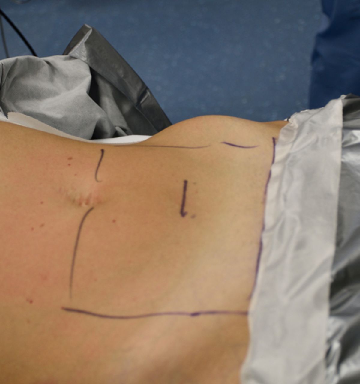
    Surgical exposure of anterior lumbosacral spine is obtained through a skin incision in lower abdominal quadrants. Our preliminary experience is based on a linear horizontal skin incision of 6 to 8 cm in length, 5 to 7 cm beneath the umbilicus.

  • Figure 2
    • Download figure
    • Open in new tab
    • Download powerpoint
    Figure 2

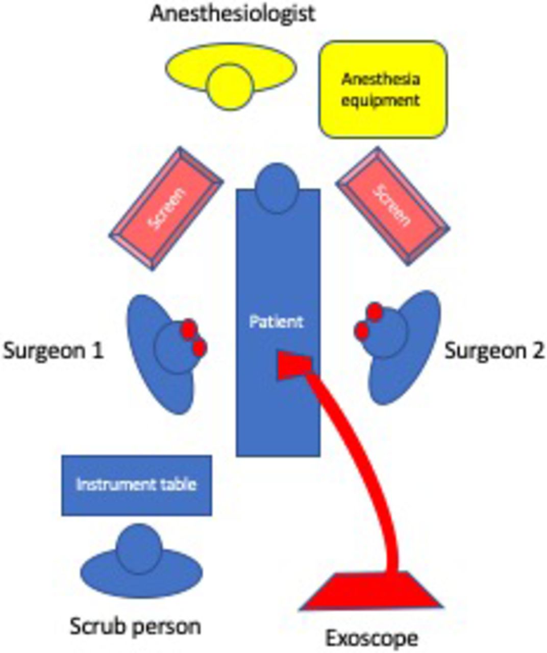
    Exoscope video-assisted telescope operating monitor (VITOM) 3D in the operating setting, kept in position by an articulated holding arm, provides an unobstructed working space. The surgical field is displayed on 2 movable 32” 3D monitors positioned on the opposite side of the surgical table respect to surgeon and assistant positions, which are counterposed and wear 3D glasses. Images displayed on the 2 screens are specular to each other to reproduce the intraoperative sight with the correct orientation for each surgeon and ease surgical maneuvers. It is possible to shift from microscopic to macroscopic vision without moving the scope or completely losing microscopic vision, if necessary.

  • Figure 3
    • Download figure
    • Open in new tab
    • Download powerpoint
    Figure 3

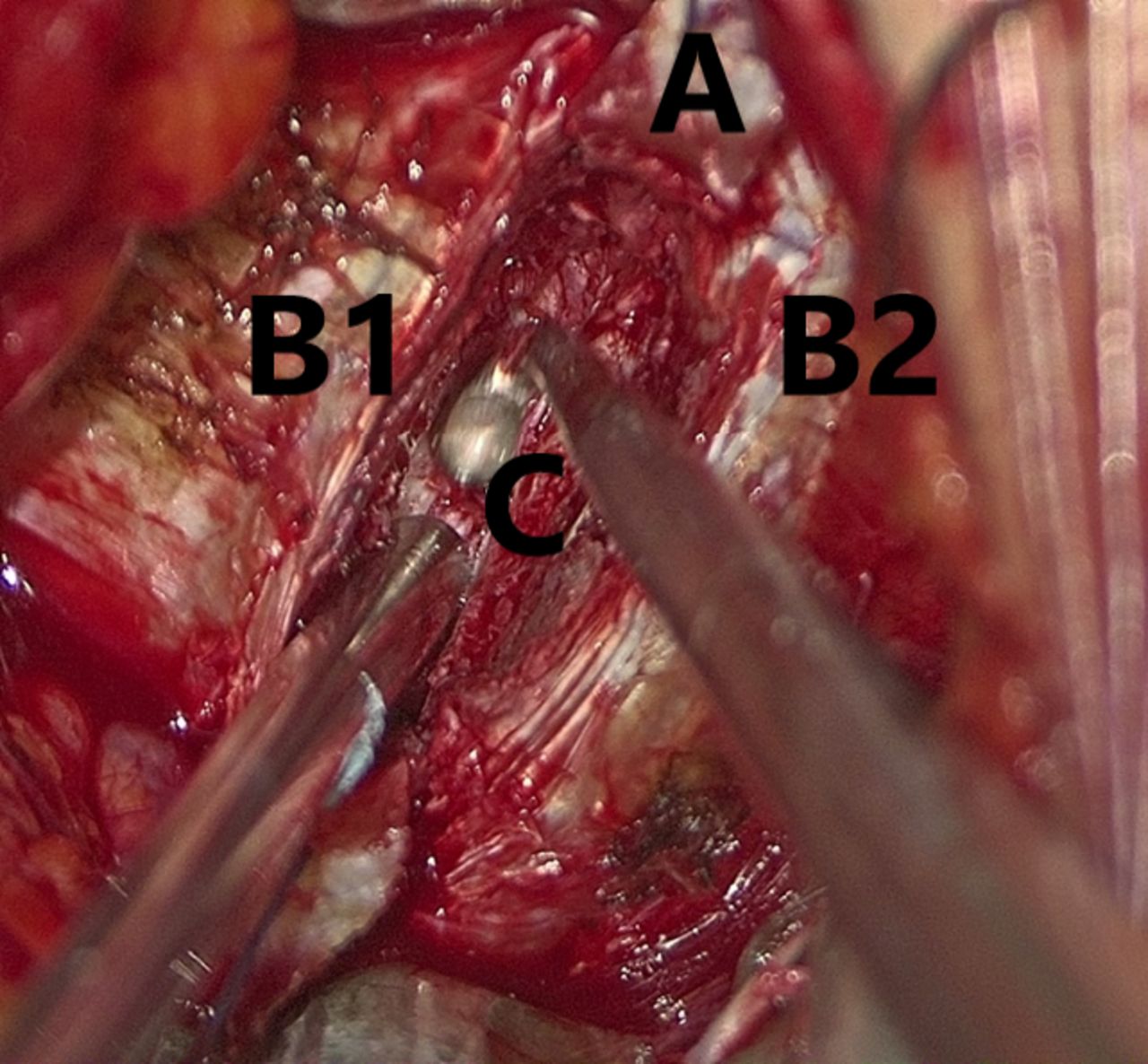
    After incision and lateral retraction of anterior longitudinal ligament (A), discectomy and curetting of upper (B1) and lower (B2) endplates is performed under exoscopic magnification. The intervertebral space (C) is then prepared for insertion of the cage.

  • Figure 4
    • Download figure
    • Open in new tab
    • Download powerpoint
    Figure 4

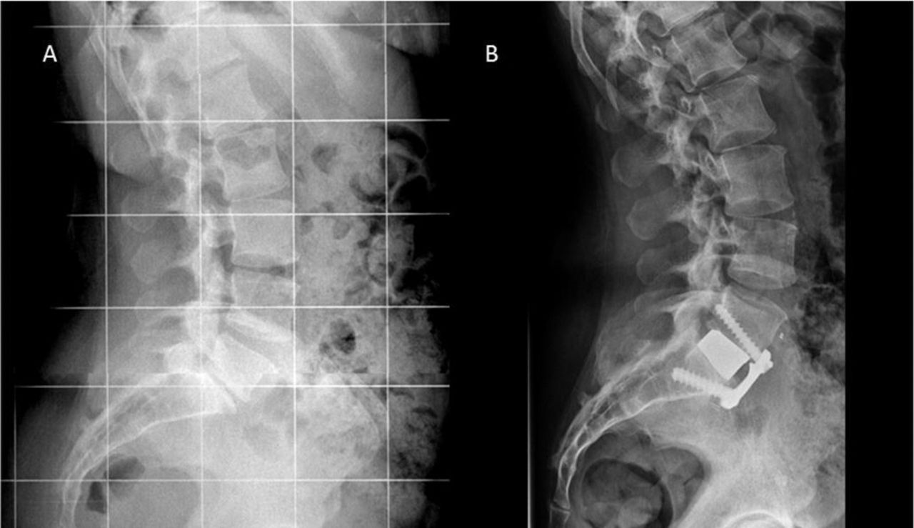
    L5-S1 anterior lumbar interbody fusion (ALIF): X-rays presurgery (A) and postsurgery (B). Anterior access to the intervertebral space allows an easier insertion of angulated cages, when compared with posterior approaches, thus generating a satisfactory restoration of lumbar lordosis and spino-pelvic balance. Moreover, the restoration of disc height obtains increase of neuroforaminal volume and consequent relief of radicular symptoms due to indirect decompression.

PreviousNext
Back to top

In this issue

International Journal of Spine Surgery
Vol. 14, Issue 6
1 Dec 2020
  • Table of Contents
  • Index by author

Print
Download PDF
Article Alerts
Sign In to Email Alerts with your Email Address
Email Article

Thank you for your interest in spreading the word on International Journal of Spine Surgery.

NOTE: We only request your email address so that the person you are recommending the page to knows that you wanted them to see it, and that it is not junk mail. We do not capture any email address.

Enter multiple addresses on separate lines or separate them with commas.
Clinical Application of a High Definition Three-Dimensional Exoscope in Anterior Lumbar Interbody Fusion: Technical Note
(Your Name) has sent you a message from International Journal of Spine Surgery
(Your Name) thought you would like to see the International Journal of Spine Surgery web site.
CAPTCHA
This question is for testing whether or not you are a human visitor and to prevent automated spam submissions.
Citation Tools
Clinical Application of a High Definition Three-Dimensional Exoscope in Anterior Lumbar Interbody Fusion: Technical Note
MANUELA D'ERCOLE, ELENA SERCHI, MATTEO ZANELLO, TOMMASO TUFO, CARMELO STURIALE
International Journal of Spine Surgery Dec 2020, 14 (6) 1003-1008; DOI: 10.14444/7150

Citation Manager Formats

  • BibTeX
  • Bookends
  • EasyBib
  • EndNote (tagged)
  • EndNote 8 (xml)
  • Medlars
  • Mendeley
  • Papers
  • RefWorks Tagged
  • Ref Manager
  • RIS
  • Zotero
Share
Clinical Application of a High Definition Three-Dimensional Exoscope in Anterior Lumbar Interbody Fusion: Technical Note
MANUELA D'ERCOLE, ELENA SERCHI, MATTEO ZANELLO, TOMMASO TUFO, CARMELO STURIALE
International Journal of Spine Surgery Dec 2020, 14 (6) 1003-1008; DOI: 10.14444/7150
Twitter logo Facebook logo Mendeley logo
  • Tweet Widget
  • Facebook Like
  • Google Plus One

Jump to section

  • Article
    • ABSTRACT
    • INTRODUCTION
    • MATERIALS AND METHODS
    • RESULTS
    • DISCUSSION
    • CONCLUSIONS
    • Footnotes
    • REFERENCES
  • Figures & Data
  • Info & Metrics
  • PDF

Related Articles

  • No related articles found.
  • PubMed
  • Google Scholar

Cited By...

  • No citing articles found.
  • Google Scholar

More in this TOC Section

  • High Uptake Detection for Spinal Degenerative Changes: A Comparison Between Bone Scintigraphy and Single Photon Emission Computed Tomography Combined With High-Resolution Computed Tomography
  • Accuracy of Pedicle Screw Placement Using the ExcelsiusGPS Robotic Navigation Platform: An Analysis of 728 Screws
  • A Network Meta-Analysis Comparing the Efficacy and Safety of Pedicle Screw Placement Techniques Using Intraoperative Conventional, Navigation, Robot-Assisted, and Augmented Reality Guiding Systems
Show more New Technology

Similar Articles

Keywords

  • anterior lumbar interbody fusion
  • video-assisted telescope operating monitor
  • minimally invasive spine surgery
  • exoscopy

Content

  • Current Issue
  • Latest Content
  • Archive

More Information

  • About IJSS
  • About ISASS
  • Privacy Policy

More

  • Subscribe
  • Alerts
  • Feedback

Other Services

  • Author Instructions
  • Join ISASS
  • Reprints & Permissions

© 2025 International Journal of Spine Surgery

International Journal of Spine Surgery Online ISSN: 2211-4599

Powered by HighWire