
Main menu

  • Home
  • Content
    • Current Issue
    • Advance Online Publication
    • Archive
  • About Us
    • About ISASS
    • About the Journal
    • Author Instructions
    • Editorial Board
    • Reviewer Guidelines & Publication Criteria
  • More
    • Advertise
    • Subscribe
    • Alerts
    • Feedback
  • Join Us
  • Reprints & Permissions
  • Sponsored Content
  • Other Publications
    • ijss

User menu

  • My alerts

Search

  • Advanced search
International Journal of Spine Surgery
  • My alerts
International Journal of Spine Surgery

Advanced Search

  • Home
  • Content
    • Current Issue
    • Advance Online Publication
    • Archive
  • About Us
    • About ISASS
    • About the Journal
    • Author Instructions
    • Editorial Board
    • Reviewer Guidelines & Publication Criteria
  • More
    • Advertise
    • Subscribe
    • Alerts
    • Feedback
  • Join Us
  • Reprints & Permissions
  • Sponsored Content
  • Follow ijss on Twitter
  • Visit ijss on Facebook
Research ArticleMinimally Invasive Surgery

Full-Endoscopic Oblique Lateral Lumbar Interbody Fusion: A Technical Note With 1-Year Follow-Up

Zhen-Zhou Li, Jin-Chang Wang, Zheng Cao, Hong-Liang Zhao, Kai-Uwe Lewandrowski and Anthony Yeung
International Journal of Spine Surgery June 2021, 15 (3) 504-513; DOI: https://doi.org/10.14444/8072
Zhen-Zhou Li
1Department of Orthopedic Surgery, the Fourth Medical Center of Chinese PLA General Hospital, Beijing, China
MD
  • Find this author on Google Scholar
  • Find this author on PubMed
  • Search for this author on this site
Jin-Chang Wang
1Department of Orthopedic Surgery, the Fourth Medical Center of Chinese PLA General Hospital, Beijing, China
MD
  • Find this author on Google Scholar
  • Find this author on PubMed
  • Search for this author on this site
Zheng Cao
1Department of Orthopedic Surgery, the Fourth Medical Center of Chinese PLA General Hospital, Beijing, China
MD
  • Find this author on Google Scholar
  • Find this author on PubMed
  • Search for this author on this site
Hong-Liang Zhao
1Department of Orthopedic Surgery, the Fourth Medical Center of Chinese PLA General Hospital, Beijing, China
MD
  • Find this author on Google Scholar
  • Find this author on PubMed
  • Search for this author on this site
Kai-Uwe Lewandrowski
2Center for Advanced Spine Care of Southern Arizona and Surgical Institute of Tucson, Arizona; Visiting Professor Department of Orthopaedics, Fundación Universitaria Sanitas, Bogotá, D.C., Colombia; and Department of Neurosurgery, Universidade Federal do Estado do Rio de Janeiro, Rio de Janeiro, Brazil
MD
  • Find this author on Google Scholar
  • Find this author on PubMed
  • Search for this author on this site
Anthony Yeung
3Desert Institute for Spine Care, Phoenix, Arizona; Executive Director International Intradiscal Therapy Society, Phoenix, Arizona
MD
  • Find this author on Google Scholar
  • Find this author on PubMed
  • Search for this author on this site
  • Article
  • Figures & Data
  • Info & Metrics
  • PDF
Loading

Article Figures & Data

Figures

  • Tables
  • Figure 1
    • Download figure
    • Open in new tab
    • Download powerpoint
    Figure 1

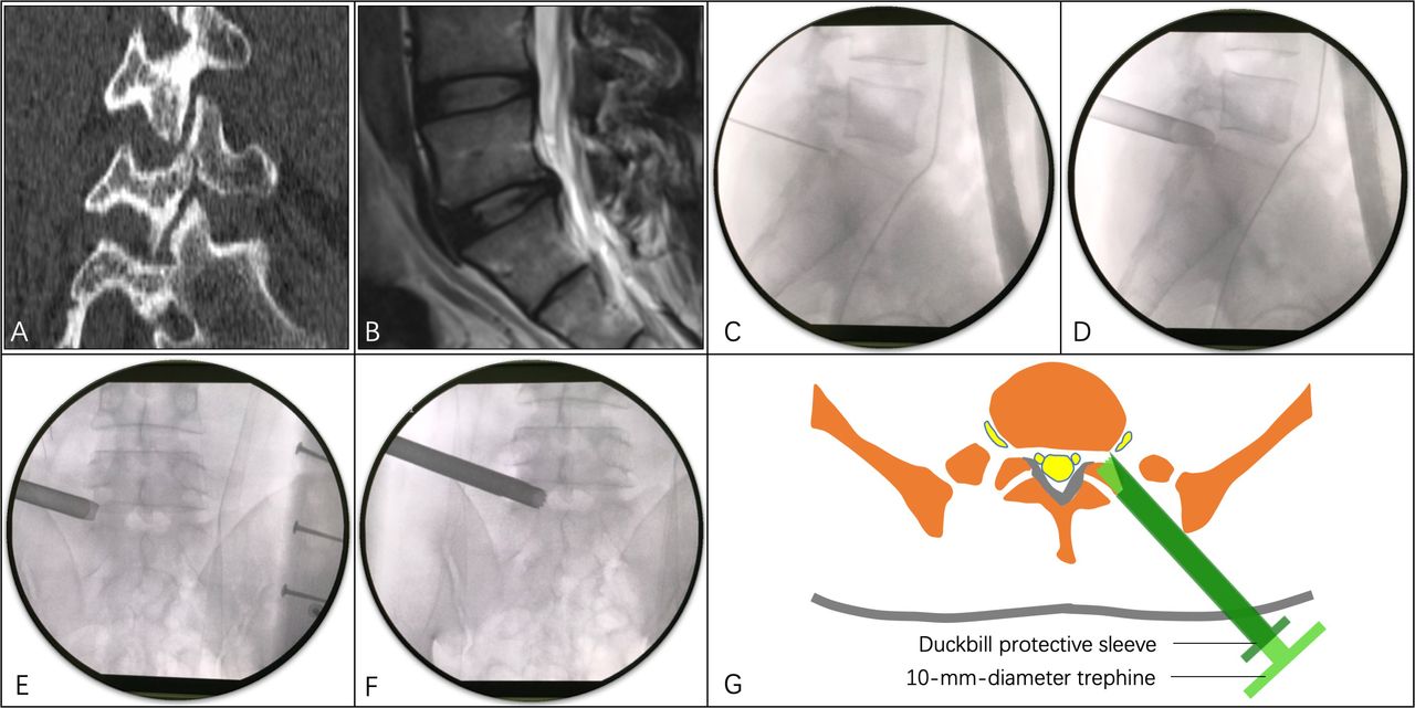
    Case 1: a 32-year-old male with discogenic low back pain and spondylolysis. (A) Preoperative computed tomography sagittal reconstruction showed L5 spondylolysis. (B) Preoperative sagittal T2-weighted magnetic resonance imaging showed a degenerative, bulging L5-S1 disc. (C) Percutaneous puncture to the intervertebral foramen under fluoroscopy guidance. The needle tip was located at the posterior upper edge of the lower vertebral body in the lateral view. The direction was parallel to the intervertebral space, located on the central line of the pedicle under the anteroposterior fluoroscopy. A guidewire was introduced along the puncture needle. (D, E) A beveled working sleeve was placed along the soft tissue dilator, and the position of the protective sleeve was confirmed again. (F) A 10-mm-diameter trephine was used in the protective sleeve to remove the lateral and ventral bone from the superior articular process and use it for interbody bone grafting. (G) The schematic diagram showed the working zone of the protective sleeve and trephine. The beveled tip of the protective sleeve protected the exiting nerve root, and the traversing nerve root protected by the ligamentum flavum.

  • Figure 2
    • Download figure
    • Open in new tab
    • Download powerpoint
    Figure 2

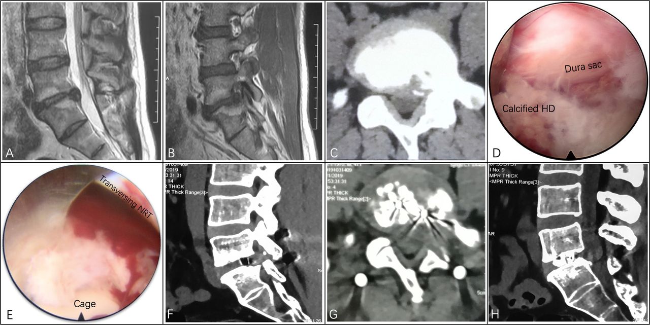
    Case 2: a 56-year-old male with L5-S1 Modic changes and lateral recess and foraminal stenosis. (A, B) Preoperative lumbar magnetic resonance imaging showed Modic changes in the upper and lower endplates of the L5-S1 disc and left foraminal stenosis. (C) The preoperative axial computed tomography (CT) scan showed calcified herniated disc compression on the left S1 nerve root. (D) Full-endoscopic exposure of calcified herniated disc. (E) The full-endoscopic view showed the positional relationship between the decompressed nerve root and the fusion cage. (F) Postoperative sagittal CT reconstruction showed left foraminal decompression. (G) Postoperative axial CT showed sufficient decompression of left lateral recess and foramen. (H) Postoperative sagittal CT reconstruction showed L5-S1 interbody fusion. Orientation of the field of view under full endoscope: left, cephalad side; right, caudal side; upper, medial side; lower, lateral side (left side). HD indicates herniated disc; and NRT, nerve root.

  • Figure 3
    • Download figure
    • Open in new tab
    • Download powerpoint
    Figure 3

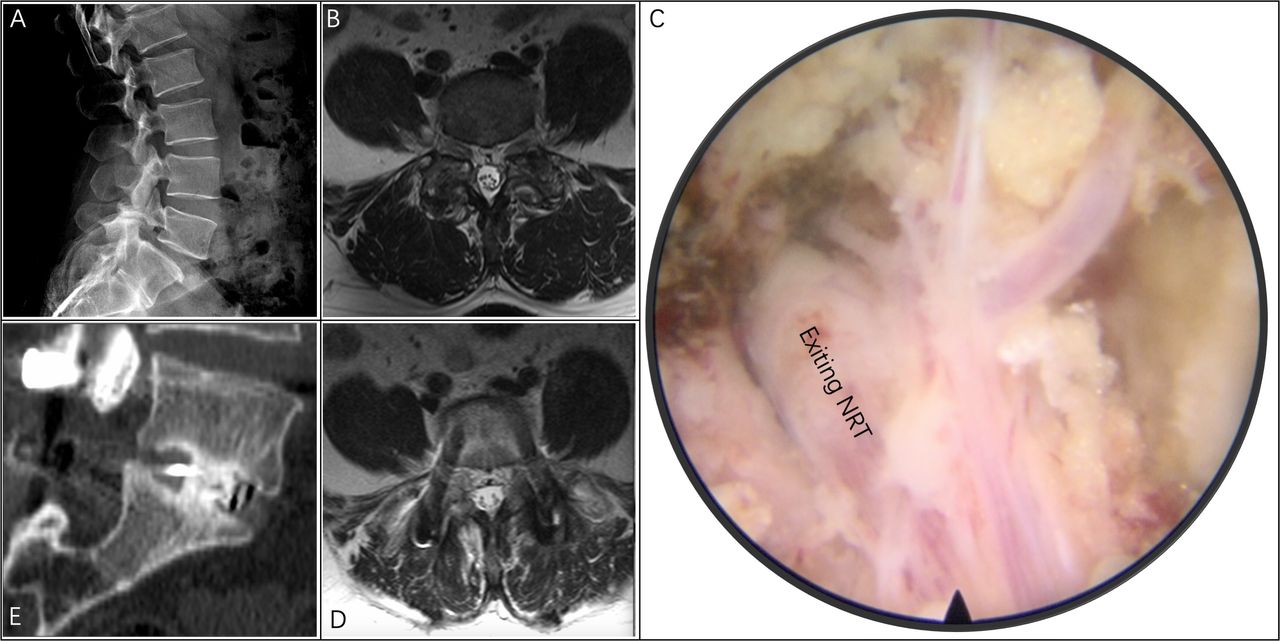
    Case 3: a 62-year-old male with isthmic spondylolisthesis. (A) Lateral radiography showed isthmic spondylolisthesis at the L5 level. (B) Preoperative axial T2-weighted magnetic resonance imaging (MRI) showed bilateral foraminal stenosis. (C) Full-endoscopic view of decompressed left L5 nerve root. (D) Three-month postoperative axial T2-weighted MRI showed bilateral foraminal decompression. (E) One-year postoperative sagittal computed tomography reconstruction showed an anatomical reduction of spondylolisthesis and solid interbody fusion. Orientation of the field of view under full endoscope: left, cephalad side; right, caudal side; upper, dorsomedial side; lower, ventrolateral side (left side). NRT indicates nerve root.

  • Figure 4
    • Download figure
    • Open in new tab
    • Download powerpoint
    Figure 4

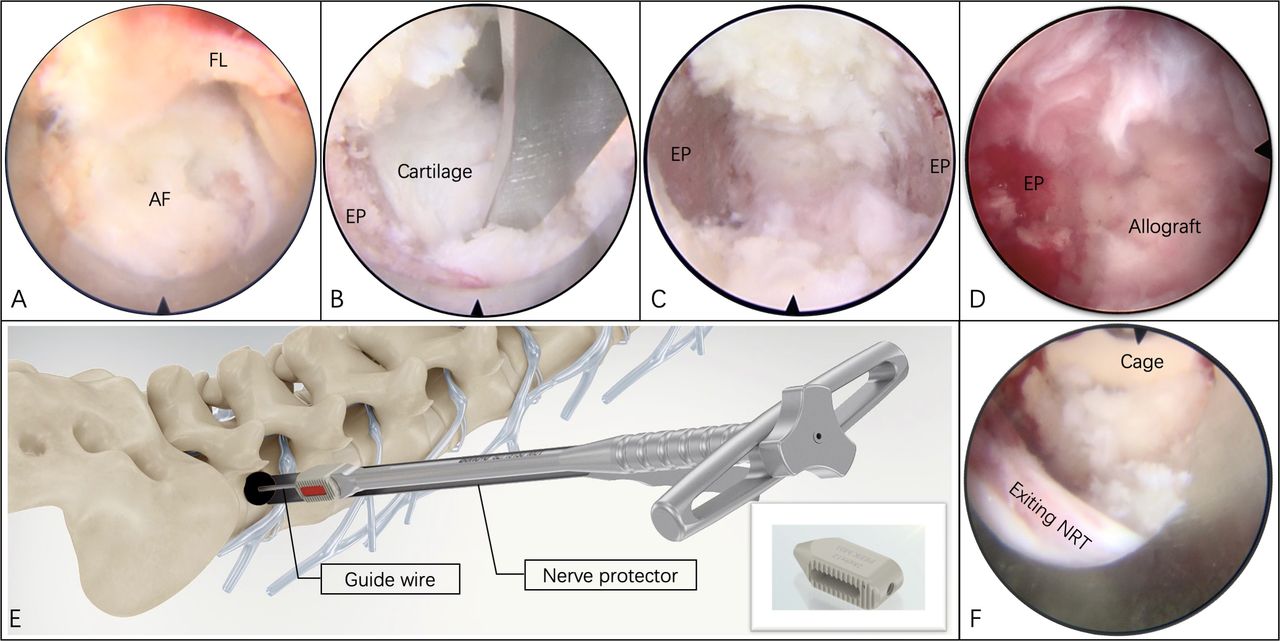
    Case 1, continued: surgical technique steps. (A) Rotate the working sheath under full-endoscopic surveillance and protect the exiting nerve root with the tongue end of the working sheath. (B) Use a curette to scrape the cartilage endplate of the upper and lower vertebral bodies under a full endoscope. (C) Prepare the bony endplates to expose the subchondral bone. (D) Place the allograft and rh-BMP-2 into the anterior half of the intervertebral space under press-fit. (E) Insert a guidewire into the intervertebral space the beveled endoscopic working sheath to protect the exiting nerve root by rotating it out of the way. Place a trial for sizing. Insert the bullet-shaped interbody fusion cage filled with autogenous bone and rhBMP-2 over the guidewire. (F) Explore the finished interbody fusion to directly visualize the exiting nerve root and define its relationship with the fusion cage. Orientation of the field of view under full endoscope: left, cephalad side; right, caudal side; upper, medial side; lower, lateral side (left side). FL indicates flavum ligament; AF, annulus fibrosis; EP, endplate; and NRT, nerve root.

  • Figure 5
    • Download figure
    • Open in new tab
    • Download powerpoint
    Figure 5

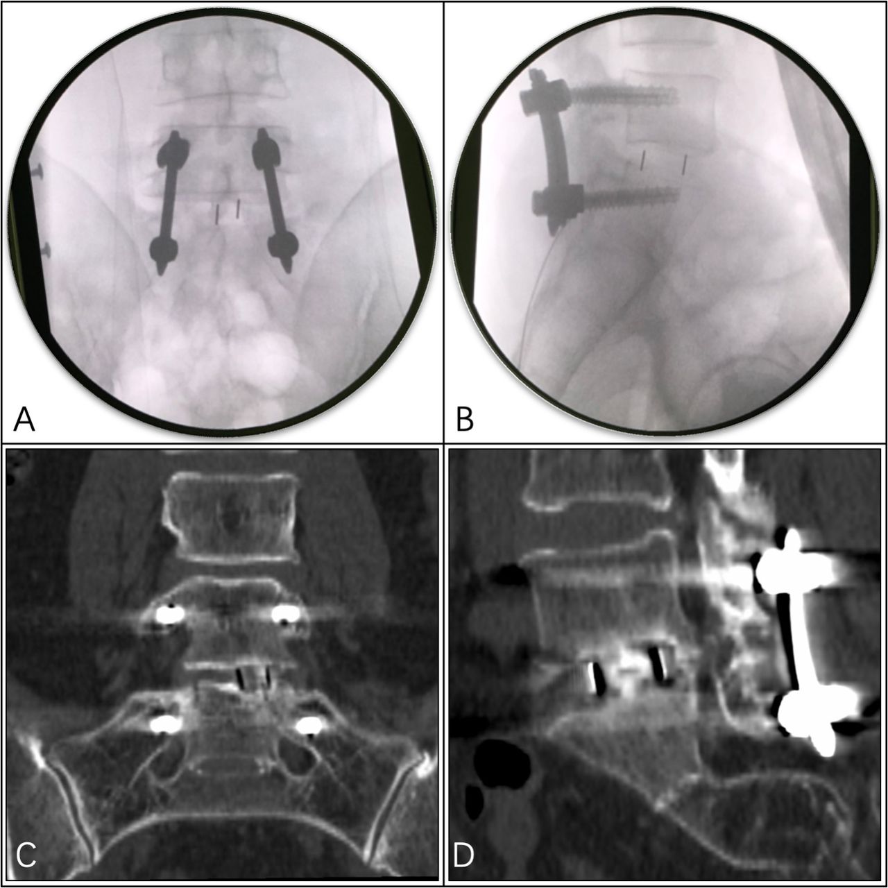
    Case 1, continued. (A, B) Anteroposterior and lateral fluoroscopic view showed the position of cage and pedicle system. (C, D) One-year postoperative coronal and sagittal computed tomography reconstruction showed solid interbody fusion.

Tables

  • Figures
  • Table 1
  • Table 2
PreviousNext
Back to top

In this issue

International Journal of Spine Surgery
Vol. 15, Issue 3
1 Jun 2021
  • Table of Contents
  • Index by author

Print
Download PDF
Article Alerts
Sign In to Email Alerts with your Email Address
Email Article

Thank you for your interest in spreading the word on International Journal of Spine Surgery.

NOTE: We only request your email address so that the person you are recommending the page to knows that you wanted them to see it, and that it is not junk mail. We do not capture any email address.

Enter multiple addresses on separate lines or separate them with commas.
Full-Endoscopic Oblique Lateral Lumbar Interbody Fusion: A Technical Note With 1-Year Follow-Up
(Your Name) has sent you a message from International Journal of Spine Surgery
(Your Name) thought you would like to see the International Journal of Spine Surgery web site.
CAPTCHA
This question is for testing whether or not you are a human visitor and to prevent automated spam submissions.
Citation Tools
Full-Endoscopic Oblique Lateral Lumbar Interbody Fusion: A Technical Note With 1-Year Follow-Up
Zhen-Zhou Li, Jin-Chang Wang, Zheng Cao, Hong-Liang Zhao, Kai-Uwe Lewandrowski, Anthony Yeung
International Journal of Spine Surgery Jun 2021, 15 (3) 504-513; DOI: 10.14444/8072

Citation Manager Formats

  • BibTeX
  • Bookends
  • EasyBib
  • EndNote (tagged)
  • EndNote 8 (xml)
  • Medlars
  • Mendeley
  • Papers
  • RefWorks Tagged
  • Ref Manager
  • RIS
  • Zotero
Share
Full-Endoscopic Oblique Lateral Lumbar Interbody Fusion: A Technical Note With 1-Year Follow-Up
Zhen-Zhou Li, Jin-Chang Wang, Zheng Cao, Hong-Liang Zhao, Kai-Uwe Lewandrowski, Anthony Yeung
International Journal of Spine Surgery Jun 2021, 15 (3) 504-513; DOI: 10.14444/8072
Twitter logo Facebook logo Mendeley logo
  • Tweet Widget
  • Facebook Like
  • Google Plus One

Jump to section

  • Article
    • ABSTRACT
    • INTRODUCTION
    • METHODS
    • RESULTS
    • DISCUSSION
    • Footnotes
    • REFERENCES
  • Figures & Data
  • Info & Metrics
  • PDF

Related Articles

  • No related articles found.
  • PubMed
  • Google Scholar

Cited By...

  • Research topics and trends of lumbar spondylolisthesis: a bibliometric study with machine-learning based text mining
  • The History of Endoscopic Posterior Lumbar Surgery
  • Editors Introduction: State-of-the-Art Techniques in Endoscopic Spine Surgery
  • Google Scholar

More in this TOC Section

  • Comparative Review of Lateral and Oblique Lumbar Interbody Fusion: Technique, Outcomes, and Complications
  • Key Considerations in Surgical Decision-Making on the Side of Approach for Lumbar Lateral Transpsoas Interbody Fusion Techniques
  • A Spine Surgeon’s Learning Curve With the Minimally Invasive L5 to S1 Lateral ALIF Surgical Approach: Perioperative Outcomes and Technical Considerations
Show more Minimally Invasive Surgery

Similar Articles

Keywords

  • lumbar interbody fusion
  • full-endoscopic
  • minimally invasive surgery
  • lumbar instability
  • low back pain
  • lumbar spondylolisthesis
  • lumbar spinal stenosis

Content

  • Current Issue
  • Latest Content
  • Archive

More Information

  • About IJSS
  • About ISASS
  • Privacy Policy

More

  • Subscribe
  • Alerts
  • Feedback

Other Services

  • Author Instructions
  • Join ISASS
  • Reprints & Permissions

© 2025 International Journal of Spine Surgery

International Journal of Spine Surgery Online ISSN: 2211-4599

Powered by HighWire