


Article Figures & Data
Figures
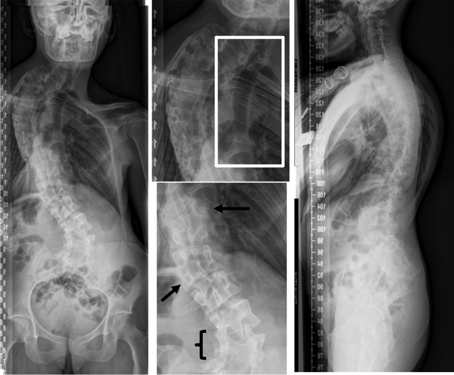
- Figure 1
A 24-year-old woman with chronic pulmonary disease. She underwent previous surgery during childhood with a T4-T7 early short fusion due to hemivertebrae. Along the years, she developed a crankshaft phenomenon showing a residual Cobb angle of 100°. Patient-reported outcome measure scores were as follows: Numerical Rating Scale back, 5; Oswestry Disability Index, 27%; Scoliosis Research Society 22-item self-image, 2; and 36-item Short Form Health Survey physical component summary, 45. Surgical indication was established for a T6-T8 vertebral column resection with T2-L3 post instrumentation and added thoracoplasty.
- Figure 2
A 29-year-old woman with cardiopathy (previous cardiac surgery). She had a T12 hemivertebra producing a T10-L1 coronal curve with 67° Cobb angle, and a thoracolumbar rotational kyphosis with a sagittal T10-L2 of 47°, well aligned. Patient-reported outcome measures scores were as follows: Numerical Rating Scale back, 6; Core Outcome Measurement Index back, 6; Oswestry Disability Index, 29%; Scoliosis Research Society 22-item survey (SRS-22) pain, 2.8; SRS-22 self-image, 2.2; and 36-item Short Form Health Survey physical component summary, 41.3. Surgery was indicated and a T9-L3 posterior fusion with T12 asymmetric osteotomy was planned.
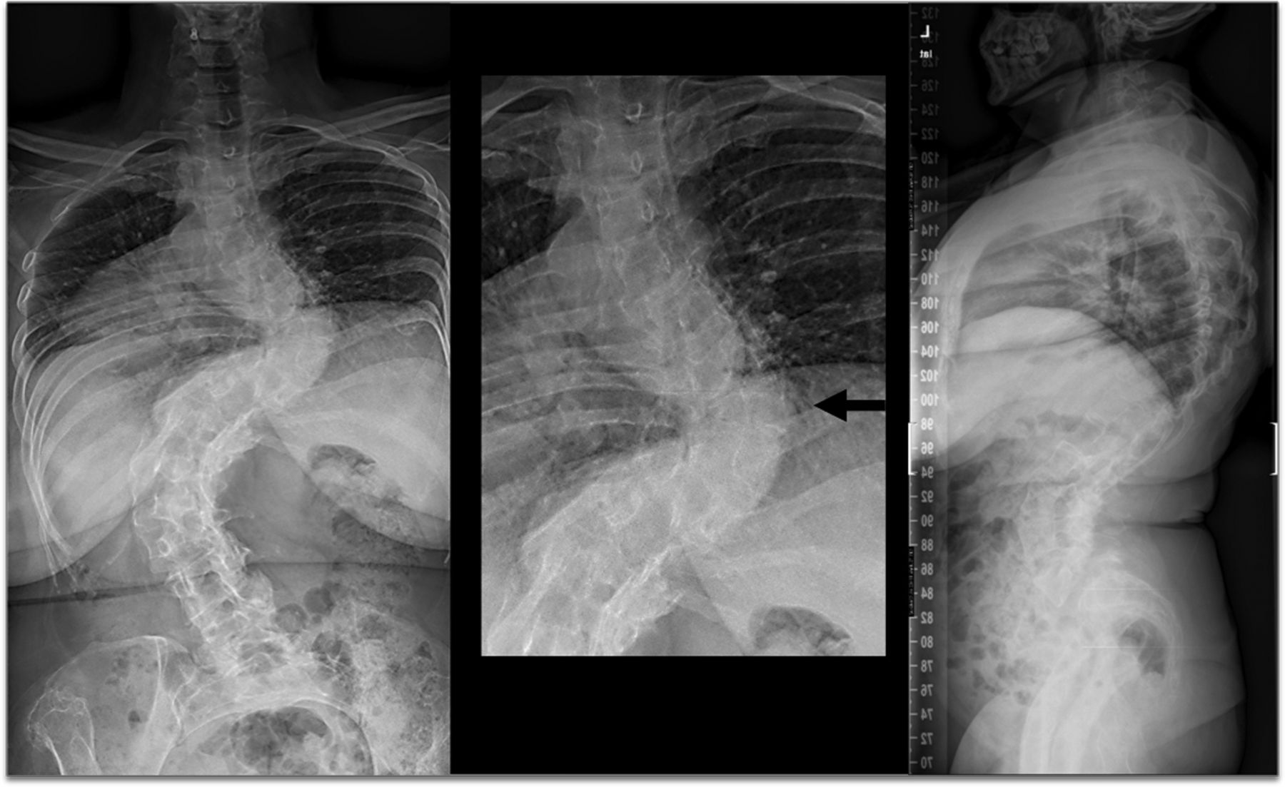
- Figure 3
A 26-year-old man with chronic pulmonary disease. He suffered a T7 hemivertebra and ipsilateral T2-T5 rib fusion. Coronal Cobb angle from T1-T5 was 35° and from T5-T9 was 61°. Patient-reported outcome measure scores were as follows: Numerical Rating Scale back, 0; Core Outcome Measurement Index back, 2; Oswestry Disability Index, 2%; Scoliosis Research Society 22-item survey (SRS-22) pain, 5; SRS-22 self-image, 3.6; and 36-item Short Form Health Survey physical component summary, 58.8. He followed conservative treatment.
- Figure 4
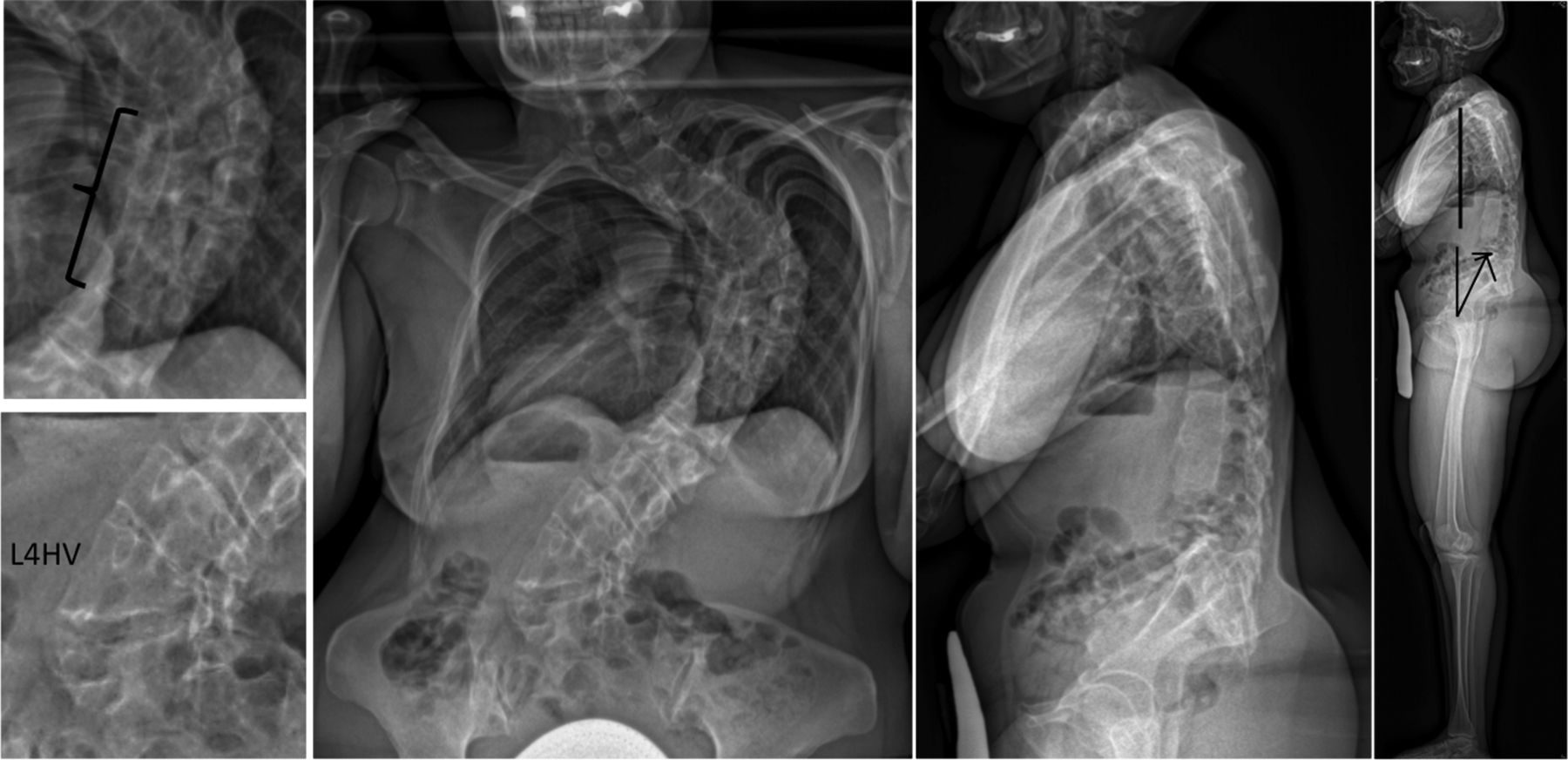
A 34-year-old woman with no comorbidities. Radiographs showed multiple anomalies: T10 right hemivertebra, L1 left hemivertebra, T4-T10 ribs fusions, and L4-L5 left bar. Coronal Cobb angles were T1-T9 73° and L3-L5 54°. Patient-reported outcome measure scores were as follows: Numerical Rating Scale back, 4; Core Outcome Measurement Index back, 3; Oswestry Disability Index, 18%; Scoliosis Research Society 22-item survey (SRS-22) pain, 3.2; SRS-22 self-image, 3.2; and 36-item Short Form Health Survey physical component summary, 42.7. Conservative treatment was indicated.
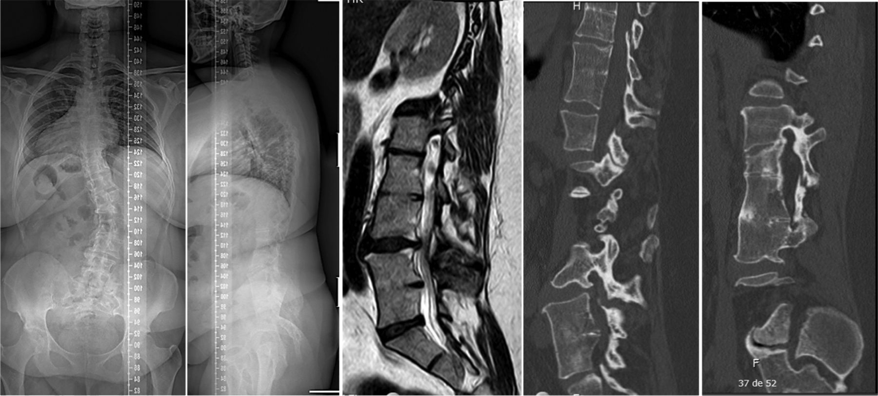
- Figure 5
A 39-year-old woman without comorbidities. Radiographs showed multiple segmentation defects at T10-T11, L2-L3, and L4-L5 drawing a lumbar flat back with correct global sagittal alignment. Patient reported outcome measure scores were as follows: Numerical Rating Scale back, 9; Core Outcome Measurement Index back, 7; Oswestry Disability Index, 58%; Scoliosis Research Society 22-item survey (SRS-22) pain, 1.2; SRS-22 self-image, 3; and 36-item Short Form Health Survey physical component summary, 21. Cobb angle T12-L4 42°. Surgical treatment was proposed for an L4 PSO and T11-iliac instrumentation.
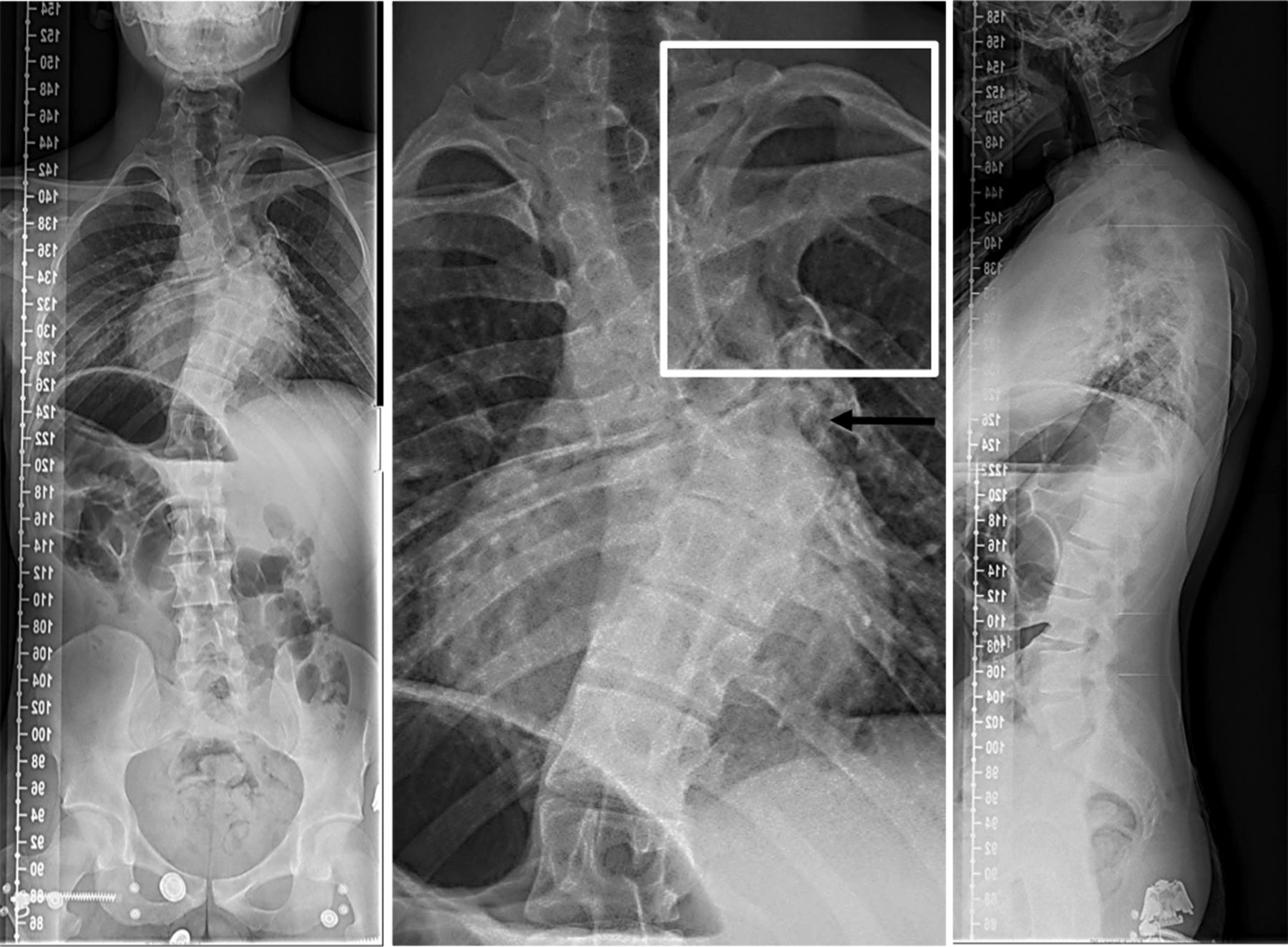
- Figure 6
A 40-year-old woman with no comorbidities. She presented a T7-T10 right incarcerated hemivertebrae and contralateral bar, and an L4 hemivertebrae. Coronal T6-T12 Cobb angle was 90° and L1-L5 Cobb angle was 66°. Patient-reported outcome measure scores were as follows: Numerical Rating Scale back, 6; Oswestry Disability Index, 33%; Scoliosis Research Society 22-item survey (SRS-22) pain, 2; SRS-22 self-image, 3; and 36-item Short Form Health Survey physical component summary, 40. Pelvic parameters of pelvic incidence, 45°; sacral slope, 20°; and pelvic tilt, 25° were compensating but well balanced. She was planned for a T8 vertebral column resection, surrounding Smith-Peterson osteotomies and T2-Iliac posterior instrumentation.
- Figure 7
A 60-year-old woman with hypertension. She had a T8 hemivertebra with coronal T6-T10 Cobb angle of 87° and T11-L3 of 90°. Patient-reported outcome measure scores were as follows: Numerical Rating Scale back, 3; Oswestry Disability Index, 18%; Scoliosis Research Society 22-item survey (SRS-22) pain, 3.7; SRS-22 self-image, 1.2; and 36-item Short Form Health Survey physical component summary, 46.4. The patient followed conservative treatment.


Tables
- Table 1
Demographic and radiographic comparisons between patients treated conservatively and those undergoing surgery.
Preoperative Data Surgical Conservative Student t/χ2 Demographic parameters Age, y 36.5 ± 10.6 38.6 ± 14.2 0.55 Gender, F/M 86.4%/13.6% 60%/40% 0.038a Height, cm 151.7 ± 10.1 157.9 ± 11.6 0.07 Weight, kg 59.2 ± 14.5 65.9 ± 16 0.15 BMI 25.9 ± 4.8 26.4 ± 5 0.76 Comorbidities 40.9% 33.3% 0.121 Previous surgery 45.5% 23.3% 0.093 Deformity type 1 0.746 Scoliosis 77.3% 73.3% Kyphosis 22.7% 26.7% Deformity type 2 0.811 Simple HV 64% 56% Multiple HV 30% 39% Segment defects 6% 5% Deformity Location 0.166 Proximal thoracic 4.5% 16.7% Main thoracic 36.4% 46.7% Thoracolumbar 31.8% 10% Lumbar 27.3% 26.7% Radiographic parameters Main Cobb, ° 59.3 ± 33.4 64.8 ± 27.1 0.52 Coronal balance, mm −9.3 ± 36.9 −0.8 ± 26.3 0.42 SVA, mm 14.2 ± 42.8 19.6 ± 56.1 0.73 Global tilt, ° 22.9 ± 15.3 18.6 ± 14.6 0.36 T10-L2 kyphosis, ° 13.7 ± 31.2 25.4 ± 35 0.24 PI-LL mismatch, ° −0.7 ± 26 −2.9 ± 24.3 0.765 LL, ° 49.2 ± 27.4 57.3 ± 23.6 0.265 PI, ° 47.8 ± 13.9 51.2 ± 16.5 0.458 T-DAR 8.9 ± 4.8 9.9 ± 4.6 0.464 Data presented as mean ± SD and percentages.
aStatistically significant.
BMI, body mass index; HV, hemivertebrae; LL, lumbar lordosis; PI, pelvic incidence; SVA, sagittal vertical axis; T-DAR, total deformity angular ratio.
- Table 2
PROMs comparisons between patients treated conservatively and those undergoing surgery.
Surgical Conservative Preoperative Data Mean ± SD Median (IQR) Mean ± SD Median (IQR) Student
t testMann-Whitney U test PROMs NRS back pain 5.2 ± 3.2 5.6 ± 3 0.61 NRS leg pain 3.6 ± 3.2 4 (0–6) 2.6 ± 3.3 0.5 (0–4.25) 0.233 COMI back 6.7 ± 2.4 3.8 ± 2.4 0.004a COMI neck 3.3 ± 2.1 2.6 ± 2.4 0.67 ODI (%) 34.6 ± 19.2 26.9 ± 15.9 0.15 SRS-22 SRS-22 function 3.1 ± 0.8 3 (2.5–4) 3.6 ± 1 4 (3–4) 0.089 SRS-22 pain 2.9 ± 1.2 3.1 ± 1 0.56 SRS-22 self-image 2.5 ± 0.9 2 (2–3) 3 ± 0.9 3 (2–4) 0.047a SRS-22 mental health 2.9 ± 1 3 (2–4) 3.5 ± 0.8 3.5 (3–4) 0.083 SRS-22 satisfaction 3.2 ± 1.3 3.5 (2.25–4) 3.6 ± 1 4 (3–4) 0.533 SRS-22 subtotal 2.82 ± 0.7 3 (2–3) 3.4 ± 0.8 4 (3–4) 0.020a SRS-22 total 2.9 ± 0.7 3 (2–3) 3.4 ± 0.8 4 (3–4) 0.014a SF-36 SF-36 function 38.6 ± 8.9 45.1±9.2 0.023a SF-36 physical role 33.7 ± 12.4 42.6 ± 12.8 0.026a SF-36 pain 38.3 ± 11.9 41 ± 10.4 0.43 SF-36 vitality 46.5 ± 8 47.9 ± 9.4 0.59 SF-36 social function 34.9 ± 8.5 37 (27–37) 43.7 ± 12.4 47 (32–57) 0.022a SF-36 emotional role 40.9 ± 13 42.5 ± 11.3 0.67 SF-36 mental health 43.8 ± 10.2 44.8 ± 9 0.72 SF-36 general health 41.9 ± 10.4 43.2 ± 11.3 0.68 SF-36 PCS 36.7 ± 10.4 43.3 ± 10.8 0.048a SF-36 MCS 44.2 ± 11.4 45.1 ± 10.9 0.79 Parametric data presented as mean ± SD. Nonparametric data presented as median (IQR).
aStatistically significant.
COMI, Core Outcome Measurement Index; IQR, interquartile range; MCS, mental component summary; NRS, Numerical Rating Scale; ODI, Oswestry Disability Index; PCS, physical component summary; PROM, patient-reported outcome measures; SF-36, 36-item Short Form Health Survey; SRS-22, Scoliosis Research Society 22-item questionaire.
In this issue


Jump to section
Related Articles
Cited By...
- No citing articles found.