
Main menu

  • Home
  • Content
    • Current Issue
    • Advance Online Publication
    • Archive
  • About Us
    • About ISASS
    • About the Journal
    • Author Instructions
    • Editorial Board
    • Reviewer Guidelines & Publication Criteria
  • More
    • Advertise
    • Subscribe
    • Alerts
    • Feedback
  • Join Us
  • Reprints & Permissions
  • Sponsored Content
  • Other Publications
    • ijss

User menu

  • My alerts

Search

  • Advanced search
International Journal of Spine Surgery
  • My alerts
International Journal of Spine Surgery

Advanced Search

  • Home
  • Content
    • Current Issue
    • Advance Online Publication
    • Archive
  • About Us
    • About ISASS
    • About the Journal
    • Author Instructions
    • Editorial Board
    • Reviewer Guidelines & Publication Criteria
  • More
    • Advertise
    • Subscribe
    • Alerts
    • Feedback
  • Join Us
  • Reprints & Permissions
  • Sponsored Content
  • Follow ijss on Twitter
  • Visit ijss on Facebook
Research ArticleLumbar Spine

Endoscopic Lateral Lumbar Interbody Fusion: Technical Note and Case Series

Ricardo Casal Grau, Francisco Javier Sánchez Benitez de Soto, Patrick Barhouse, Christian Schroeder, Owen P. Leary, Patricia Zadnik Sullivan and Albert E. Telfeian
International Journal of Spine Surgery February 2024, 18 (1) 101-109; DOI: https://doi.org/10.14444/8572
Ricardo Casal Grau
1 Casal Dots SLU and Asepeyo Hospital, Madrid, Spain
2 Asepeyo Traumatology and Rehabilitation Monographic Hospital, Coslada, Madrid, Spain
MD
  • Find this author on Google Scholar
  • Find this author on PubMed
  • Search for this author on this site
Francisco Javier Sánchez Benitez de Soto
1 Casal Dots SLU and Asepeyo Hospital, Madrid, Spain
2 Asepeyo Traumatology and Rehabilitation Monographic Hospital, Coslada, Madrid, Spain
MD
  • Find this author on Google Scholar
  • Find this author on PubMed
  • Search for this author on this site
Patrick Barhouse
3 Department of Neurosurgery, Rhode Island Hospital, The Warren Alpert Medical School of Brown University, Providence, RI, USA
BS
  • Find this author on Google Scholar
  • Find this author on PubMed
  • Search for this author on this site
Christian Schroeder
3 Department of Neurosurgery, Rhode Island Hospital, The Warren Alpert Medical School of Brown University, Providence, RI, USA
BS
  • Find this author on Google Scholar
  • Find this author on PubMed
  • Search for this author on this site
Owen P. Leary
3 Department of Neurosurgery, Rhode Island Hospital, The Warren Alpert Medical School of Brown University, Providence, RI, USA
BS
  • Find this author on Google Scholar
  • Find this author on PubMed
  • Search for this author on this site
Patricia Zadnik Sullivan
3 Department of Neurosurgery, Rhode Island Hospital, The Warren Alpert Medical School of Brown University, Providence, RI, USA
MD
  • Find this author on Google Scholar
  • Find this author on PubMed
  • Search for this author on this site
Albert E. Telfeian
3 Department of Neurosurgery, Rhode Island Hospital, The Warren Alpert Medical School of Brown University, Providence, RI, USA
MD, PHD
  • Find this author on Google Scholar
  • Find this author on PubMed
  • Search for this author on this site
  • For correspondence: ATelfeian@Lifespan.org
  • Article
  • Figures & Data
  • Info & Metrics
  • PDF
Loading

Article Figures & Data

Figures

  • Tables
  • Figure 1
    • Download figure
    • Open in new tab
    • Download powerpoint
    Figure 1

    Operative images demonstrating endoscopic lateral lumbar interbody fusion procedure. (A) The patient is positioned in the lateral position with the surgeon demonstrated holding the endoscope and performing the discectomy with fluoroscopic and endoscopic visualization. (B) The endoscope and endoscopic grasper are used to retrieve iliac crest graft for packing the interbody titanium cage. (C) The titanium cage being packed with iliac crest autograft. (D and E) The titanium cage delivery over a nitinol blunt-tip wire is demonstrated, with zoomed-in details shown in (D) and zoomed-out views presented in (E).

  • Figure 2
    • Download figure
    • Open in new tab
    • Download powerpoint
    Figure 2

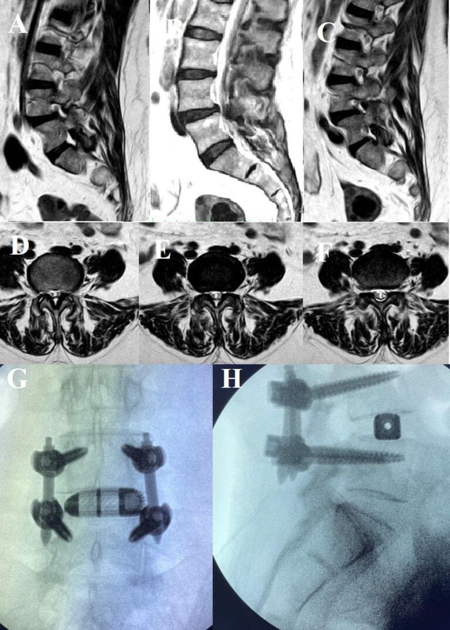
    Endoscopic lateral lumbar fusion of L4-5. (A–C) Sagittal T2 magnetic resonance images (MRIs) from right (A), midline (B), and left (C), demonstrating grade 1 spondylolisthesis and severe left foraminal narrowing. (D–F) Axial T2 MRI of the lumbar spine demonstrating severe L4-5 foraminal narrowing in axial images from the bottom of the L4 endplate (D), through the L4-5 disc (E), and through the top of the L5 endplate (F). (G and H) Anteroposterior and lateral fluoroscopic images demonstrate the final position of the lateral interbody fusion device and pedicle screw instrumentation.

  • Figure 3
    • Download figure
    • Open in new tab
    • Download powerpoint
    Figure 3

    Step-by-step endoscopic lateral lumbar fusion (ELLIF) of L4-5. (A–R) Anteroposterior (AP) and lateral fluoroscopic intraoperative images of the key operative ELLIF procedure. (A) AP fluoroscopic image demonstrates the beveled tubular retractor placed through the psoas, and the endoscopic grasper is shown performing the discectomy. (B) AP fluoroscopic image demonstrating the endoscopic curette preparing the endplate for arthrodesis. (C and D) Joimax EndoLIF titanium interbody cage is introduced into the disc space over a nitinol wire. (E and F) Lateral fluoroscopic images demonstrate the placement of the interbody cage with the wire (E) and then with the wire removed (F).(G and H) AP fluoroscopic images of the Jamshidi needle used for placement of the K-wires in the L4 pedicles (G) and L5 pedicles (H). (I) AP fluoroscopic image of the wire placement. (J–M) Lateral fluoroscopic images demonstrate the endoscopic right lumbar L4-5 lateral recess stenosis decompression and discectomy. (J) Endoscopic Shrill drill shown decompressing the lateral recess. (K) Endoscopic Kerrison rongeur shown completing the lateral recess stenosis decompression. (L and M) Lateral fluoroscopic images demonstrating the endoscopic grasper beginning (L) and completing (M) the discectomy as part of the lateral recess decompression. (N and O) AP fluoroscopic images demonstrate iliac crest graft harvesting. (N) Wire cannulates the iliac crest. (O) Crown reamer after placement over dilator shown harvesting iliac crest though ELLIF incision. (P and Q) Lateral fluoroscopic images demonstrate percutaneous pedicle screw placement over the wires. (R) Final AP fluoroscopic image of the fusion construct.

  • Figure 4
    • Download figure
    • Open in new tab
    • Download powerpoint
    Figure 4

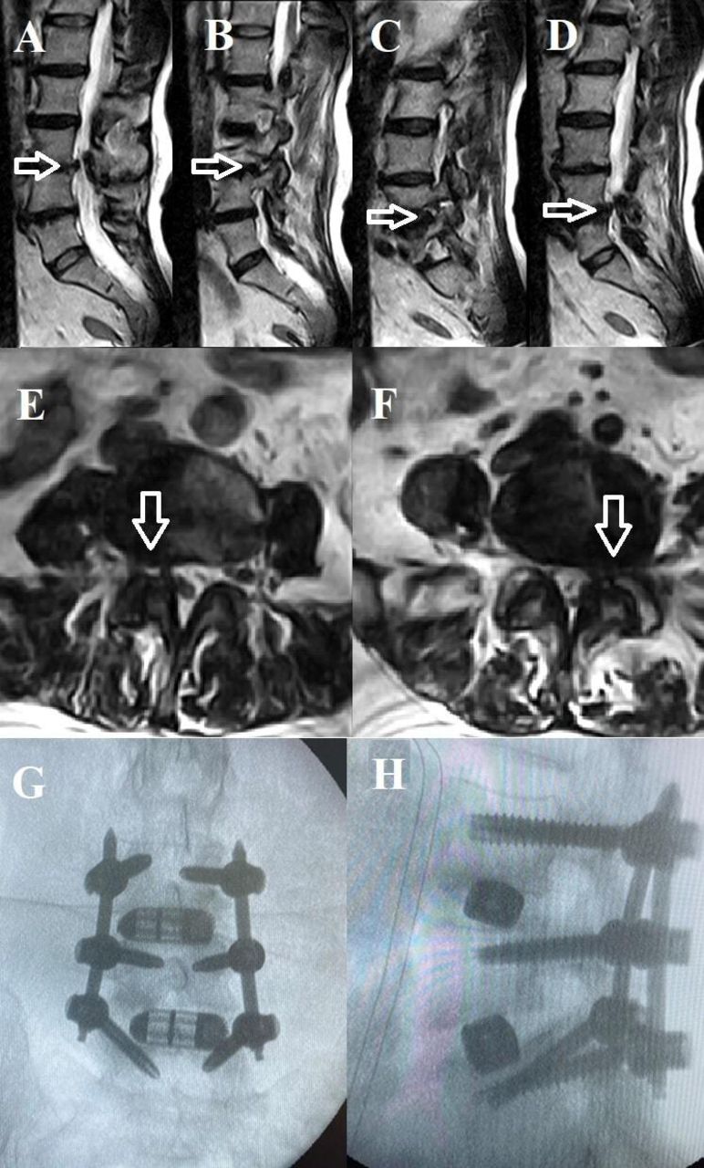
    Endoscopic lateral lumbar fusion of L3-5. Sagittal T2 magnetic resonance image (MRI) reveals severe right L3-4 foraminal narrowing and endplate changes at L3-4 (A is paracentral and B is foraminal) (open arrows) as well as severe left L4-5 foraminal narrowing and endplate changes at L4-5 (C is foraminal and D is paracentral) (open arrows). (E and F) Axial T2 MRIs of the right L3-4 foraminal narrowing (E, open arrow) and the left L4-5 foraminal narrowing (F, open arrow). (G and H) AP and lateral fluoroscopic images demonstrate the final position of the lateral interbody fusion devices and pedicle screw instrumentation.

  • Figure 5
    • Download figure
    • Open in new tab
    • Download powerpoint
    Figure 5

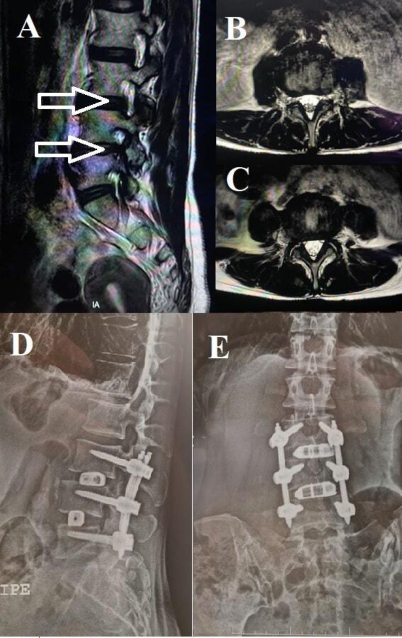
    Endoscopic lateral lumbar fusion of L3-5. (A) Sagittal T2 magnetic resonance image (MRI) demonstrates moderate right L3-4 and severe right L4-5 foraminal narrowing (open arrows). (B and C) Axial T2 MRI demonstrates moderate bilateral L3-4 (B) and severe bilateral L4-5 (C) foraminal narrowing. (D and E) Anteroposterior and lateral fluoroscopic images demonstrate the final position of the lateral interbody fusion devices and pedicle screw instrumentation.

Tables

  • Figures
    • View popup
    Table 1

    Summary of postoperative changes in VAS and ODI scores at 1-month and 2-year follow-up.

    CaseVASODI
    Preoperative1 mo2 y% ChangePreoperative1 mo2 y% Change
    184275%32221069%
    2104280%42191076%
    3106190%4628589%
    Mean9.34.71.782%40238.378%
    • Abbreviations: ODI, Oswestry Disability Index; VAS, visual analog scale.

    • View popup
    Table 2

    Summary of postoperative recovery.

    CaseTime in Hospital, hReturn to Normal Daily Activity, dComplications
    14860None
    22440None
    33645None
    Mean3648NA
    • Abbreviation: NA, not applicable.

PreviousNext
Back to top

In this issue

International Journal of Spine Surgery
Vol. 18, Issue 1
1 Feb 2024
  • Table of Contents
  • Index by author

Print
Download PDF
Article Alerts
Sign In to Email Alerts with your Email Address
Email Article

Thank you for your interest in spreading the word on International Journal of Spine Surgery.

NOTE: We only request your email address so that the person you are recommending the page to knows that you wanted them to see it, and that it is not junk mail. We do not capture any email address.

Enter multiple addresses on separate lines or separate them with commas.
Endoscopic Lateral Lumbar Interbody Fusion: Technical Note and Case Series
(Your Name) has sent you a message from International Journal of Spine Surgery
(Your Name) thought you would like to see the International Journal of Spine Surgery web site.
CAPTCHA
This question is for testing whether or not you are a human visitor and to prevent automated spam submissions.
Citation Tools
Endoscopic Lateral Lumbar Interbody Fusion: Technical Note and Case Series
Ricardo Casal Grau, Francisco Javier Sánchez Benitez de Soto, Patrick Barhouse, Christian Schroeder, Owen P. Leary, Patricia Zadnik Sullivan, Albert E. Telfeian
International Journal of Spine Surgery Feb 2024, 18 (1) 101-109; DOI: 10.14444/8572

Citation Manager Formats

  • BibTeX
  • Bookends
  • EasyBib
  • EndNote (tagged)
  • EndNote 8 (xml)
  • Medlars
  • Mendeley
  • Papers
  • RefWorks Tagged
  • Ref Manager
  • RIS
  • Zotero
Share
Endoscopic Lateral Lumbar Interbody Fusion: Technical Note and Case Series
Ricardo Casal Grau, Francisco Javier Sánchez Benitez de Soto, Patrick Barhouse, Christian Schroeder, Owen P. Leary, Patricia Zadnik Sullivan, Albert E. Telfeian
International Journal of Spine Surgery Feb 2024, 18 (1) 101-109; DOI: 10.14444/8572
Twitter logo Facebook logo Mendeley logo
  • Tweet Widget
  • Facebook Like
  • Google Plus One

Jump to section

  • Article
    • Abstract
    • Introduction
    • Materials and Methods
    • Results
    • Discussion
    • Conclusion
    • Footnotes
    • References
  • Figures & Data
  • Info & Metrics
  • PDF

Related Articles

  • No related articles found.
  • PubMed
  • Google Scholar

Cited By...

  • Prone Position for Preoperative Planning in Lumbar Endoscopic and Minimally Invasive Fusion Procedures: Insights From a Magnetic Resonance Imaging Study
  • Google Scholar

More in this TOC Section

  • Comparison of Stand-Alone Anterior Lumbar Interbody Fusion, 360° Anterior Lumbar Interbody Fusion, and Arthroplasty for Recurrent Lumbar Disc Herniation: Focus on Nerve Decompression and Painful Spinal Instability Resolution
  • Recovery Trajectories After Lumbar Fusion Stratified by Baseline Patient-Reported Outcomes Measurement Information System Physical Function Disability Levels
  • Association Between Nonsteroidal Anti-inflammatory Drugs Use and Surgical Outcomes Following Posterior Lumbar Fusion: A Medical Claims Database Analysis
Show more Lumbar Spine

Similar Articles

Keywords

  • endoscopic spine; lateral lumbar interbody fusion
  • radiculopathy
  • minimally invasive spine
  • indirect decompression

Content

  • Current Issue
  • Latest Content
  • Archive

More Information

  • About IJSS
  • About ISASS
  • Privacy Policy

More

  • Subscribe
  • Alerts
  • Feedback

Other Services

  • Author Instructions
  • Join ISASS
  • Reprints & Permissions

© 2025 International Journal of Spine Surgery

International Journal of Spine Surgery Online ISSN: 2211-4599

Powered by HighWire