


Article Figures & Data
Figures
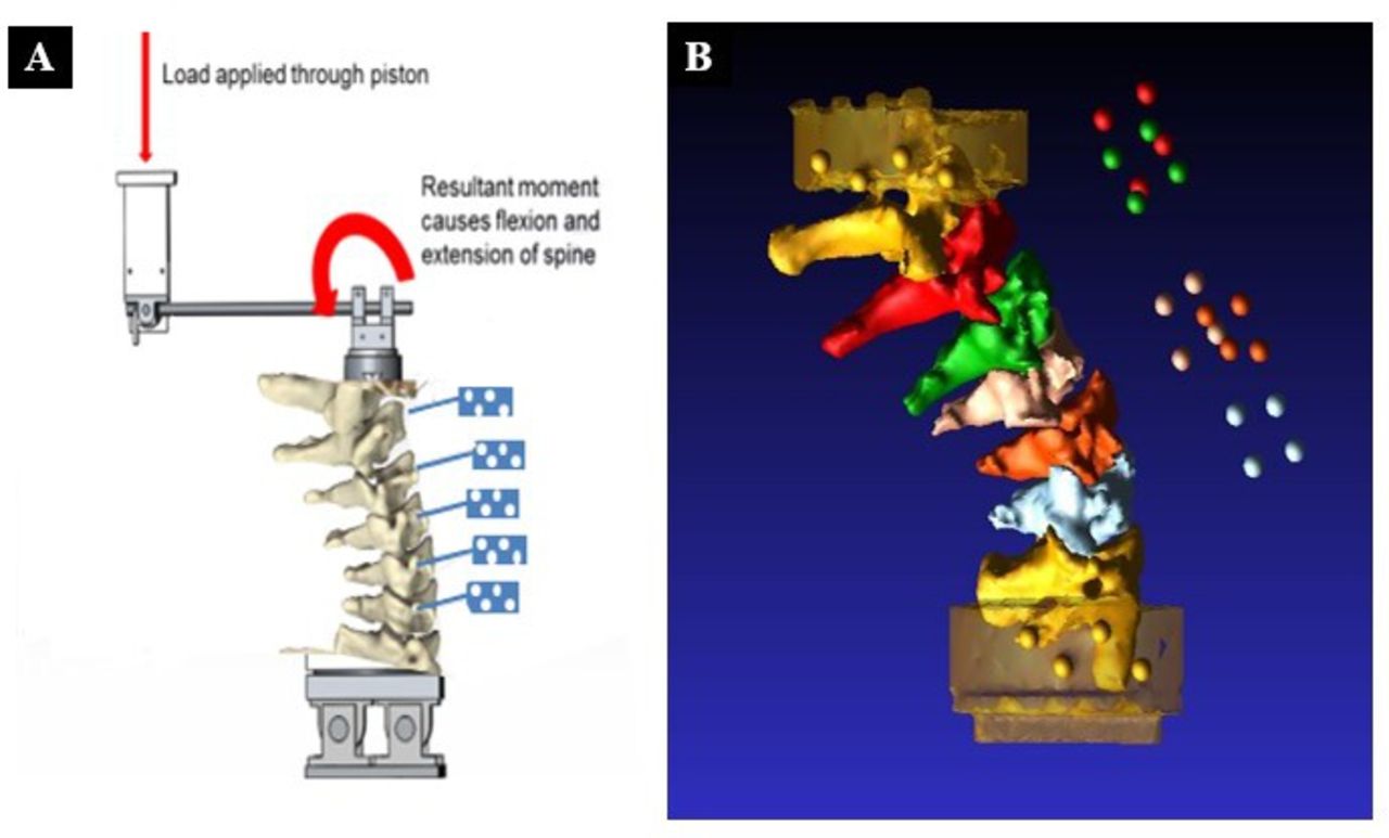
- Figure 1
(A) Schema of the test set-up showing the cervical spine specimen and motion capture markers (in blue). (B) Computed tomography reconstruction of a spine showing vertebrae.
- Figure 2
Photographs of the testing apparatus with servo-hydraulic loading frame demonstrating hydraulic actuator mounted on crosshead connected to the mounting fixture in a lateral (A) and anterior (B) view.
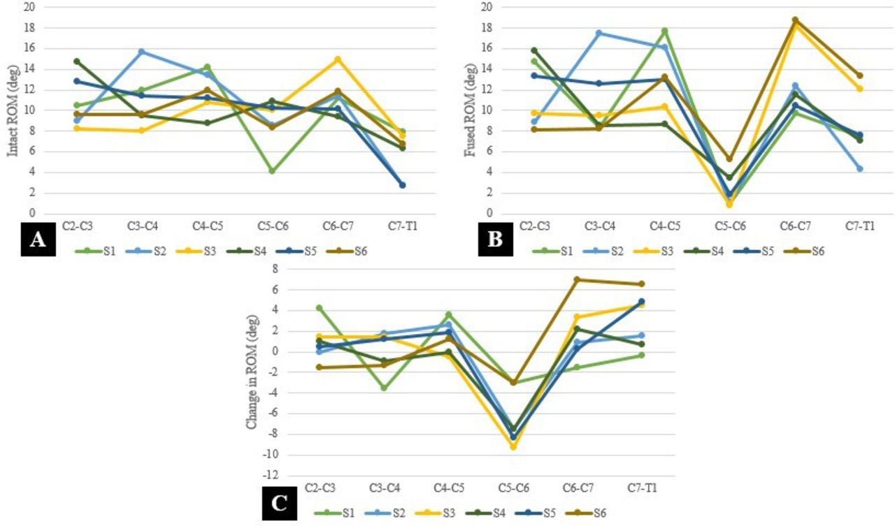
- Figure 3
(A) Preoperative range of motion (ROM) of each segment per specimen. (B) Postoperative ROM after fusion of C5-C6 of each segment per specimen. (C) Characteristic changes in motion in all 6 specimens after C5-C6 anterior cervical discectomy and fusion at each motion segment.


Tables
Motion Segment Average Change in Motion (°) P Value C2–C3 +1.0 0.28 C3–C4 +0.2 0.80 C4–C5 +1.4 0.07 C5–C6 −6.8 0.002 C6–C7 +2.0 0.15 C7–T1 +3.0 0.04 Boldface indicates statistically significant differences of P < 0.05.
- Table 2
Segmental total motion for each specimen (C2–T1) in the intact state and fused state after anterior cervical discectomy with cage placement and plate fixation at C5–C6.a
Motion Segment Specimen 1 Specimen 2 Specimen 3 Specimen 4 Specimen 5 Specimen 6 Intact Fused Delta Intact Fused Delta Intact Fused Delta Intact Fused Delta Intact Fused Delta Intact Fused Delta C2–C3 (°) 10 15 4 9 9 0 8 10 1 15 16 1 13 13 0 10 8 −2 C3–C4 (°) 12 8 −4 16 17 1 8 9 1 9 9 0 11 12 2 10 8 −2 C4–C5 (°) 14 18 4 13 16 3 11 10 −1 9 9 0 11 13 2 12 13 1 C5–C6 (°) 4 1 −3 9 1 −8 10 1 −9 11 3 −8 10 2 −8 8 5 −3 C6–C7 (°) 11 10 −1 11 12 1 15 18 3 9 12 3 10 10 0 12 19 7 C7–T1 (°) 8 8 0 3 4 1 8 12 4 6 7 1 3 8 5 7 13 6 ↵a Gray shading indicates motion that exceeded the error of the testing system.
- Table 3
Cumulative scores of radiographic grading (Rydman et al) of the extent of degeneration of cervical spine cadaveric specimens at each motion segment from C2 to T1.
Motion Segment Specimen 1 Specimen 2 Specimen 3 Specimen 4 Specimen 5 Specimen 6 Disc Facet Disc Facet Disc Facet Disc Facet Disc Facet Disc Facet C2–C3 1 0 0 2 0 0 0 0 0 1 0 0 C3–C4 1 0 4 1 0 0 0 0 0 1 0 0 C4–C5 3 0 3 2 0 3 0 0 1 3 0 0 C6–C7 5 1 4 1 0 0 0 0 2 3 0 1 C7–T1 3 0 1 0 0 0 0 0 0 0 0 0 Note: Disc degeneration: Height loss 0–4 points, anterior osteophytes 0–3 points, and endplate sclerosis 0–2 points. Total score: 0 = no degeneration, 1–3 = mild degeneration, 4–6 = moderate degeneration, and 7–9 = severe degeneration. Facet degeneration: Joint space narrowing (0–1 points), osteophytes (0–1 points), and irregularity of articular surface (0–1 point). Total score: 0 = no degeneration, 1 = mild degeneration, 2 = moderate degeneration, and 3 = severe degeneration.
In this issue


Jump to section
Related Articles
Cited By...
- No citing articles found.