


Article Figures & Data
Figures
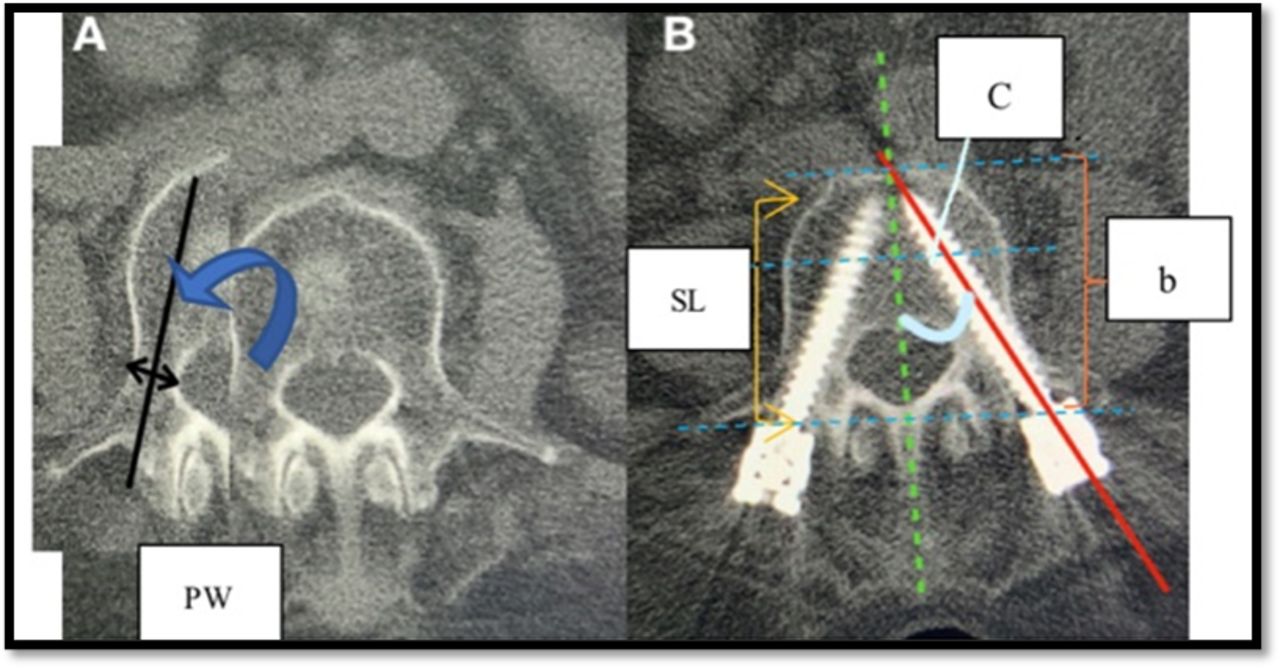
- Figure 1
Postoperative thin-slice computed tomography performed at 12 months. (A) PW = pedicle width, (B) b = body length, SL = screw length (% SD = [SL/b] × 100), and C = angle of medial convergence.
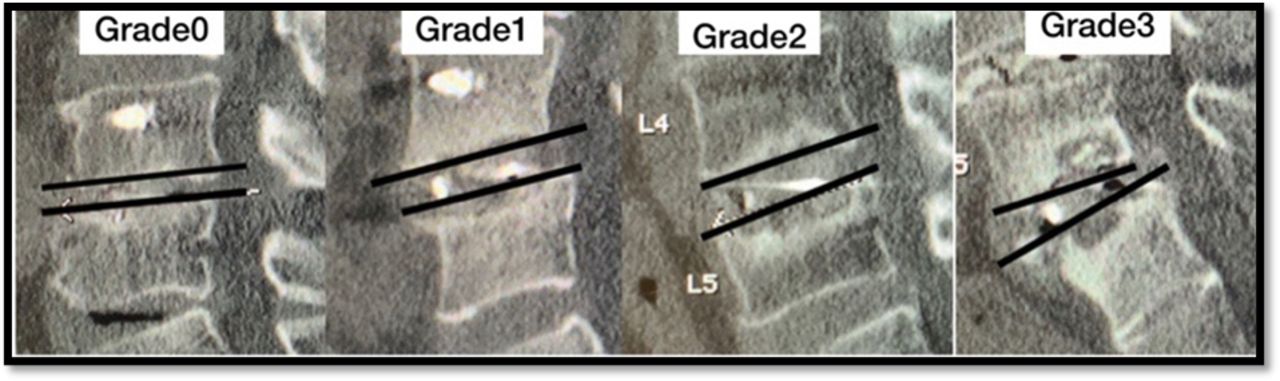
- Figure 2
Cage subsidence grading (0–3).
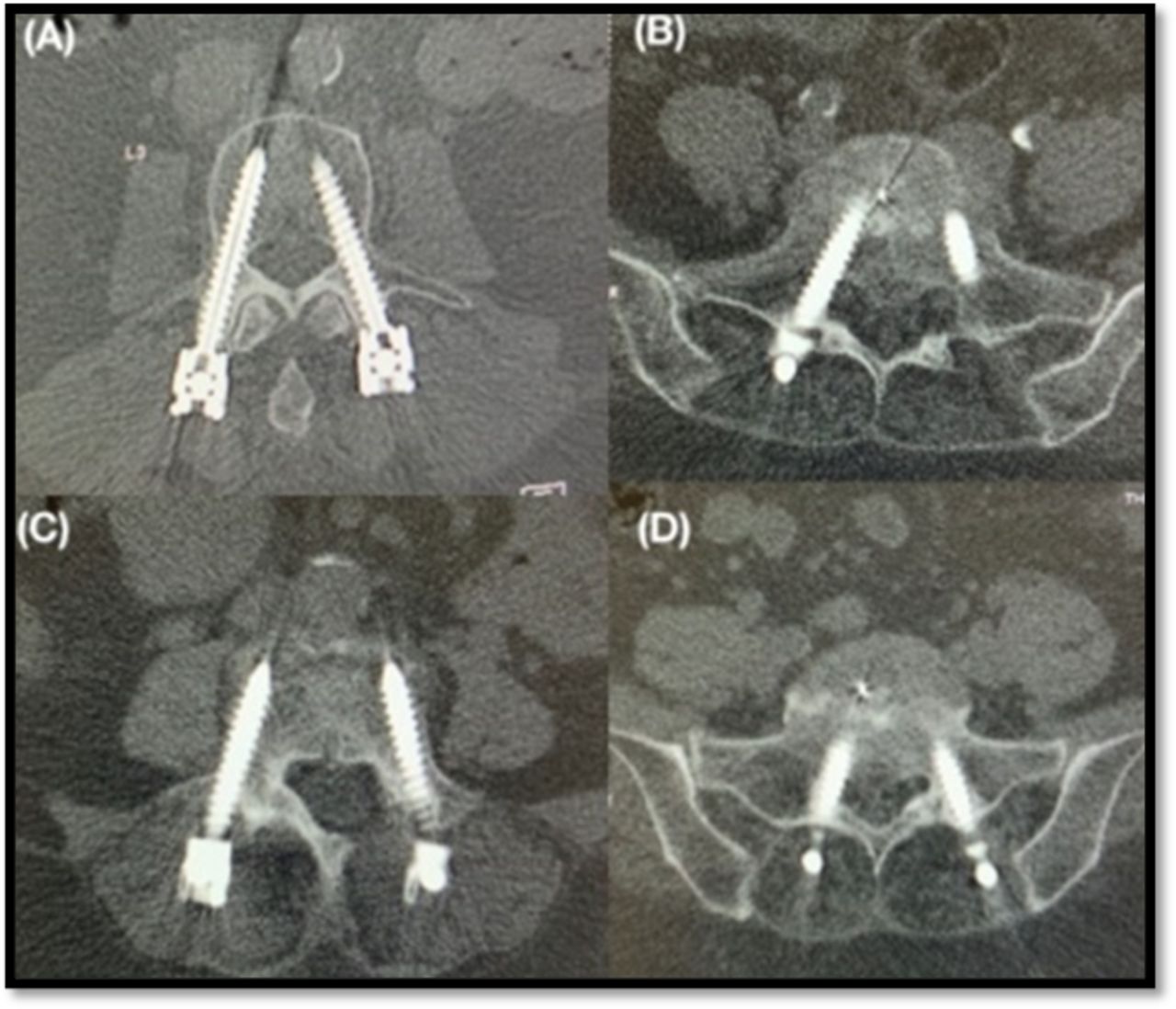
- Figure 3
The grading system (Gertzbein). (A) Grade 0: screw within pedicle, (B) Grade 1: screw thread breach of pedicle <2 mm, (C) Grade 2: significant breach 2–4 mm, and (D) Grade 3: significant breach <4 mm.
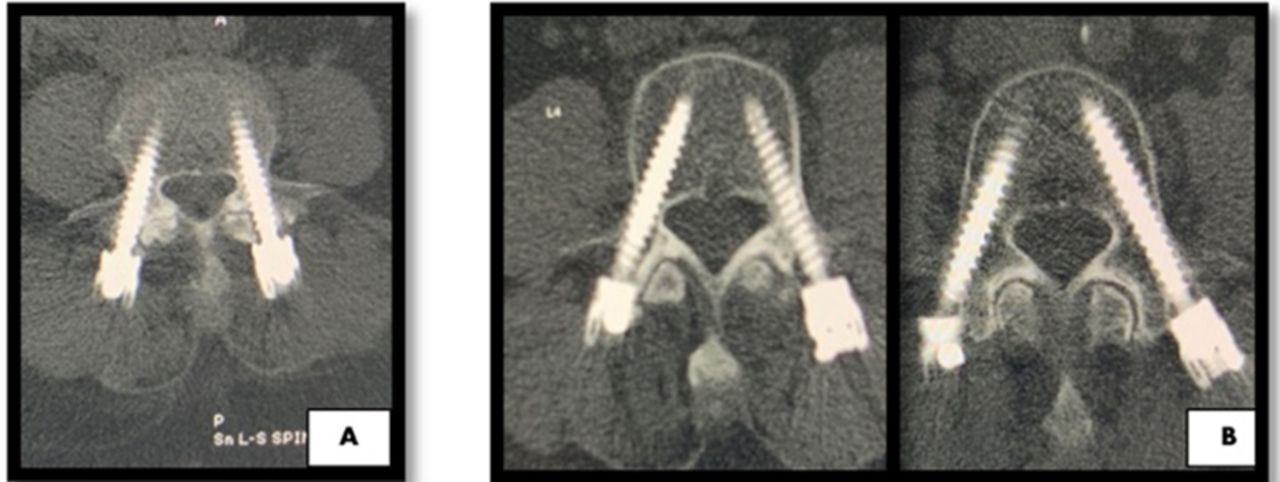
- Figure 4
Facet joint violation. (A) Intra-articular pedicle screw that penetrates the facet joint transversely, interposed between the superior and inferior facet at the cephalad level. (B) Screw did not transverse the facet joint; extra-articular screw.


Tables
Patient Data FLUO MIS-TLIF (N = 61) NAV MIS-TLIF (N = 36) P Value Age 65.67 ± 9.23 65.89 ± 9.69 0.913 Sex Female 41 (67.2%) 23 (63.9%) 0.825 Male 20 (32.8%) 13 (36.1%) 0.825 Body mass index 25.22 ± 2.96 24.86 ± 3.31 0.660 Diagnosis Spondylolisthesis 24 (39.3%) 22 (61.1%) 0.058 Spinal stenosis 7 (11.5%) 2 (5.6%) 0.477 Spinal canal stenosis 17 (27.9%) 8 (22.2%) 0.634 Herniated nucleus pulposus 12 (19.7%) 3 (8.3%) 0.159 Degenerative scoliosis 0 (0%) 1 (2.8%) 0.371 Degenerative disc disease 1 (1.6%) 0 (0%) 1.000 Level L3-4 3 (4.9%) 2 (5.6%) 0.745 L3-5 7 (11.5%) 6 (16.6%) 0.703 L4-5 38 (62.3%) 20 (55.6%) 0.612 L4-S1 6 (9.8%) 4 (11.1%) 0.800 L5-S1 7 (11.5%) 4 (11.1%) 0.258 Blood loss (mL) 171.03 ± 157.75 124.44 ± 147.42 0.157 Operative time (min) 173.91 ± 52.93 163.64 ± 52.06 0.360 Length of stay (d) 5.2 ± 2.41 5.03 ± 2.46 0.751 Radiation dose (mGy) 38.26 ± 5.66 11.49±2.13 < 0.05 Data presented as mean ± SD or n (%).
FLUO, fluoroscopic-guided; MIS-TILF, minimally invasive transforaminal lumbar interbody fusion; NAV, 3-dimensional computer navigation.
Raidographic Parameters FLUO MIS-TLIF (N = 61) NAV MIS-TLIF (N = 36) P Value Screw-to-pedicle ratio 0.7 ± 0.13 0.66 ± 0.09 0.079 % Screw depth 85.18 ± 7.54 89.04 ± 6.07 0.011a Angle of medial convergence 18.44 ± 11.55 27.7±3.93 <0.001a % Screw penetration (n = 270) (n = 172) Anterior 47 (17.4%) 16 (9.7%) 0.027a Lateral 5 (1.9%) 4 (2.4%) 0.684 Medial 10 (3.7%) 0 (0%) 0.012a Superior 1 (0.4%) 1 (0.6%) 0.724 Cage size 23.61 ± 1.93 24.78 ± 1.74 0.003a Cage location Anterior 24 (44.4%) 30 (55.6%) 0.471 Posterior 49 (74.2%) 17 (25.8%) 0.471 Cage subsidence (<2 mm) 39 (63.9%) 23 (63.9%) 0.996 Fusion 55 (90.2%) 33 (91.7%) 0.481 Data presented as mean ± SD or n (%).
aStatistically significant (P < 0.05)
FLOU, fluoroscopic-guided; MIS-TILF, minimally invasive transforaminal lumbar interbody fusion; NAV, 3-dimensional computer navigation.
Clinical Scores FLUO MIS-TLIF NAV MIS-TLIF P Value Mean ± SE Mean ± SE VAS of back score Pre-OP 6.48 ± 0.43 7.14 ± 0.45 0.312 1 mo 1.13 ± 0.23 0.78 ± 0.26 0.325 3 mo 0.72 ± 0.2 0.48 ± 0.22 0.454 6 mo 0.83 ± 0.25 0.43 ± 0.23 0.361 1 y 0.63 ± 0.2 0.11 ± 0.11 0.025a VAS leg score Pre-OP 6.9 ± 0.38 7.56 ± 0.39 0.261 1 mo 1.32 ± 0.27 1.14 ± 0.23 0.649 3 mo 1.11 ± 0.29 0.58 ± 0.21 0.143 6 mo 1.07 ± 0.29 0.13 ± 0.12 < 0.001a 1 y 0.78 ± 0.26 0.11 ± 0.11 0.019a ODI Pre-OP 49.07 ± 2.06 53.83 ± 1.91 0.123 1 mo 11.86 ± 1.74 11.69 ± 1.89 0.949 3 mo 8.26 ± 2 4.39 ± 1.73 0.200 6 mo 6.86 ± 1.86 0.67 ± 0.67 0.003a 1 y 5.21 ± 1.69 0.49 ± 0.49 0.009a Satisfaction score 1 mo 8.81 ± 0.23 9.31 ± 0.14 0.068 3 mo 8.82 ± 0.23 9.42 ± 0.16 0.081 6 mo 8.93 ± 0.23 9.35 ± 0.15 0.302 1 y 9.16 ± 0.18 9.39 ± 0.16 0.495 aStatistically significant (P < 0.05).
FLUO, fluoroscopic-guided; MIS-TILF, minimally invasive transforaminal lumbar interbody fusion; NAV, 3-dimensional computer navigation.
In this issue


Jump to section
Related Articles
Cited By...
- No citing articles found.