


Article Figures & Data
Figures
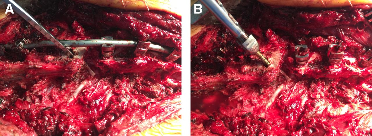
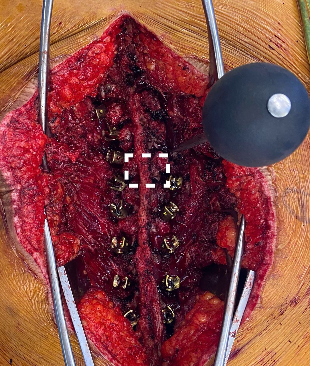
- Figure 1
(A) Intraoperative photograph of fusion mass screw cannulation lateral to medial immediately inferior to a chevron posterior column osteotomy using a Lenke probe. (B) Intraoperative photograph of polyaxial screw placement into the fusion mass.
- Figure 2
Axial computed tomography image showing low-grade breach (<2 mm) into spinal canal of a f fusion mass screw placed at T12, Case 4.
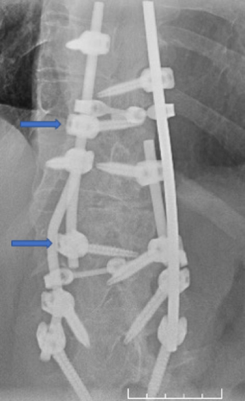
- Figure 3
Fusion mass screws (arrows) assisting in osteotomy closure and providing an additional anchor to save a distal fusion level below a 3-column osteotomy.
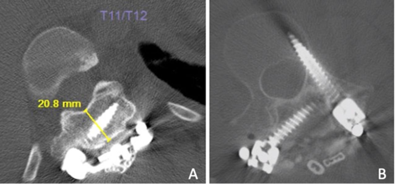
- Figure 4
(A) Axial computed tomography (CT) image showing fusion mass screws (FMSs) placement at T12 through a thick fusion mass bed measuring 21.8 mm. (B) CT image showing right-sided FMS placement with a concurrent pedicle screw placement at L2.
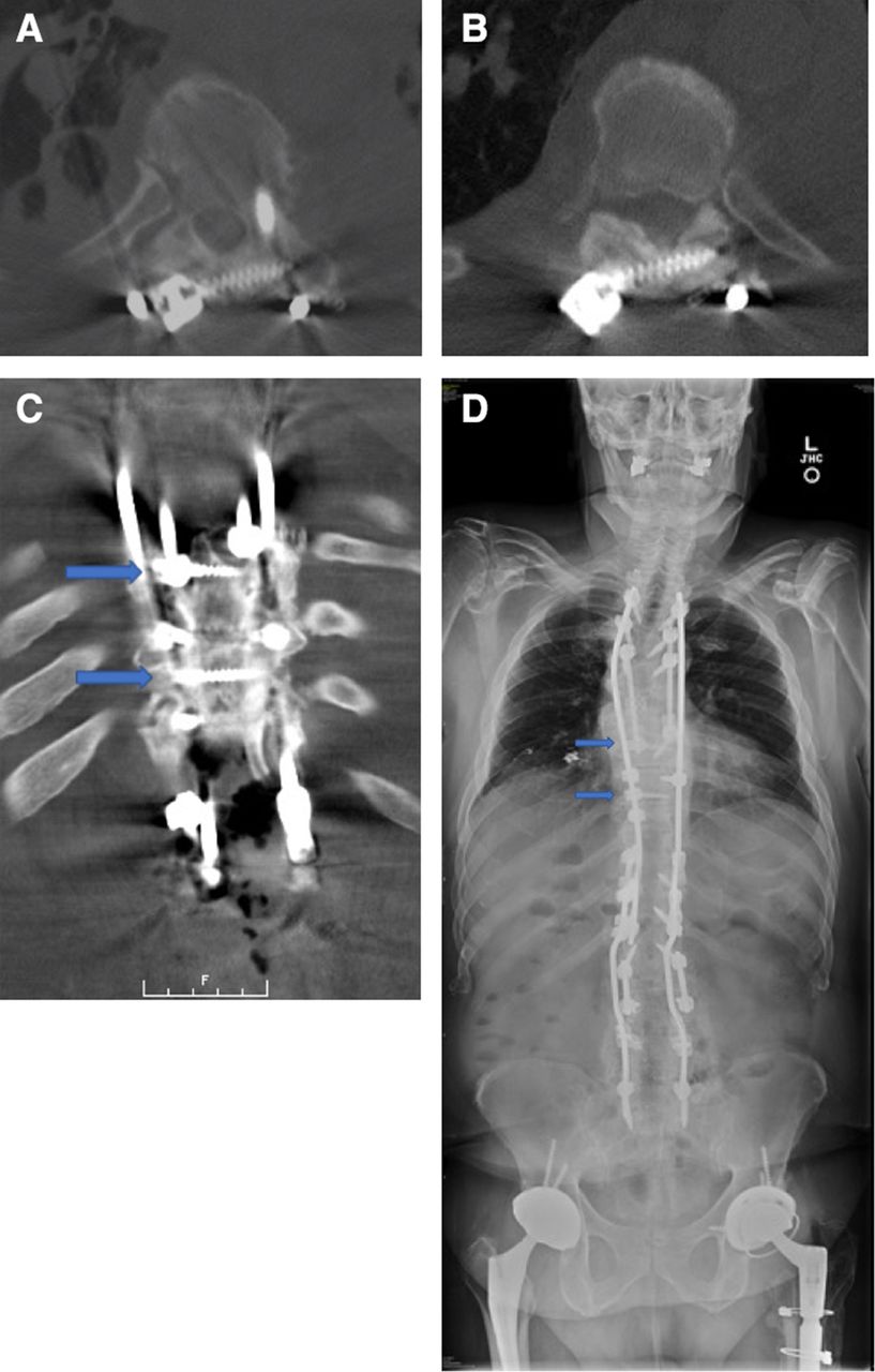
- Figure 5
(A-C) CT and (D) full-length standing radiograph images demonstrating fusion mass screw placement at T8 and T10 (arrows) distal to the vertebral column resection to assist in osteotomy closure. Concurrent pedicle screw placement is also shown at T8.
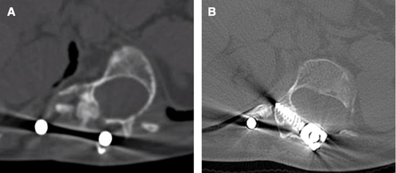
- Figure 6
(A) Axial computed tomography images of Patient 6 demonstrating pedicular dysplasia in the setting of neurofibromatosis-1 and juvenile onset scoliosis. (B) Fusion mass screw placed at T12. Pedicle screw placement was avoided due to dysplasia and obscured landmarks intraoperatively. The pedicles were 2.2 and 3.9 mm in width at this level.
- Figure 7
Intraoperative photograph demonstrating identification of the pedicle screw starting point using a quadrangulation technique as described by Kim et al.8


Tables
- Table 1
Clinical data for 6 adult spinal deformity patients treated with FMS fixation during revision spine surgery.
Case Age (y), Gender Diagnosis Levels Fused FMS Levels Indication for FMS Placement Complications Follow-Up (y) 1 29, M Juvenile onset scoliosis, sagittal imbalance T2-L4 T12, L2 Pedicular dysplasia, pedicle compromise from prior instrumentation None 2.4 2 66, M Adult spinal deformity, osteomyelitis, fusion mass fracture at T6 and T7 T3-S1 T8, T10 Assist in closure of 3-column osteotomy Pneumothorax 1.7 3 58, F Adolescent idiopathic scoliosis, fusion mass fracture at T11, sagittal imbalance C2-pelvis T9, T10, T11 Fusion mass fracture, assist in closure of 3-column osteotomy None 2.1 4 57, F T12 burst fracture and T12 paraparesis, charcot arthropathy at L3-L4 and L4-L5 T9-Pelvis T12, L1 Augment fixation at prior corpectomy site Anaphylaxis, FMS low-grade breach into spinal canal at T12 (Figure 2) 1.4 5 57, F Adolescent idiopathic scoliosis, pseudarthrosis at L3-L4, distal junctional failure at L4-S1. T1-Pelvis L2, L5 Augment pedicle screw fixation None 2.3 6 28, F Juvenile onset scoliosis, neurofibromatosis-1, pseudarthrosis at T12-L1 T6-L4 T9, T10, T11, T12 Pedicular dysplasia Dural tear 3.1 Abbreviation: FMS, fusion mass screw.
- Table 2
Comparison of fusion mass thickness, BMD, and screw length in 6 adult spinal deformity patients treated with FMS fixation during revision spine surgery.
Case Level Fusion Mass Thickness (mm) Fusion Mass BMD (HU) Screw Length (mm) Complications Related to FMS Placement FMS Loosening, Breakage, or Pseudarthrosis at Latest Follow-Up 1 T12 23.7 315 40 None None 1 L2 24.4 475 35 None None 2 T8 15.8 628 40 None None 2 T10 13.3 544 40 None None 3 T9 8.6 574 40 None None 3 T10 10.4 496 35 None None 3 T11 12.8 378 40 None None 4 T12 18.7 500 35 Spinal canal breach None 4 L1 19.4 378 35 None None 5 L2 11.1 455 40 None None 5 L5 13.9 372 40 None None 6 T9 10.7 156 30 None None 6 T10 14.3 284 30 None None 6 T11 19.6 215 25 None None 6 T12 16.5 188 25 None None Mean ± SD _ 15.5 ± 4.8 397 ± 144 35 ± 5.5 _ _ Abbreviations: BMD, bone mineral density; CT, computed tomography; FMS, fusion mass screw; HU, Hounsfield units.
Note: There was no evidence of FMS loosening, breakage, or pseudarthrosis on CT at an mean of 2.2 years of follow-up (range: 1.4 to 3.1 years). On postoperative CT, 1 out of 15 screws had a low-grade breach (<2 mm) into the canal (Case 4 at T12). There were otherwise no complications related to FMS placement.
In this issue


Jump to section
Related Articles
Cited By...
- No citing articles found.