


Article Figures & Data
Figures
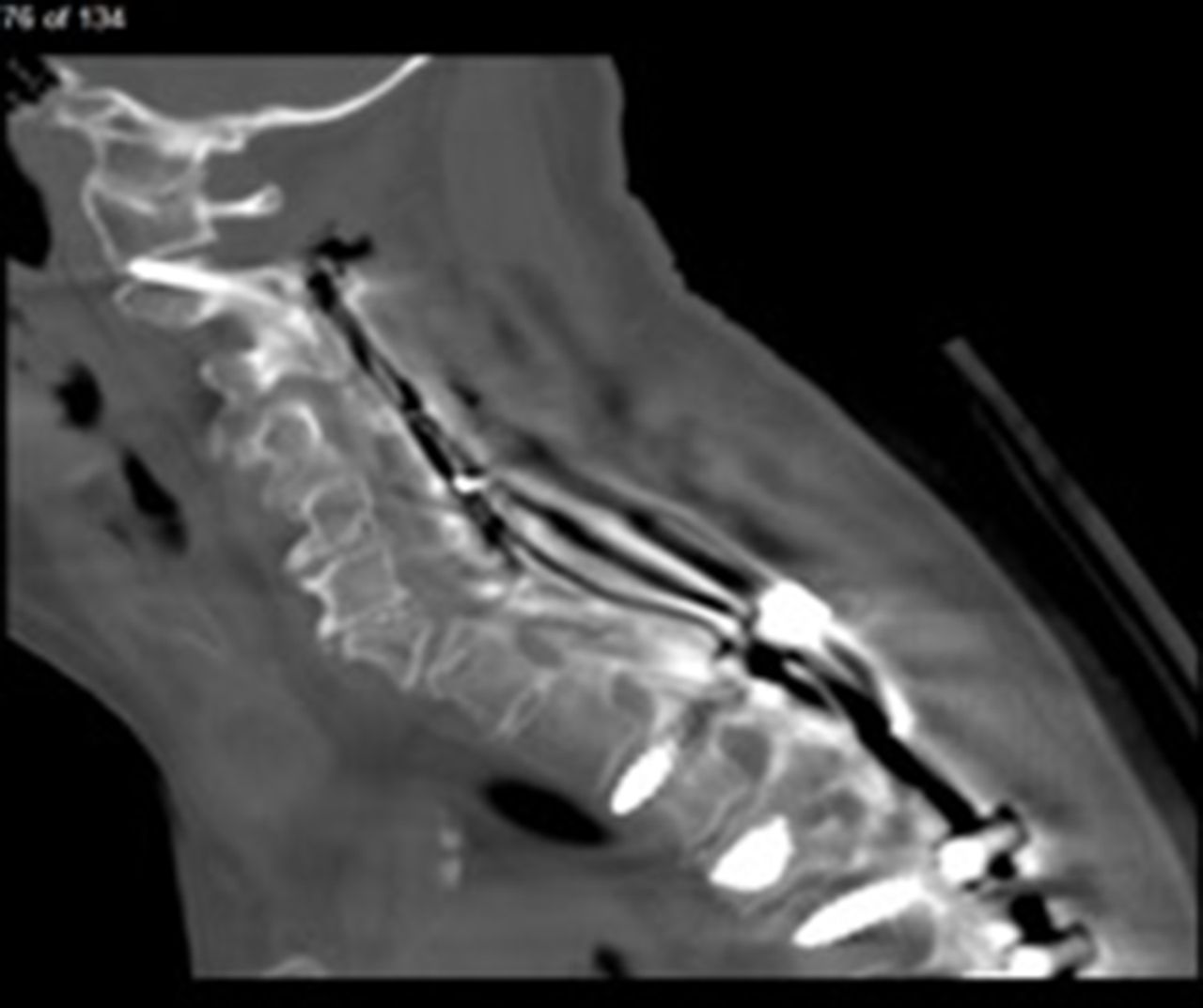
- Figure 1
Sagittal computed tomography image of C1-C2 facet joint penetration.
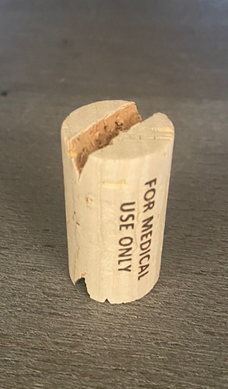
- Figure 2
A wine cork placed in the mouth to help visualize the C2 pedicles on the intraoperative open mouth view.
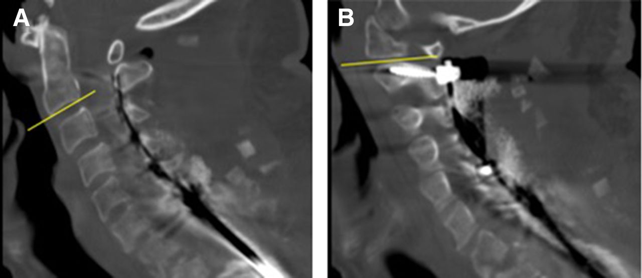
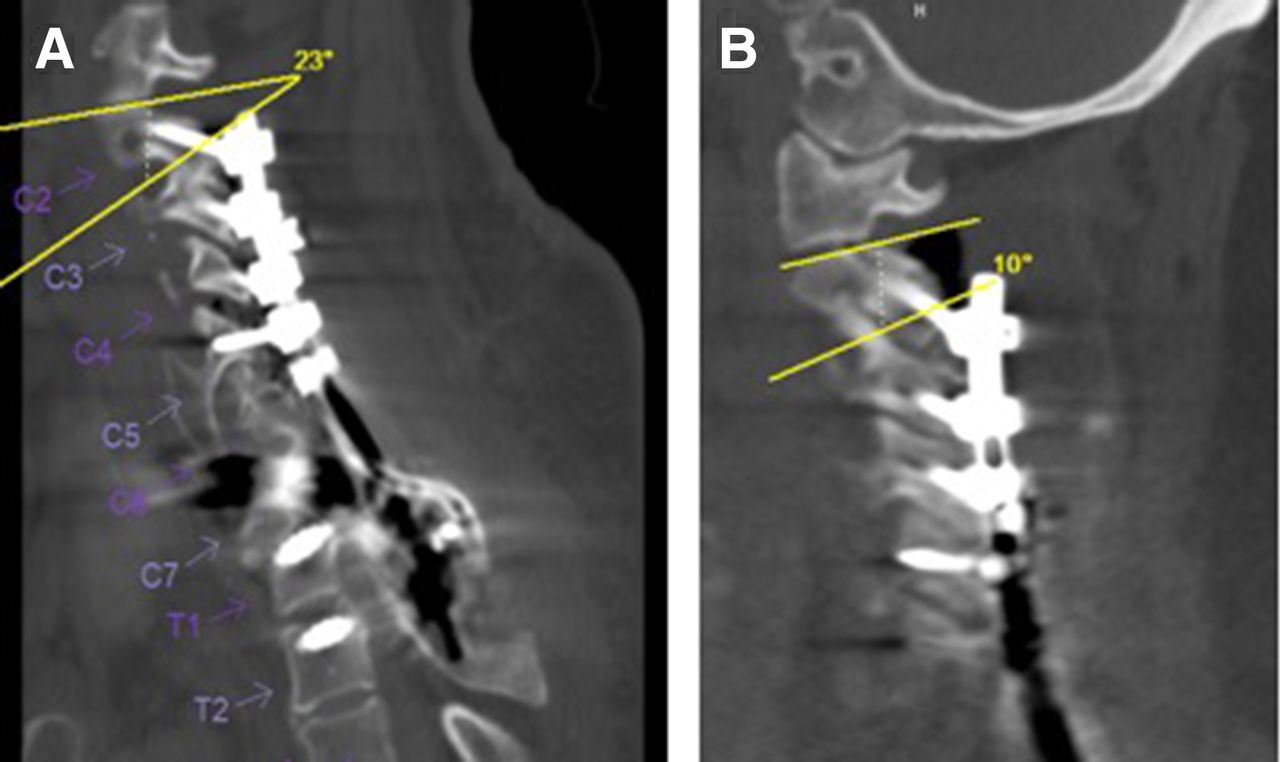
- Figure 3
Measurement of the sagittal angle of the C1-C2 facet joint. The inferior endplate of C2 represents the reference line (A). A second line approximates the slope of the C1-C2 facet joint (B). The sagittal screw angles were measured using a similar method.
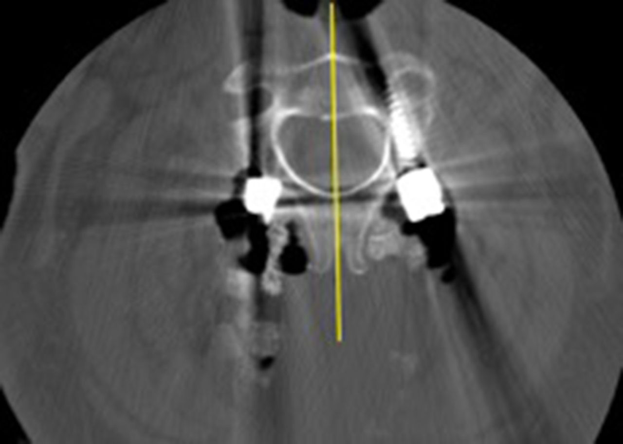
- Figure 4
The axial angle of each pedicle screw was measured relative to a line bisecting the C2 vertebra and aligned with the spinous processes to account for any vertebral rotation.
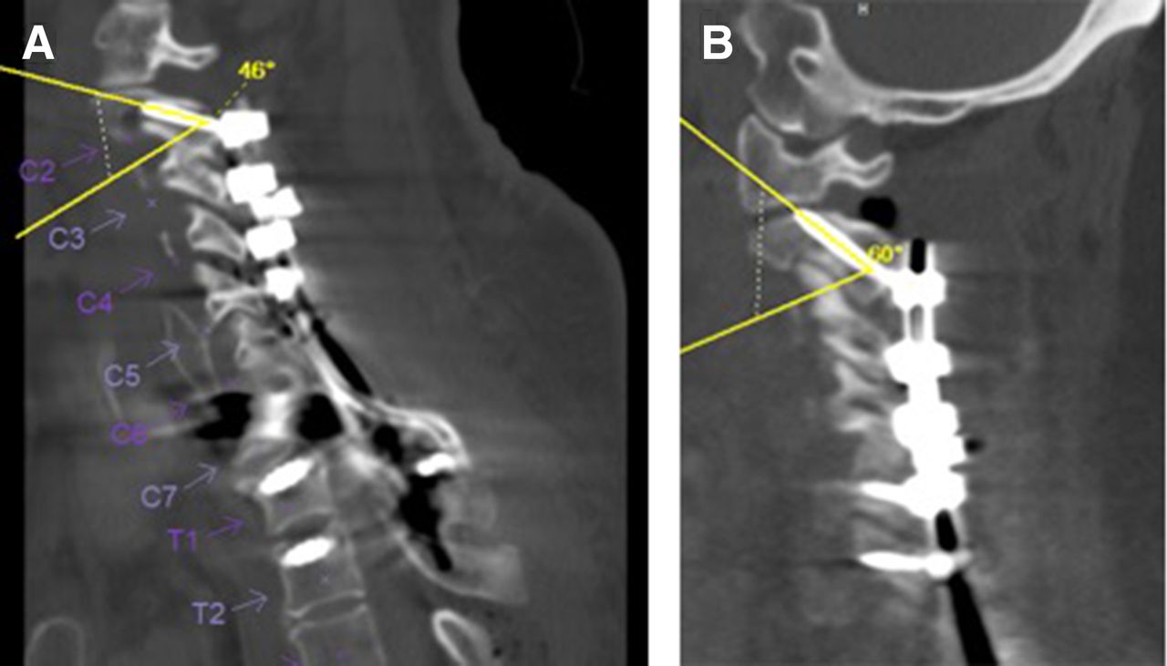
- Figure 5
Computed tomography images demonstrating a lower sagittal facet joint angle in the facet joint penetration (FJP) group (B) compared with the non-FJP group (A).
- Figure 6
Computed tomography images demonstrating a higher sagittal screw angle in the facet joint penetration (FJP) group (B) compared with the non-FJP group (A).
- Figure 7
Computed tomography images demonstrating increased screw length in the facet joint penetration (FJP) group (B, 38 mm) compared with the non-FJP group (A, 26 mm). Both patients had similar sagittal facet joint angles, sagittal screw angles, and axial screw angles (19° vs 21°, 51° vs 53°, and 20° vs 21°, respectively).
- Figure 8
Intraoperative fluoroscopic open mouth view demonstrating C1-C2 facet joint penetration.


Tables
- Table 1
Comparison of postoperative CT measurements between patients with and without FJP.
Postoperative CT Measurement Non-FJP Screw Placement (n = 60) FJP Screw Placement (n = 8) P Value Sagittal angle of the C1-C2 facet joint 20.8° ± 5.5° 13.9° ± 4.4° <0.01 Sagittal angle of the C2 pedicle screw 50.1° ± 6.1° 55.5° ± 6.4° 0.02 Axial angle of the C2 pedicle screw 17.9° ± 7.7° 13.8° ± 3.4° 0.02 C2 pedicle screw length, mm 23.5 ± 4.3 28.4 ± 3.9 <0.01 Bone mineral density of C2, Hounsfield units 397 ± 134 305 ± 72 0.06 Abbreviations: CT, computed tomography; FJP, facet joint penetration.
Note: Data represented as mean ± SD.
Computed Tomography Measurement Non-FJP Screw Placement (n = 60) FJP Screw Placement (n = 8) P Value Width, mm 7.5 ± 2.1 7.4 ± 1.1 0.88 Length, mm 32.2 ± 2.9 31.7 ± 1.3 0.59 Height, mm 7.7 ± 1.4 7.4 ± 1.1 0.48 Transverse angle 43.0° ± 3.4° 42.3° ± 2.5° 0.55 Abbreviation: FJP, facet joint penetration.
Note: All data are represented as mean ± SD.
In this issue


Jump to section
Related Articles
Cited By...
- No citing articles found.