
Main menu

  • Home
  • Content
    • Current Issue
    • Advance Online Publication
    • Archive
  • About Us
    • About ISASS
    • About the Journal
    • Author Instructions
    • Editorial Board
    • Reviewer Guidelines & Publication Criteria
  • More
    • Advertise
    • Subscribe
    • Alerts
    • Feedback
  • Join Us
  • Reprints & Permissions
  • Sponsored Content
  • Other Publications
    • ijss

User menu

  • My alerts

Search

  • Advanced search
International Journal of Spine Surgery
  • My alerts
International Journal of Spine Surgery

Advanced Search

  • Home
  • Content
    • Current Issue
    • Advance Online Publication
    • Archive
  • About Us
    • About ISASS
    • About the Journal
    • Author Instructions
    • Editorial Board
    • Reviewer Guidelines & Publication Criteria
  • More
    • Advertise
    • Subscribe
    • Alerts
    • Feedback
  • Join Us
  • Reprints & Permissions
  • Sponsored Content
  • Follow ijss on Twitter
  • Visit ijss on Facebook
Research ArticleCervical Spine

Combining Laminoplasty With Artificial Disc Replacement for the Treatment of Cervical Spondylotic Myelopathy With Congenital Cervical Stenosis

Zhao-Quan Liu, Cheng-Ta Hsieh, Chih-Ta Huang, Szu-Kai Hsu, Jing-Jing Fang and Chih-Ju Chang
International Journal of Spine Surgery May 2023, 8475; DOI: https://doi.org/10.14444/8475
Zhao-Quan Liu
1 Division of Neurosurgery, Department of Surgery, Cathay General Hospital, Taipei City, Taiwan
MD
  • Find this author on Google Scholar
  • Find this author on PubMed
  • Search for this author on this site
Cheng-Ta Hsieh
1 Division of Neurosurgery, Department of Surgery, Cathay General Hospital, Taipei City, Taiwan
2 Division of Neurosurgery, Department of Surgery, Sijhih Cathay General Hospital, New Taipei City, Taiwan
3 School of Medicine, National Tsing Hua University, Hsinchu, Taiwan
MD, PHD
  • Find this author on Google Scholar
  • Find this author on PubMed
  • Search for this author on this site
Chih-Ta Huang
1 Division of Neurosurgery, Department of Surgery, Cathay General Hospital, Taipei City, Taiwan
2 Division of Neurosurgery, Department of Surgery, Sijhih Cathay General Hospital, New Taipei City, Taiwan
MD
  • Find this author on Google Scholar
  • Find this author on PubMed
  • Search for this author on this site
Szu-Kai Hsu
1 Division of Neurosurgery, Department of Surgery, Cathay General Hospital, Taipei City, Taiwan
2 Division of Neurosurgery, Department of Surgery, Sijhih Cathay General Hospital, New Taipei City, Taiwan
MD
  • Find this author on Google Scholar
  • Find this author on PubMed
  • Search for this author on this site
Jing-Jing Fang
4 Department of Mechanical Engineering, National Cheng Kung University, Tainan, Taiwan
PHD
  • Find this author on Google Scholar
  • Find this author on PubMed
  • Search for this author on this site
Chih-Ju Chang
1 Division of Neurosurgery, Department of Surgery, Cathay General Hospital, Taipei City, Taiwan
2 Division of Neurosurgery, Department of Surgery, Sijhih Cathay General Hospital, New Taipei City, Taiwan
3 School of Medicine, National Tsing Hua University, Hsinchu, Taiwan
5 Department of Medicine, School of Medicine, Fu Jen Catholic University, New Taipei City, Taiwan
MD, PHD
  • Find this author on Google Scholar
  • Find this author on PubMed
  • Search for this author on this site
  • For correspondence: miklechang5639@gmail.com
  • Article
  • Figures & Data
  • Info & Metrics
  • PDF
Loading

Article Figures & Data

Figures

  • Tables
  • Supplementary Materials
  • Figure 1
    • Download figure
    • Open in new tab
    • Download powerpoint
    Figure 1

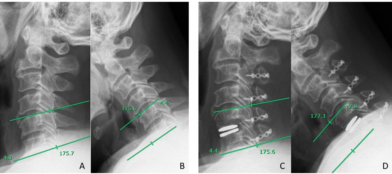
    (A) Preoperative extension radial graphic, between line means C5 and C6 (index) Cobb angle. (B) Preoperative flexion radial graphic, between line means C5 and C6 (index) Cobb angle; therefore, preoperative range of motion = 4.3° + 4. 4° = 8.7°. (C) Postoperative 6-mo extension radial graphic, C5-C6 (index) Cobb angle. (D) Postoperative 6-mo flexion radial graphic, C5-C6 (index) Cobb angle, postoperative 6-mo range of motion; 4.4° + 2.9° = 7.3°.

  • Figure 2
    • Download figure
    • Open in new tab
    • Download powerpoint
    Figure 2

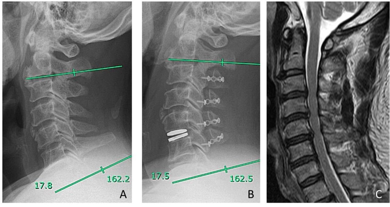
    (A) Preoperative lateral graphic, Cobb angle = 17.8°. (B) Postoperative 6-mo neutral lateral graphic, Cobb angle = 17.5°. (C) Preoperative sagittal T2-weighted magnetic resonance image.

  • Figure 3
    • Download figure
    • Open in new tab
    • Download powerpoint
    Figure 3

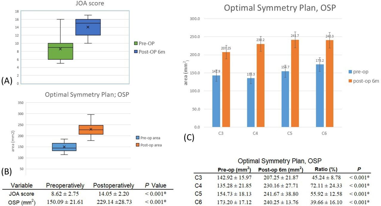
    (A) The Japanese Orthopedic Association (JOA) score significantly improved after surgery (P < 0.001). (B) The calculated spinal canal diameter significantly expanded after surgery (P < 0.001). (C) Each laminoplasty level canal expansion area. OSP, optimal symmetry plan; post-op, postoperative; pre-op, preoperative. *Statistically significant at P < 0.05.

  • Figure 4
    • Download figure
    • Open in new tab
    • Download powerpoint
    Figure 4

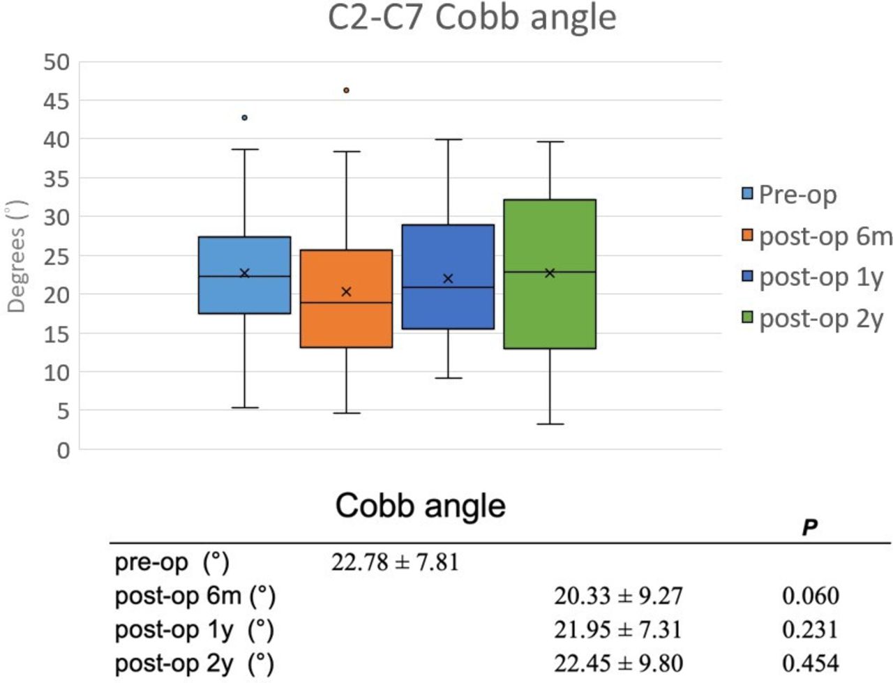
    Cobb angle at preoperative and postoperative 6 mo, 1 y, and 2 y. Each of them had no significant difference.

  • Figure 5
    • Download figure
    • Open in new tab
    • Download powerpoint
    Figure 5

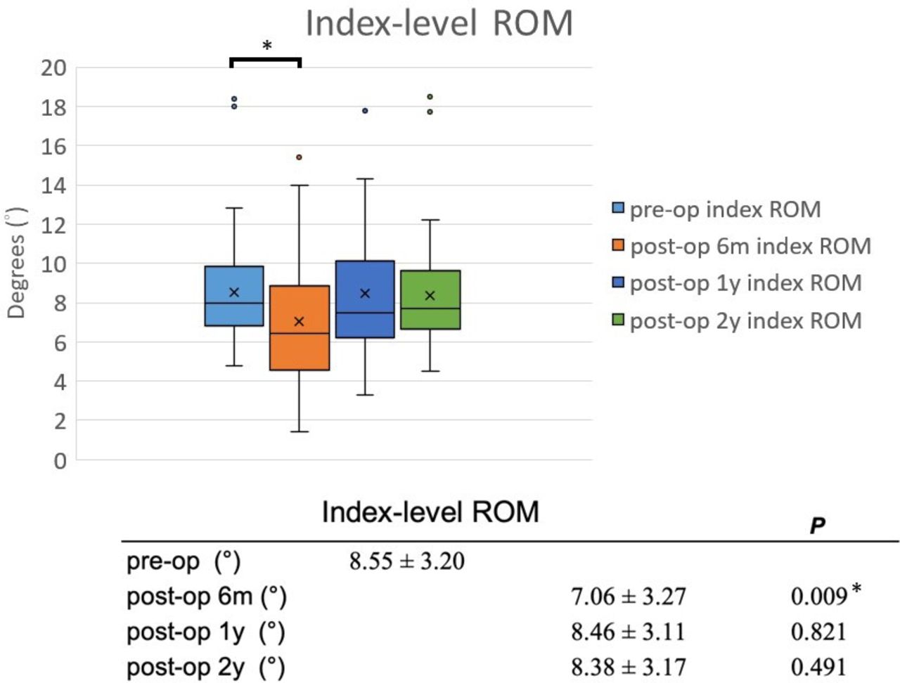
    Index-level range of motion (ROM) at preoperative and postoperative 6 mo, 1 y, and 2 y; only postoperative 6 mo had difference than preoperative; After 2-y follow-up, the index-level ROM could preserve as preoperation.

  • Figure 6
    • Download figure
    • Open in new tab
    • Download powerpoint
    Figure 6

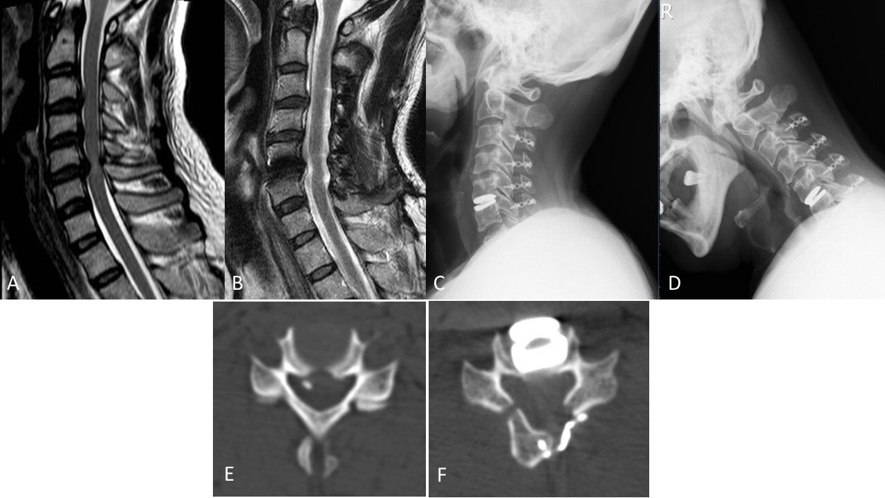
    Case 1. (A) Preoperative sagittal T2-weighted magnetic resonance image (MRI). (B) Postoperative 2-y sagittal T2-weighted MRI. (C and D) Postoperative 2-y extension and flexion view of cervical spine x-ray image: preservation of range of motion. (E) Preoperative bone window axial view at the C5-C6 level. (F) Postoperative 6-mo bone window axial view at C5-C6 level with an obviously expanded spinal canal.

  • Figure 7
    • Download figure
    • Open in new tab
    • Download powerpoint
    Figure 7

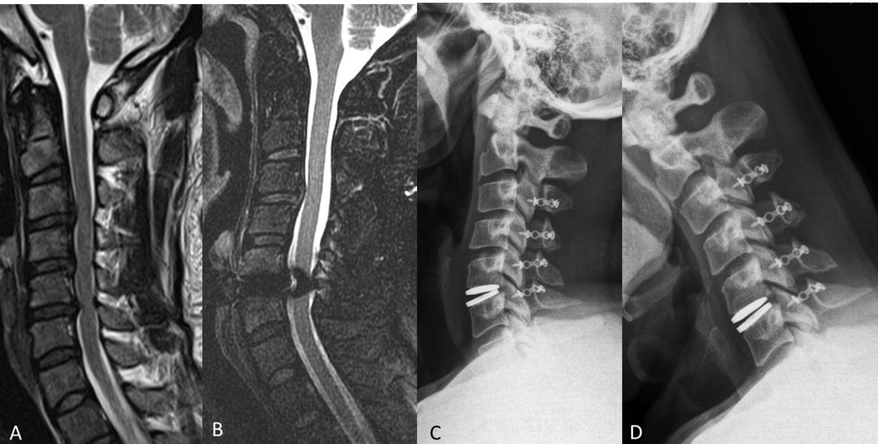
    Case 2. (A) Preoperative sagittal T2-weighted magnetic resonance imaging (MRI), C3-C4 and C5-C6 had anterior disc compression lesion. (B) Postoperative 2-y follow-up T2-weighted MRI, spinal canal expansion. (C and D) Postoperative 2-y extension and flexion x-ray images reveal preserved range of motion.

Tables

  • Figures
  • Supplementary Materials
    • View popup
    Table

    Patients’ demographics and surgery level.

    Category N = 39
    Age, y, mean ± SD49.49 ± 9.36
    Sex, n
     Male33
     Female6
    Body mass index, kg/m2, mean ± SD26.80 ± 3.00
    Smoke, n 20
    Diabetes mellitus, n 5
    No. of segments artificial disc replacement operated, n (%)
     C3-C43 (7.7%)
     C4-C513 (33.3%)
     C5-C619 (48.7%)
     C4-C5-C63 (7.7%)
     C5-C6-C71 (2.6%)
    No. of segments laminoplasty operated, n (%)
     C3-C4-C5-C626 (66.7%)
     C4-C5-C612 (30.8%)
     C3-C4-C51 (2.6%)

Supplementary Materials

  • Figures
  • Tables
  • Online Supplemental Video 1.

    Flexion and Extension.

    [8475supp001.mp4]

  • Online Supplemental Video 2.

    Rotation.

    [8475supp002.mp4]

PreviousNext
Back to top

In this issue

International Journal of Spine Surgery: 19 (S2)
International Journal of Spine Surgery
Vol. 19, Issue S2
1 Apr 2025
  • Table of Contents
  • Index by author

Print
Download PDF
Article Alerts
Sign In to Email Alerts with your Email Address
Email Article

Thank you for your interest in spreading the word on International Journal of Spine Surgery.

NOTE: We only request your email address so that the person you are recommending the page to knows that you wanted them to see it, and that it is not junk mail. We do not capture any email address.

Enter multiple addresses on separate lines or separate them with commas.
Combining Laminoplasty With Artificial Disc Replacement for the Treatment of Cervical Spondylotic Myelopathy With Congenital Cervical Stenosis
(Your Name) has sent you a message from International Journal of Spine Surgery
(Your Name) thought you would like to see the International Journal of Spine Surgery web site.
CAPTCHA
This question is for testing whether or not you are a human visitor and to prevent automated spam submissions.
Citation Tools
Combining Laminoplasty With Artificial Disc Replacement for the Treatment of Cervical Spondylotic Myelopathy With Congenital Cervical Stenosis
Zhao-Quan Liu, Cheng-Ta Hsieh, Chih-Ta Huang, Szu-Kai Hsu, Jing-Jing Fang, Chih-Ju Chang
International Journal of Spine Surgery May 2023, 8475; DOI: 10.14444/8475

Citation Manager Formats

  • BibTeX
  • Bookends
  • EasyBib
  • EndNote (tagged)
  • EndNote 8 (xml)
  • Medlars
  • Mendeley
  • Papers
  • RefWorks Tagged
  • Ref Manager
  • RIS
  • Zotero
Share
Combining Laminoplasty With Artificial Disc Replacement for the Treatment of Cervical Spondylotic Myelopathy With Congenital Cervical Stenosis
Zhao-Quan Liu, Cheng-Ta Hsieh, Chih-Ta Huang, Szu-Kai Hsu, Jing-Jing Fang, Chih-Ju Chang
International Journal of Spine Surgery May 2023, 8475; DOI: 10.14444/8475
Twitter logo Facebook logo Mendeley logo
  • Tweet Widget
  • Facebook Like
  • Google Plus One

Jump to section

  • Article
    • Abstract
    • Introduction
    • Materials and Methods
    • Results
    • Discussion
    • Conclusions
    • Footnotes
    • References
  • Figures & Data
  • Info & Metrics
  • PDF

Related Articles

  • No related articles found.
  • PubMed
  • Google Scholar

Cited By...

  • No citing articles found.
  • Google Scholar

More in this TOC Section

  • Innovation and Adversity in Spine Surgery: A Retrospective
  • Cervical Myelopathy Secondary to Bilateral Atlantoaxial Pseudoarticulations in Rheumatoid Arthritis: A Case Report
  • Incidence of Traumatic Spinal Injury Following Public Policy Update on Moped Usage in South Carolina
Show more Cervical Spine

Similar Articles

Keywords

  • cervical laminoplasty
  • artificial disc
  • range of motion
  • cervical spondylotic myelopathy
  • congenital cervical stenosis

Content

  • Current Issue
  • Latest Content
  • Archive

More Information

  • About IJSS
  • About ISASS
  • Privacy Policy

More

  • Subscribe
  • Alerts
  • Feedback

Other Services

  • Author Instructions
  • Join ISASS
  • Reprints & Permissions

© 2025 International Journal of Spine Surgery

International Journal of Spine Surgery Online ISSN: 2211-4599

Powered by HighWire