


Article Figures & Data
Figures
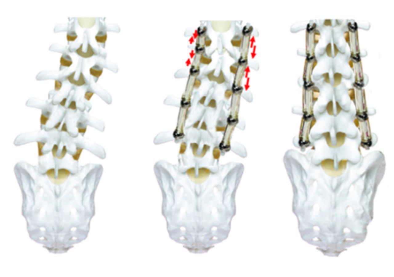
- Figure 1
If the Dynesys system is used, the residual deformity is corrected by cutting the spacers shorter than normal and providing greater torque than normal in the concave part of the deformity.
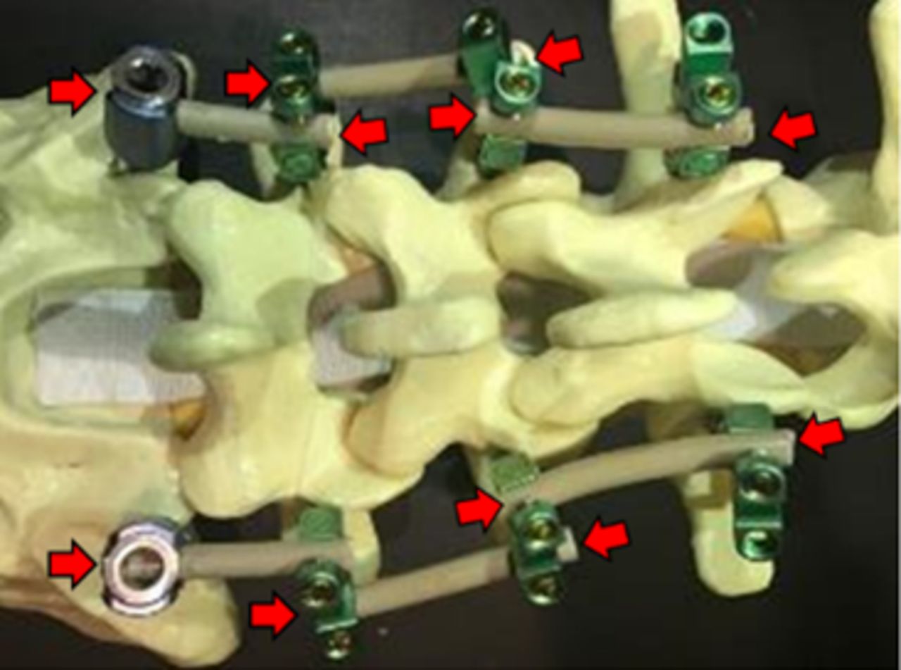
- Figure 2
When Orthrus and Peek rods are used, compressing the concave side and locking the rod to the screw in this manner is sufficient to provide additional improvement.
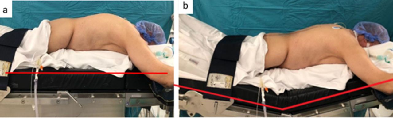
- Figure 3
In kyphotic deformities, in the second stage, (A) the table is positioned under the scope, (B) normal sagittal balance is achieved, and the rods are placed.
- Figure 4
In patients undergoing 2-stage surgery, temporary rods can be placed in the segments that were decompressed in the first stage. In this case, the risk of screw loosening in the relevant segments increases.
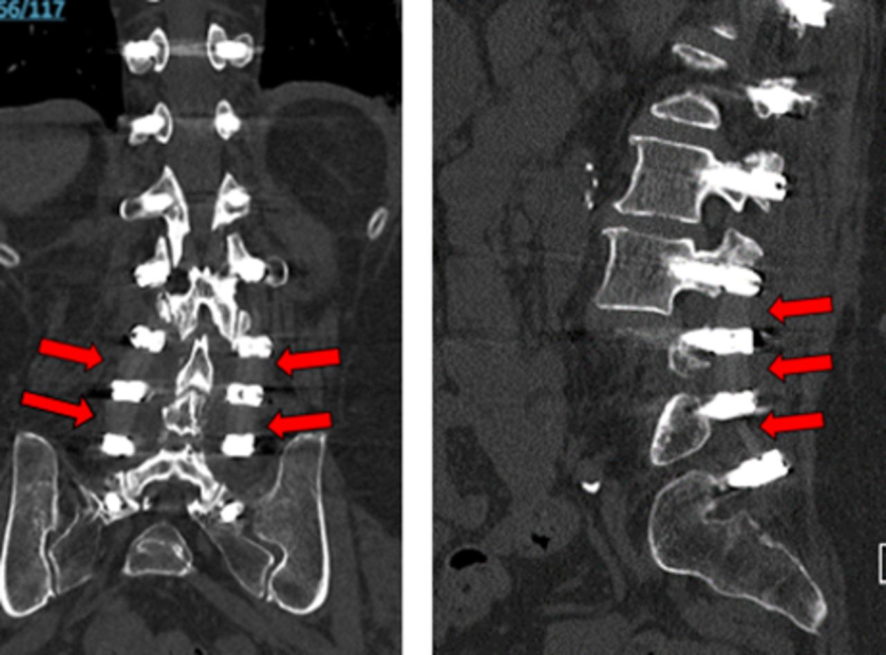
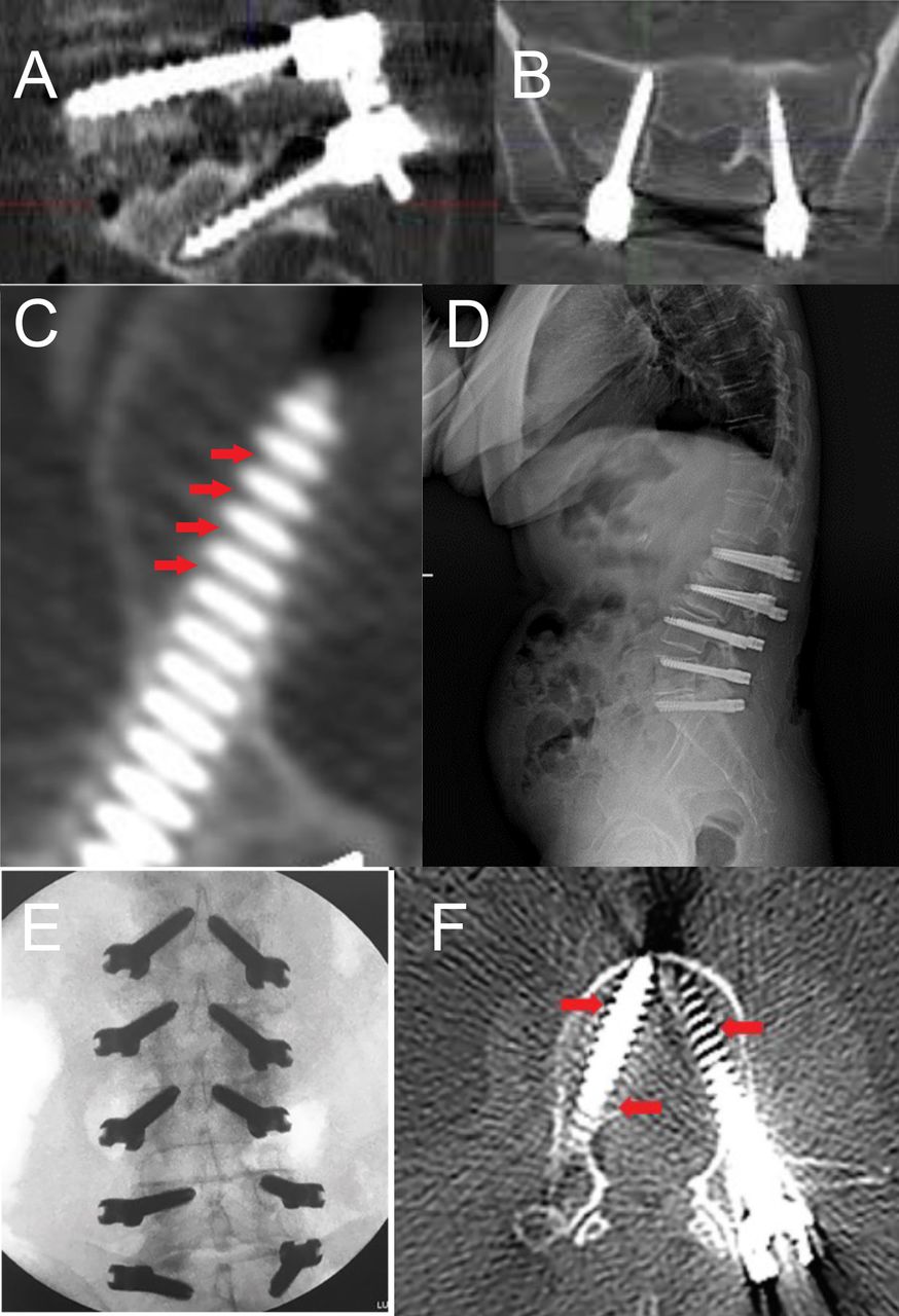
- Figure 5
Patients are evaluated with computed tomography (CT) after an average of 16–20 weeks for the determination of osteointegration of screws. If osteointegration is completed, then rods are placed and screws are connected to each other. (A) Sagittal and (B) coronal CT image of patients showing screw loosening after traditional surgery with rigid stabilization. (C) Axial CT image of the patient showing successful osteointegration after the first stage of 2-stage surgery. (D) Lateral x-ray image of the patient after the second stage stabilization surgery by Dynesys system. (E) Intraoperative coronal fluoroscopy image and (F) CT image of the patient showing osteointegration after the first stage of 2-stage surgery. Red arrows indicate areas of connection between bone and screws.
- Figure 6
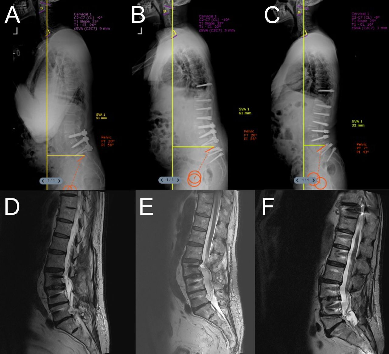
Patient who previously underwent L3-L4 interbody fusion and L3-L4-L5 stabilization due to spondylolisthesis presented with worsening kyphosis and difficulty looking forward. (A) Preoperative standing lateral x-ray image, (B) first stage postoperative standing lateral x-ray image, (C) second stage postoperative standing lateral x-ray image, (D) sagittal magnetic resonance image (MRI) after initial L3-L4-L5 stabilization, (E) preoperative sagittal MRI showing proximal junctional kyphosis and Pfirrmann grade 4 intervertebral disc degeneration, and (F) second stage postoperative sagittal MRI. The Dynesis system was used for dynamic stabilization. Two-stage surgery was performed because the patient was osteoporotic (T score = −2.5 preop, –1.5 before the second stage). The patient showed significant improvement in both spinopelvic parameters and clinical findings after surgery.
- Figure 7
Radiological images of a patient with neurological claudication and walking difficulties. Decompression and dynamic stabilization with the Orthrus system were performed. (A) Preoperative standing lateral x-ray image and (B) Postoperative standing lateral x-ray image showing sagittal imbalance and related pelvic parameters. (C and E) Preoperative sagittal magnetic resonance images and (D and F) preoperative axial magnetic resonance imaging (MRI), showing Schizas grade D and Lee grade 3 spinal stenosis. The patient showed significant increase in walking distance without difficulty.
- Figure 8
Radiological images of a patient with lower back pain and walking difficulties. Decompression and dynamic stabilization with Dynesys system were performed. (A) Preoperative sagittal magnetic resonance image showing Pfirrmann grade 4 degenerative intervertebral disc changes. (B) Preoperative lateral x-ray image showing sagittal imbalance and pelvic parameters. (C) Postoperative lateral x-ray image showing improved sagittal balance.
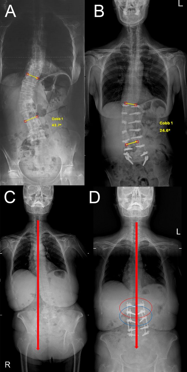
- Figure 9
Radiological images of 2 different patients with severe lower back pain and walking difficulties showing decompensated coronal imbalance. Both of the patients were decompressed and dynamically stabilized with Orthrus system. (A and C) Preoperative and (B and D) postoperative anteroposterior x-ray images showing significant improvement in coronal balance of the patients.
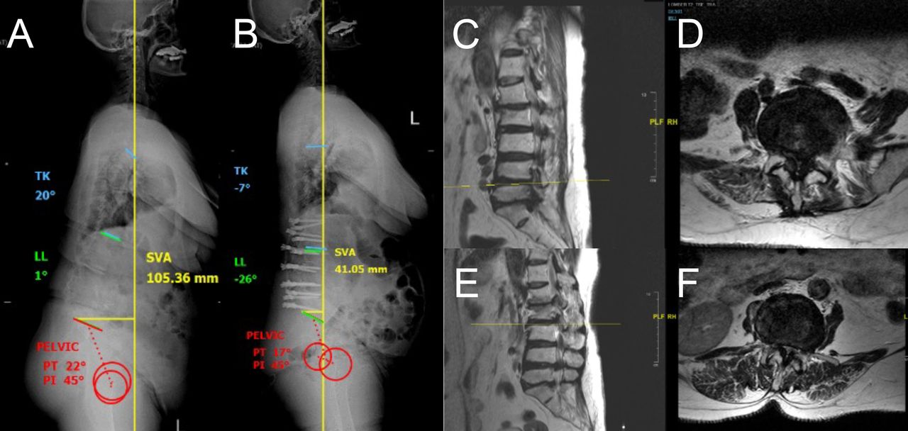
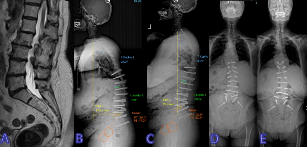
- Figure 10
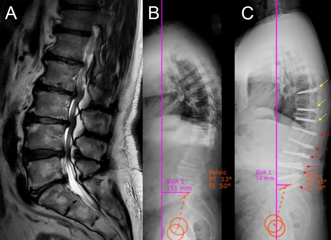
Two-stage surgery: A 69-year-old woman, who underwent an operation for L4-5 stenosis 10 years ago, developed low back pain during the process and started to lean forward gradually. (A) Preoperative magnetic resonance image (MRI); (B) lateral x-ray image after the first stage, before placement of the rods; (C) lateral x-ray image after placement of the rods; (D) antero-posterior x-ray image after the first stage, before placement of the rods; (E) antero-posterior x-ray image after placement of the rods. The patient showed significant improvement in both spinopelvic parameters and clinical findings after surgery.


Tables
- Table 1
Details of the system used, patient age and gender, number of levels operated, Schizas and Lee grades of spinal stenosis, Pfirrmann grade of intervertebral disc degeneration, number of stages of surgery, and preoperative T scores.
Patient Number System Age, y Gender Level Schizas Grade Lee Grade Pfirrmann Grade Stage Preop T Score 1 Dynesys 48 F T12-S1 B 1 4 2 -2.5 2 Dynesys 65 F T10-S1 C 2 4 2 -2.5 3 Dynesys 51 M L2-Iliac B 1 3 1 -1.5 4 Dynesys 63 F T9-L5 D 3 5 1 -1.5 5 Dynesys 47 M T12-Iliac B 1 4 1 -1.5 6 Dynesys 54 F L3-Iliac A3 0 4 1 -1.5 7 Dynesys 41 F L2-Iliac C 2 3 1 -1 8 Dynesys 77 F T12-L5 D 3 5 1 -1.5 9 Dynesys 65 F T10-Iliac D 3 5 1 -2.5 10 Dynesys 79 F L2-Iliac A2 0 4 1 -2.5 11 Dynesys 66 F L2-S1 B 1 4 2 -2.5 12 Dynesys 68 F T10-S1 C 2 4 1 -2.5 13 Dynesys 44 M L2-Iliac A3 0 2 1 -1 14 Dynesys 68 F T10-Iliac D 3 4 1 -1.5 15 Dynesys 69 F T6-S1 D 3 4 2 -2.5 16 Dynesys 61 M L1-S1 D 3 5 1 -1.5 17 Dynesys 72 M T12-Iliac A2 0 4 1 -2.5 18 Dynesys 78 F L1-S1 B 1 4 1 -1 19 Dynesys 67 M T10-Iliac B 1 5 2 -2.5 20 Orthrus 68 F T12-S1 C 2 5 1 -1.5 21 Orthrus 61 F T11-S1 A3 0 3 1 -1.5 22 Orthrus 77 F L2-Iliac B 1 4 1 -1.5 23 Orthrus 65 M L2-S1 D 3 4 1 -1.5 24 Orthrus 52 M T11-L5 C 2 4 1 -1 25 Orthrus 67 M L1-S1 D 3 4 1 -1.5 Abbreviations: F, female; M, male; Preop, preoperative.
Characteristic n (%) Age, y, mean ± SD 62.92 ± 10.80 Gender Female 17 (68.0) Male 8 (32.0) System Dynesys 19 (76.0) Orthrus 6 (24.0) VAS ODI Patient number Preop 6-mo Postop 12-mo Postop 24-mo Postop Preop 6-mo Postop 12-mo Postop 24-mo Postop 1 7 3 1 1 72 26 6 8 2 7 3 1 0 66 26 16 10 3 6 2 0 0 56 28 12 8 4 7 3 2 0 66 36 16 16 5 8 1 1 1 58 16 10 10 6 7 2 0 2 72 18 12 8 7 7 3 0 1 74 26 8 6 8 5 4 2 0 72 24 26 8 9 6 4 4 4 68 56 58 52 10 8 1 1 0 68 18 8 8 11 6 3 2 1 66 16 8 12 12 7 2 0 1 72 26 8 6 13 8 2 1 2 76 28 2 2 14 7 2 2 1 62 32 6 8 15 8 2 1 1 76 32 8 2 16 6 4 1 2 76 36 4 4 17 7 3 1 1 58 26 2 20 18 7 2 0 2 76 26 6 8 19 8 1 0 1 72 28 8 2 20 7 2 1 1 58 26 8 6 21 6 1 1 0 62 26 4 4 22 7 3 1 1 58 36 4 2 23 7 1 1 0 60 16 12 18 24 6 3 2 1 72 30 2 2 25 7 2 0 0 72 16 12 8 Mean 6.88 2.36 1.04 0.96 67.52 26.96 10.64 9.52 Abbreviations: ODI, Oswestry Disability Index; Postop, postoperative; Preop, preoperative; VAS, visual analog scale.
Outcome Measure Preoperative, mean ± SD Postoperative, mean ± SD Pa 6-mo 12-mo 24-mo VAS 6.88 ± 0.78 2.36 ± 0.95 1.04 ± 0.93 0.96 ± 0.93 <0.001 ODI 67.52 ± 6.74 26.96 ± 8.68 10.64 ± 11.21 9.52 ± 10.07 <0.001 Abbreviations: ODI, Oswestry Disability Index; VAS, visual analog scale.
↵a Repeated measure analysis of variance was applied.
Outcome Measure Preoperative, mean ± SD Postoperative, mean ± SD 3-mo 6-mo 12-mo 24-mo Scoliotic Cobb angle 19.23 ± 7.68 11.29 ± 7.06 (P = 0.001) 12.16 ± 8.11 12.57 ± 7.67 12.51 ± 9.21 Thoracic kyphosis angle 27.36 ± 11.40 23.48 ± 9.61 (P = 0.013) 25.11 ± 9.62 25.26 ± 10.88 25.44 ± 10.45 SVA (mm) 75.84 ± 63.56 52.78 ± 49.37 (P = 0.047) 56.17 ± 46.95 58.46 ± 51.39 58.74 ± 53.87 PI 52.76.± 15.64 50.80 ± 12.74 (P = 0.442) PT 22.68 ± 12.76 26.08 ± 8.51 (P = 0.159) SS 30.04 ± 7.75 24.80 ± 8.98 (P = 0.008) Abbreviations: PI, pelvic incidence; PT, pelvic tilt; SS, sacral slope; SVA, sagittal vertical axis.
a Paired samples t test was applied.
In this issue


Jump to section
Related Articles
Cited By...
- No citing articles found.