


Article Figures & Data
Figures
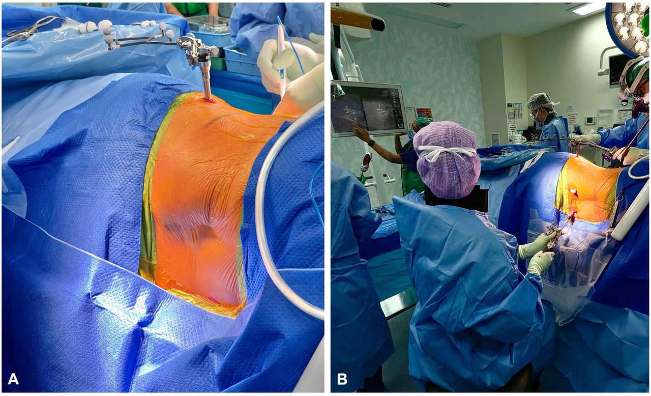
- Figure 1
Intraoperative clinical photographs. (A) Lateral decubitus patient positioning with reference frame attached to the iliac crest. (B) Use of 3-dimensional computer-assisted navigation for placement of a percutaneous pedicle screw starting with the most proximal vertebra to be instrumented.
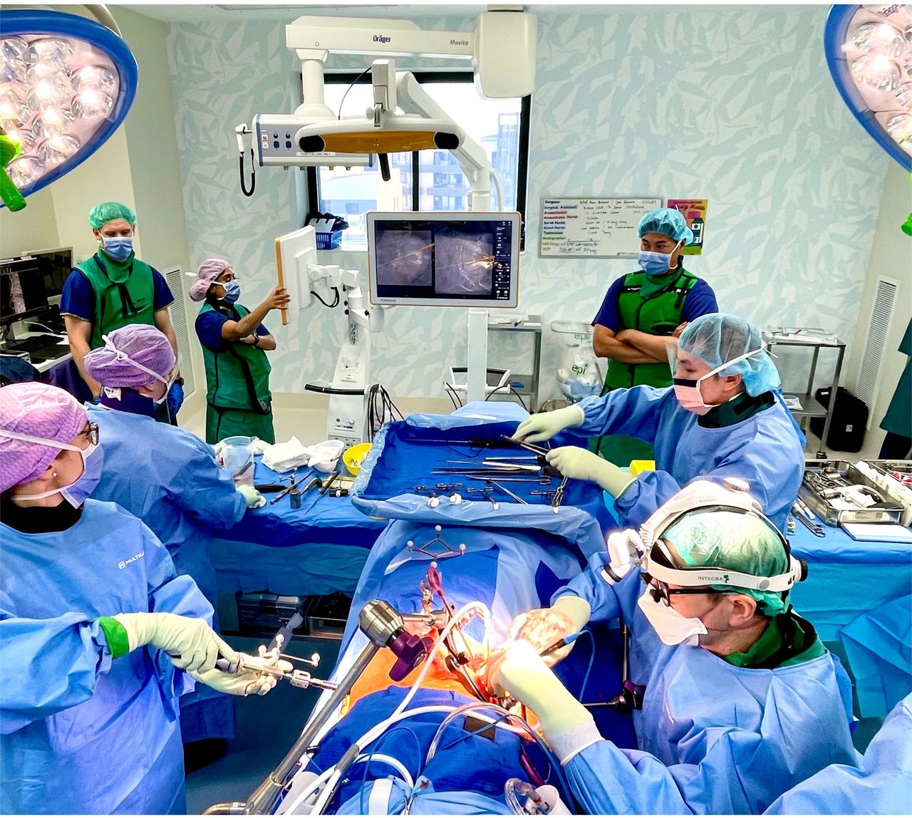
- Figure 2
Intraoperative clinical photograph demonstrating the operating room setup. The anterior exposure of the disc space is performed by the vascular surgeon on the right side. Simultaneously, the spine surgeon performs the percutaneous pedicle screw placement with 3-dimensional computer-assisted navigation, seen in the background. Each surgeon has their own scrub nurse.
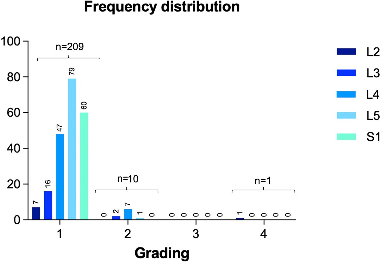
- Figure 3
Frequency distribution of pedicle screws according to grading (Grades I–IV).
- Figure 4
Illustrative case with axial (A) and coronal (B) postoperative computed tomography images demonstrating a Grade IV lateral breach (orange line: 4.8 mm) of the left L2 pedicle due to pedicle orientation. Had the “perfect” pedicle trajectory been followed (blue line), facet joint violation would have been inevitable. The coronal view (B) demonstrates a narrow “V-shaped” pedicle on the left side.
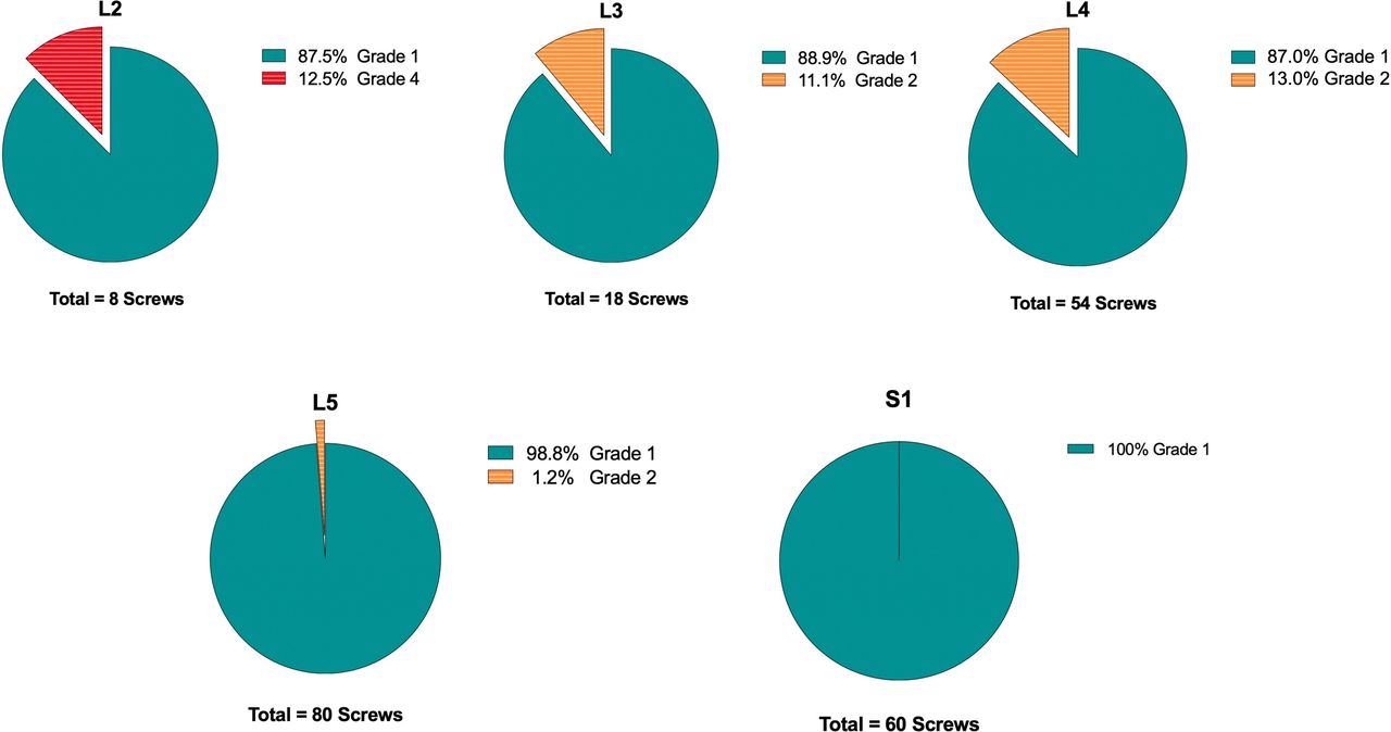
- Figure 5
Pedicle screw breaches according to level.
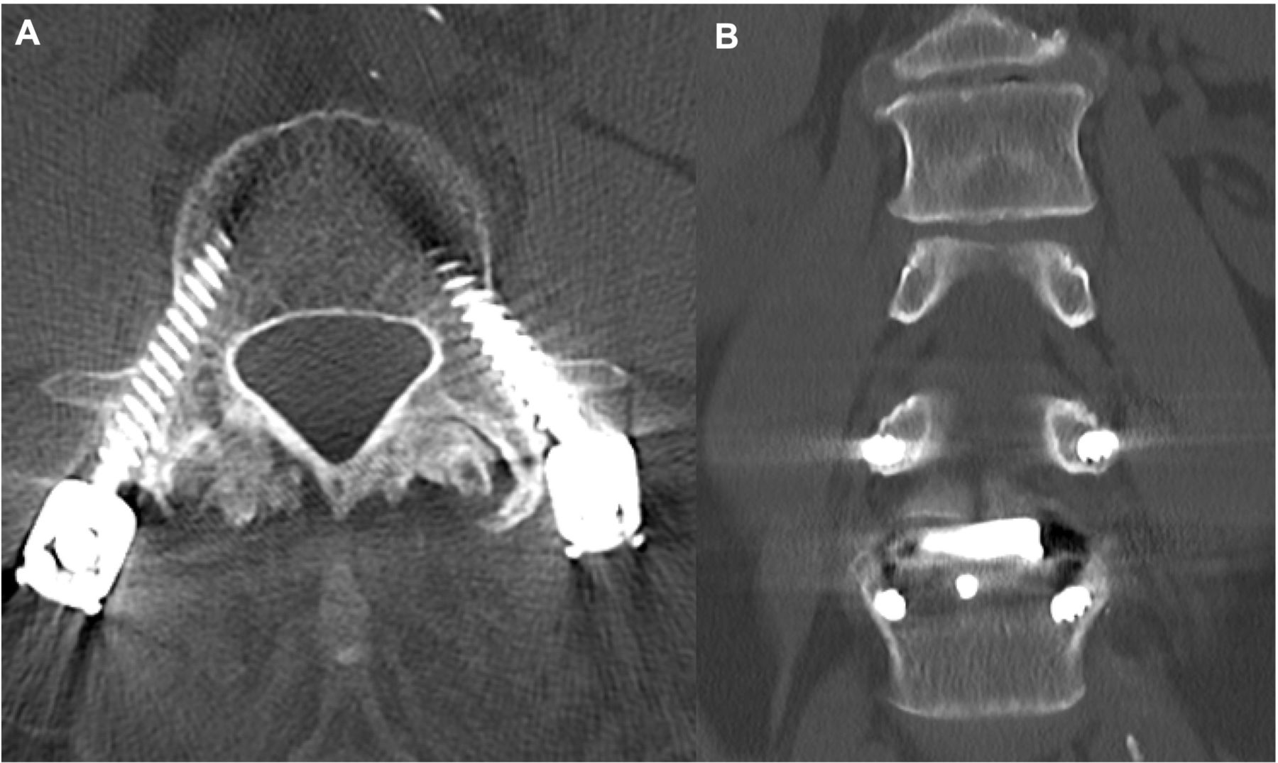
- Figure 6
Illustrative case with axial (A) and coronal (B) postoperative computed tomography images demonstrating a Grade II lateral breach of both L4 pedicles with hypertrophic facet joints at L3/4.


Tables
Characteristic Value Gender, n (%) Women 28 (63.6) Men 16 (36.4) Age, y, median (range) 64.1 (17.3–86.3) Body mass index, mean (SD) 27.62 (5.74) Smoking, n (%) Never 39 (88.6) Former 2 (4.5) Unknown 3 (6.8) Diabetes, n (%) No 40 (90.9) Yes 4 (9.1) Osteoporosis, n (%) No 24 (54.5) Yes 2 (4.5) Unknown 18 (40.9) Charlson Comorbidity Index, median (range) 2 (0–8) Outcome Measure Value Indication for surgery, n (%) Degenerative spondylolisthesis 24 (54.5) Degenerative disc disease 12 (27.3) Foraminal stenosis with radiculopathy 6 (13.6) Facet arthropathy 1 (2.3) Spondylolysis/pedicle fracture 1 (2.3) Prior surgery, n (%) No 36 (81.8) Yes 8 (18.2) Levels fused, n (%) 1 26 (59.1) 2 15 (34.1) 3 3 (6.8) Median (range) 1 (1–3) ALIF cage levels, n (%) 0 6 (13.6) 1 24 (54.5) 2 14 (31.8) LLIF cage levels, n (%) 0 34 (77.3) 1 7 (15.9) 2 3 (6.8) Cages, n (%) ALIF only 35 (79.5) LLIF only 6 (13.6) Combination of ALIF and LLIF 3 (6.8) Most commonly treated levels, n (%) L5/S1 46.2% L4/5 36.9% L3/4 10.8% L2/3 6.2% Lateral position, n (%) Left side up 36 (81.8) Right side up 8 (18.2) Neuromonitoring, n (%) Yes 44 (100.0) Operative time, min, median (range) 110 (53–293) Fluoroscopy time, sec, median (range) 98.5 (12.8–283) Estimated blood loss, mL, median (range) 100 (100–1900) Length of stay, d 2 (1–14) Abbreviations: ALIF, anterior lumbar interbody fusion; EMG, electromyography; LLIF, lateral lumbar interbody fusion.
a EMG and free-running EMG.
- Table 3
Lateral breach location as a function of pedicle orientation during instrumentation.
Pedicle Orientation n Down-side screw 7 Up-side screw 4 Total 11 - Table 4
Published rates of percutaneous pedicle screw accuracy in lateral decubitus single-position anterior-posterior surgery assessed by computed tomography.
Author Year Insertion Technique No. of Screws Breach Rate Breach Grading Breach Laterality Facet Joint Violations Blizzard et al6 2018 Fluoroscopy 300 5.1% Grade II: 84.6% Grade III: 15.4% Medial: 69.2%
Lateral: 23.1%- Ouchida et al16 2020 Computer-assisted navigation 228 1.8% Grade II: unknown
Grade III: 100%- - Okuda et al17 2023 Computer-assisted navigation 453 4.6% Grade II: 52.4%
Grade III: 27.3%Grade IV: 18.2%Medial: 14.3%
Lateral: 85.7%- Hiyama et al24 2023 Computer-assisted navigation 728 1.9% Grade II: 35.7%
Grade III: 50% Grade IV: 14.3%- - Present study 2024 Computer-assisted navigation 220 5% Grade II: 90%
Grade III: 0%
Grade IV: 10%Medial: 0 %
Lateral: 100%None a Grade II breaches were not reported in this study.
b Only screws in the lumbar spine were considered.
In this issue


Jump to section
Related Articles
Cited By...
- No citing articles found.