
Main menu

  • Home
  • Content
    • Current Issue
    • Advance Online Publication
    • Archive
  • About Us
    • About ISASS
    • About the Journal
    • Author Instructions
    • Editorial Board
    • Reviewer Guidelines & Publication Criteria
  • More
    • Advertise
    • Subscribe
    • Alerts
    • Feedback
  • Join Us
  • Reprints & Permissions
  • Sponsored Content
  • Other Publications
    • ijss

User menu

  • My alerts

Search

  • Advanced search
International Journal of Spine Surgery
  • My alerts
International Journal of Spine Surgery

Advanced Search

  • Home
  • Content
    • Current Issue
    • Advance Online Publication
    • Archive
  • About Us
    • About ISASS
    • About the Journal
    • Author Instructions
    • Editorial Board
    • Reviewer Guidelines & Publication Criteria
  • More
    • Advertise
    • Subscribe
    • Alerts
    • Feedback
  • Join Us
  • Reprints & Permissions
  • Sponsored Content
  • Follow ijss on Twitter
  • Visit ijss on Facebook
Research ArticleTDR

Radiologic Analysis of C2 to Predict Safe Placement of Pedicle Screws

International Journal of Spine Surgery January 2018, 12 (1) 30-36; DOI: https://doi.org/10.14444/5006
1Houston Methodist Hospital, Department of Orthopaedic Surgery, Houston, Texas
2University of Texas Medical School at Houston, Department of Orthopaedic Surgery, Houston, Texas
MD
  • Find this author on PubMed
2University of Texas Medical School at Houston, Department of Orthopaedic Surgery, Houston, Texas
MD
  • Find this author on PubMed
2University of Texas Medical School at Houston, Department of Orthopaedic Surgery, Houston, Texas
MD
  • Find this author on PubMed
2University of Texas Medical School at Houston, Department of Orthopaedic Surgery, Houston, Texas
MD
  • Find this author on PubMed
2University of Texas Medical School at Houston, Department of Orthopaedic Surgery, Houston, Texas
MD
  • Find this author on PubMed
2University of Texas Medical School at Houston, Department of Orthopaedic Surgery, Houston, Texas
MD
  • Find this author on PubMed
1Houston Methodist Hospital, Department of Orthopaedic Surgery, Houston, Texas
MD
  • Find this author on PubMed
  • Article
  • Figures & Data
  • Info & Metrics
  • PDF
Loading

Article Figures & Data

Figures

  • Tables
  • Figure 1
    • Download figure
    • Open in new tab
    • Download powerpoint
    Figure 1

    (a) Identification of the medial wall slice. Sagittal computed tomography reconstruction cut showing the lateral-most aspect of the spinal canal. (b) The next cut is 2 mm lateral to the lateral aspect of the spinal canal and shows the medial wall slice with complete continuity of the pedicle from the vertebral body to the posterior elements.

  • Figure 2
    • Download figure
    • Open in new tab
    • Download powerpoint
    Figure 2

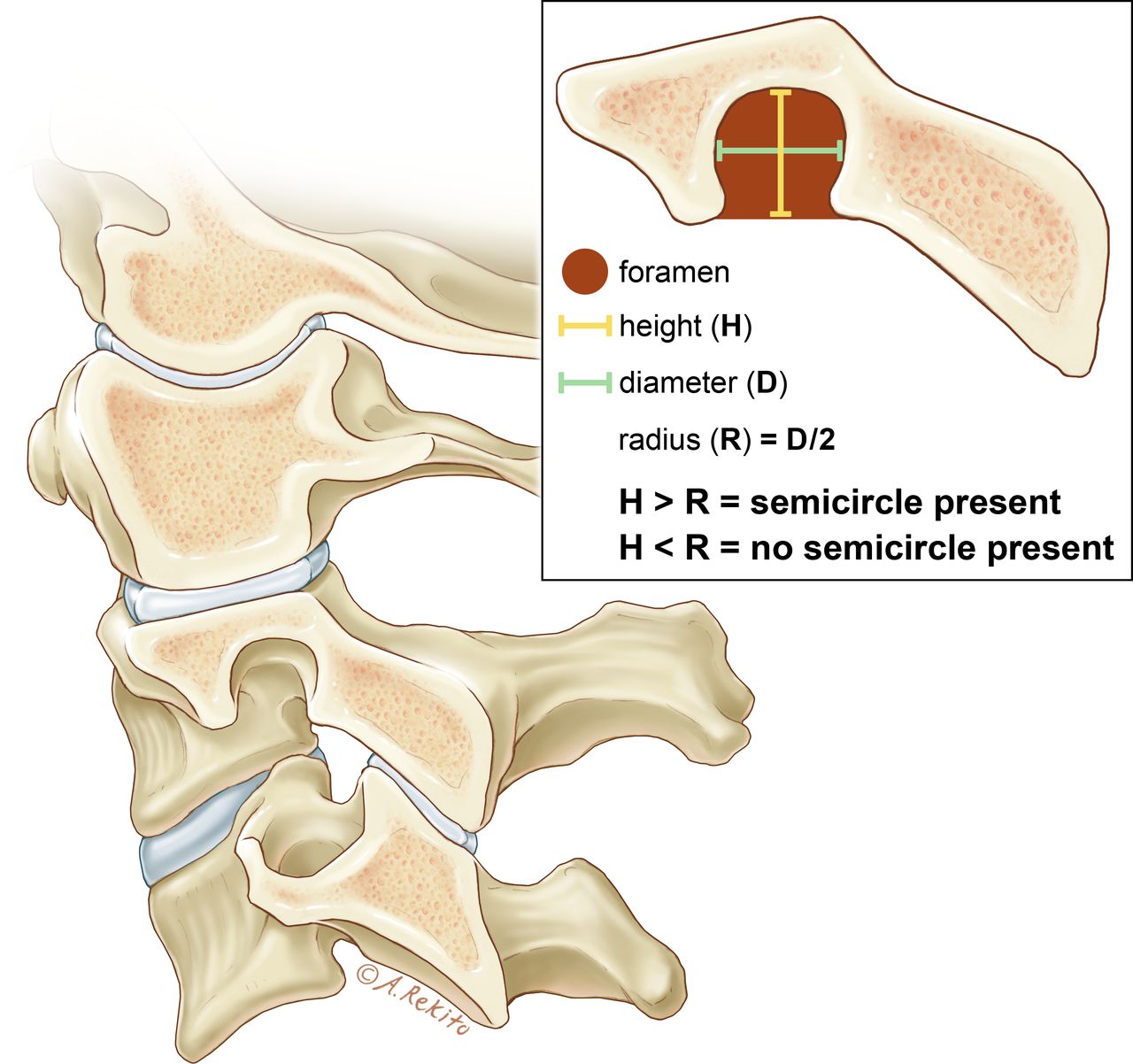
    Identification of the vertebral artery foramen cut. The first cut that shows a complete semicircular opening is deemed the vertebral artery foramen cut. A complete semicircle is considered present when the height (H) is greater than the radius (R) of the foramen.

  • Figure 3
    • Download figure
    • Open in new tab
    • Download powerpoint
    Figure 3

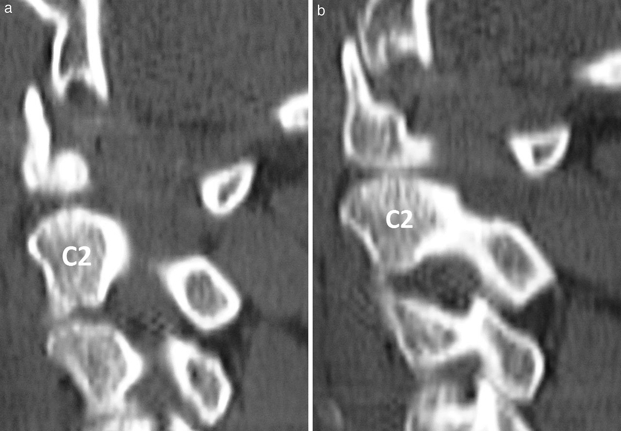
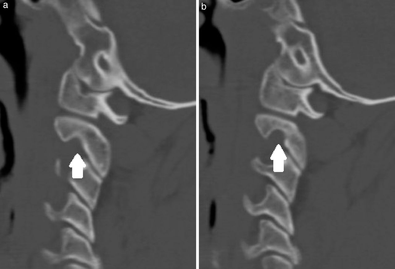
    (a) Identification of the vertebral artery foramen slice. A small portion of the superomedial aspect of the vertebral artery foramen can be seen in the C2 pedicle. (b) The next cut is 2 mm lateral and demonstrates a vertebral artery foramen with at least a full semicircular opening. This cut is deemed the vertebral artery foramen slice.

  • Figure 4
    • Download figure
    • Open in new tab
    • Download powerpoint
    Figure 4

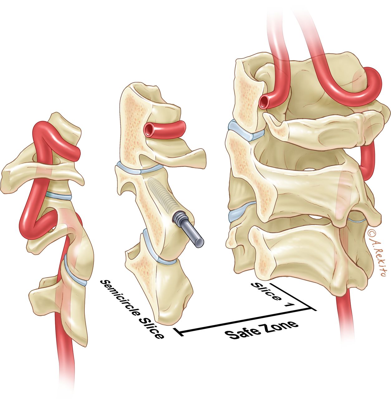
    Safe zone for C2 pedicle screw placement between slice 1 and the semicircle slice.

  • Figure 5
    • Download figure
    • Open in new tab
    • Download powerpoint
    Figure 5

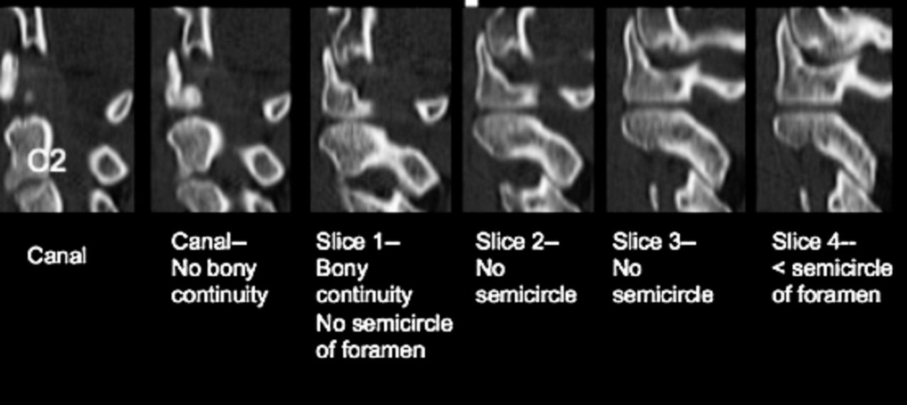
    Series of sagittal computed tomography cuts from the spinal canal through the pedicle. This patient has 4 slices of bony continuity that do not show the vertebral artery foramen. Thus, slices 1 through 4 represent the safe zone where a long pedicle screw can be placed.
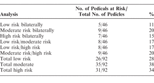
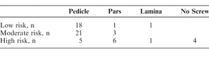
Tables

  • Figures
  • Table 1
  • Table 2
PreviousNext
Back to top

In this issue

International Journal of Spine Surgery
Vol. 12, Issue 1
1 Jan 2018
  • Table of Contents
  • Index by author

Print
Download PDF
Article Alerts
Sign In to Email Alerts with your Email Address
Email Article

Thank you for your interest in spreading the word on International Journal of Spine Surgery.

NOTE: We only request your email address so that the person you are recommending the page to knows that you wanted them to see it, and that it is not junk mail. We do not capture any email address.

Enter multiple addresses on separate lines or separate them with commas.
Radiologic Analysis of C2 to Predict Safe Placement of Pedicle Screws
(Your Name) has sent you a message from International Journal of Spine Surgery
(Your Name) thought you would like to see the International Journal of Spine Surgery web site.
CAPTCHA
This question is for testing whether or not you are a human visitor and to prevent automated spam submissions.
Citation Tools
Radiologic Analysis of C2 to Predict Safe Placement of Pedicle Screws
REX A.W. MARCO, CHRISTOPHER I. PHELPS, REBECCA C. KUO, WU ZHUGE, CLINTON W. HOWARD, VIVEK P. KUSHWAHA, DEREK T. BERNSTEIN
International Journal of Spine Surgery Jan 2018, 12 (1) 30-36; DOI: 10.14444/5006

Citation Manager Formats

  • BibTeX
  • Bookends
  • EasyBib
  • EndNote (tagged)
  • EndNote 8 (xml)
  • Medlars
  • Mendeley
  • Papers
  • RefWorks Tagged
  • Ref Manager
  • RIS
  • Zotero
Share
Radiologic Analysis of C2 to Predict Safe Placement of Pedicle Screws
REX A.W. MARCO, CHRISTOPHER I. PHELPS, REBECCA C. KUO, WU ZHUGE, CLINTON W. HOWARD, VIVEK P. KUSHWAHA, DEREK T. BERNSTEIN
International Journal of Spine Surgery Jan 2018, 12 (1) 30-36; DOI: 10.14444/5006
Twitter logo Facebook logo Mendeley logo
  • Tweet Widget
  • Facebook Like
  • Google Plus One

Jump to section

  • Article
    • Abstract
    • INTRODUCTION
    • MATERIALS AND METHODS
    • RESULTS
    • DISCUSSION
    • CONCLUSION
    • Footnotes
    • REFERENCES
  • Figures & Data
  • Info & Metrics
  • PDF

Related Articles

  • No related articles found.
  • PubMed
  • Google Scholar

Cited By...

  • No citing articles found.
  • Google Scholar

More in this TOC Section

  • Total Joint Replacement of the Lumbar Spine: The Future of Motion Preservation
  • REPRINT: Surgical Treatment for Discogenic Low-Back Pain: Lumbar Arthroplasty Results in Superior Pain Reduction and Disability Level Improvement Compared With Lumbar Fusion
  • Long-Term Results of Charité Lumbar Disc Replacement: A 17-Year Follow-Up in a Workers’ Compensation Cohort
Show more TDR

Similar Articles

Keywords

  • pedicle screw
  • vertebral artery
  • CT scan
  • vertebral artery injury
  • C2 pedicle
  • high-riding vertebral artery

Content

  • Current Issue
  • Latest Content
  • Archive

More Information

  • About IJSS
  • About ISASS
  • Privacy Policy

More

  • Subscribe
  • Alerts
  • Feedback

Other Services

  • Author Instructions
  • Join ISASS
  • Reprints & Permissions

© 2025 International Journal of Spine Surgery

International Journal of Spine Surgery Online ISSN: 2211-4599

Powered by HighWire