


Article Figures & Data
Figures
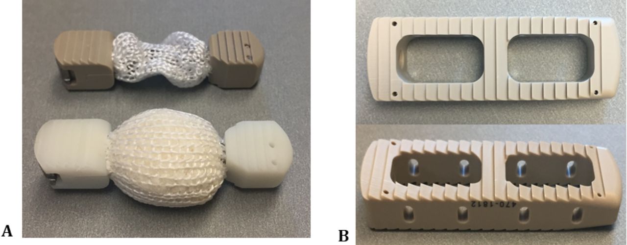
- Figure 1
Representative images showing the P+EPM device (A) and the MPLC (B) device.
- Figure 2
Procedure used to determine the contact footprint in ImageJ. First, the region of interest was selected (left), the image was then placed under threshold (middle), and the resultant area was selected and measured (right). The total footprint was determined by selecting the entire ROI (region of interest; left), then the graft footprint was measured by cropping the central area occupied by the graft (right). Implant footprint could then be determined as the difference between the total footprint and the graft footprint.
- Figure 3
Measured bone graft areas under direct loading for 1100 N and 2000 N for the P+EPM versus MPLC. The measured areas of loaded bone graft for the P+EPM were significantly greater than the MPLC, P < .001. Very few samples tested at both loads for the MPLC did not detect direct loading of the bone graft material. There was 1 sample for the 1100 N and 2 samples for the 2000 N that detected bone graft loading less than 0.5 cm2.
- Figure 4
Central bone graft loading pressure maps measured for filled P+EPM in flat Grade 15 pcf blocks; under 1100 N load with pressures ranging from 3.0 MPa to 9.0 MPa (A), and under 2000 N load with pressures ranging from 3.4 MPa to 9.6 MPa (B), and a representative pressure profile at 1100 N along the longitudinal axis of the P+EPM device. The pressure distribution through the P+EPM identifies that maximum pressures of 9.6 MPa were measured within the bone graft region (bracket) with a mean pressure graft loading of 6.7 MPa (C).
- Figure 5
Evidence of central bone graft loading was not present in the central pores of the MPLC device in flat Grade 15 pcf blocks, as the flat blocks present the best case for potential loading of the bone graft region. The pressure maps at the central bone graft region of the MPLC registered as 0 MPa for filled devices; under 1100 N load (A) and under 2000 N load, where 0 MPa of pressure indicating the bone graft material in the central pores of the MPLC were not in direct contact of the vertebral endplates, nor directly loaded (B), and a representative pressure profile at 1100 N and also observed for 2000 N, along the longitudinal axis of the P+EPM device (brackets indicate graft regions where loading of the graft material was not present). The pressure distribution through the MPLC measured within the bone graft cores did not measure any pressure (0 MPa) indicating that the bone graft was not directly loaded under 1100 N and 2000 N (C).


Tables

In this issue


Jump to section
Related Articles
Cited By...
- No citing articles found.