
Main menu

  • Home
  • Content
    • Current Issue
    • Advance Online Publication
    • Archive
  • About Us
    • About ISASS
    • About the Journal
    • Author Instructions
    • Editorial Board
    • Reviewer Guidelines & Publication Criteria
  • More
    • Advertise
    • Subscribe
    • Alerts
    • Feedback
  • Join Us
  • Reprints & Permissions
  • Sponsored Content
  • Other Publications
    • ijss

User menu

  • My alerts

Search

  • Advanced search
International Journal of Spine Surgery
  • My alerts
International Journal of Spine Surgery

Advanced Search

  • Home
  • Content
    • Current Issue
    • Advance Online Publication
    • Archive
  • About Us
    • About ISASS
    • About the Journal
    • Author Instructions
    • Editorial Board
    • Reviewer Guidelines & Publication Criteria
  • More
    • Advertise
    • Subscribe
    • Alerts
    • Feedback
  • Join Us
  • Reprints & Permissions
  • Sponsored Content
  • Follow ijss on Twitter
  • Visit ijss on Facebook
Research ArticleCervical Spine

Upper Cervical Spine Injuries: Profile and Management of 120 Cases

Geraldo de Sá Carneiro-Filho, Lívio Pereira de Macêdo, Lidemarcks Irineu Andrade, Brenno Franklynn Alves-Sá, Luan Amaral Magalhães Sousa, Saul Cavalcanti de Medeiros Quirino, Juliano Rodrigues Chaves, Deoclides Lima Bezerra-Júnior, Nivaldo S. Almeida and Hildo Rocha Cirne Azevedo-Filho
International Journal of Spine Surgery December 2022, 16 (6) 1001-1008; DOI: https://doi.org/10.14444/8321
Geraldo de Sá Carneiro-Filho
1 Chief of Spine Section - Departament of Neurosurgery, Hospital da Restauração, Recife, Pernambuco, Brazil
MD
  • Find this author on Google Scholar
  • Find this author on PubMed
  • Search for this author on this site
Lívio Pereira de Macêdo
2 Hospital da Restauração, Recife, Pernambuco, Brazil
MD
  • Find this author on Google Scholar
  • Find this author on PubMed
  • Search for this author on this site
  • For correspondence: livio21@gmail.com
Lidemarcks Irineu Andrade
3 Department of Neurosurgery, Hospital da Restauração, Recife, Pernambuco, Brazil
MD
  • Find this author on Google Scholar
  • Find this author on PubMed
  • Search for this author on this site
Brenno Franklynn Alves-Sá
3 Department of Neurosurgery, Hospital da Restauração, Recife, Pernambuco, Brazil
MD
  • Find this author on Google Scholar
  • Find this author on PubMed
  • Search for this author on this site
Luan Amaral Magalhães Sousa
4 Universidade Católica de Pernambuco, Recife, Pernambuco, Brazil
MD
  • Find this author on Google Scholar
  • Find this author on PubMed
  • Search for this author on this site
Saul Cavalcanti de Medeiros Quirino
5 Department of Neurosurgery - Spine Surgery, Hospital da Restauração, Recife, Pernambuco, Brazil
MD
  • Find this author on Google Scholar
  • Find this author on PubMed
  • Search for this author on this site
Juliano Rodrigues Chaves
5 Department of Neurosurgery - Spine Surgery, Hospital da Restauração, Recife, Pernambuco, Brazil
MD
  • Find this author on Google Scholar
  • Find this author on PubMed
  • Search for this author on this site
Deoclides Lima Bezerra-Júnior
5 Department of Neurosurgery - Spine Surgery, Hospital da Restauração, Recife, Pernambuco, Brazil
MD
  • Find this author on Google Scholar
  • Find this author on PubMed
  • Search for this author on this site
Nivaldo S. Almeida
3 Department of Neurosurgery, Hospital da Restauração, Recife, Pernambuco, Brazil
MD
  • Find this author on Google Scholar
  • Find this author on PubMed
  • Search for this author on this site
Hildo Rocha Cirne Azevedo-Filho
3 Department of Neurosurgery, Hospital da Restauração, Recife, Pernambuco, Brazil
MSc, PhD, FRCS
  • Find this author on Google Scholar
  • Find this author on PubMed
  • Search for this author on this site
  • Article
  • Figures & Data
  • Info & Metrics
  • PDF
Loading

Article Figures & Data

Figures

  • Tables
  • Figure 1
    • Download figure
    • Open in new tab
    • Download powerpoint
    Figure 1

    Postoperative standard x-ray images showing a patient who benefited from craniocervical fusion.
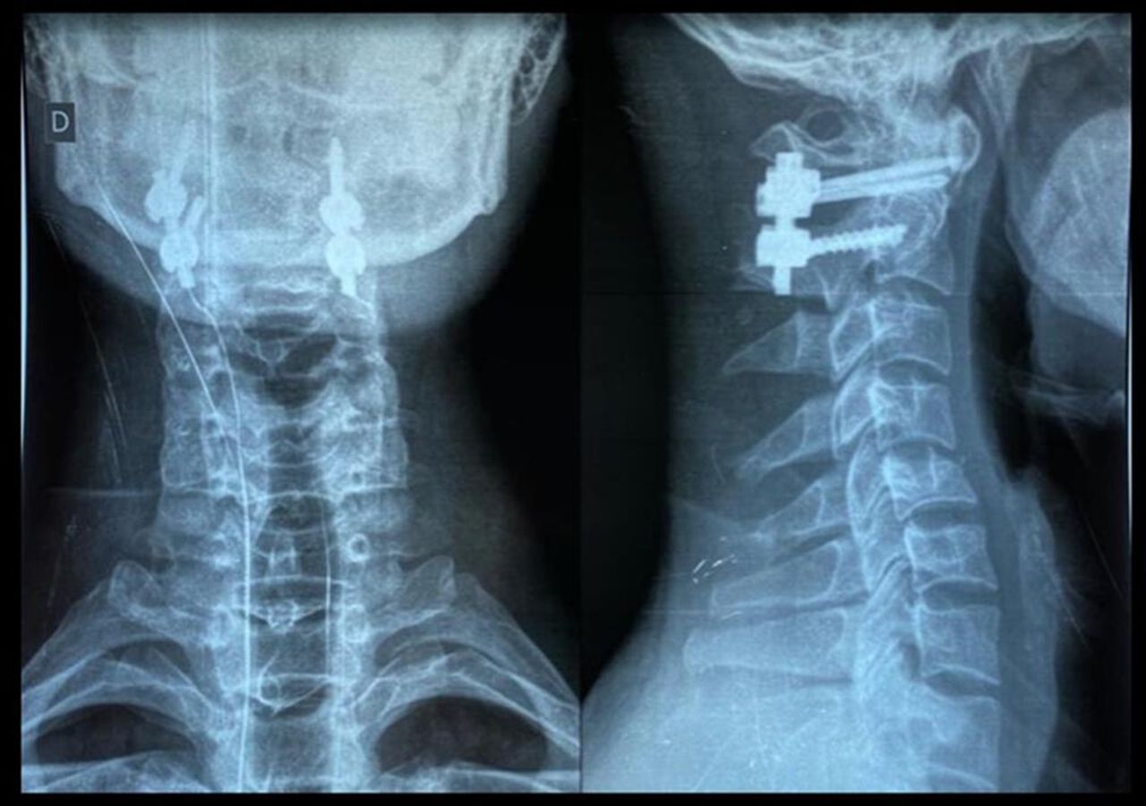
  • Figure 2
    • Download figure
    • Open in new tab
    • Download powerpoint
    Figure 2

    Postoperative standard x-ray images showing a patient who underwent a C1-C2 posterior arthrodesis (Goel-Harms technique).

  • Figure 3
    • Download figure
    • Open in new tab
    • Download powerpoint
    Figure 3

    Intraoperative fluoroscopy showing a type II odontoid fracture. The patient who underwent a transarticular C1-C2 posterior arthrodesis (Magerl technique).

  • Figure 4
    • Download figure
    • Open in new tab
    • Download powerpoint
    Figure 4

    Preoperative computed tomography images showing a patient with type II odontoid fracture presenting a split in the odontoid process. The patient underwent a C1-C2 posterior arthrodesis (Goel-Harms technique), which is shown on a postoperative standard x-ray image.

  • Figure 5
    • Download figure
    • Open in new tab
    • Download powerpoint
    Figure 5

    Postoperative computed tomography images showing a patient with type II odontoid fracture who benefited from anterior odontoid screwing.

  • Figure 6
    • Download figure
    • Open in new tab
    • Download powerpoint
    Figure 6

    Postoperative standard x-ray images showing a patient with type II odontoid fracture who benefited from anterior fixation of the odontoid process with 2 screws.

  • Figure 7
    • Download figure
    • Open in new tab
    • Download powerpoint
    Figure 7

    Preoperative and postoperative standard x-ray images showing a patient with hangman’s fracture who benefited from C2-C3 discectomy and interbody fusion.

  • Figure 8
    • Download figure
    • Open in new tab
    • Download powerpoint
    Figure 8

    Intraoperative view showing a case of anterior C2-C3 discectomy and interbody fusion. Recurrent laryngeal nerve was identified and preserved (right).

  • Figure 9
    • Download figure
    • Open in new tab
    • Download powerpoint
    Figure 9

    Postoperative standard x-ray images showing a patient with hangman’s fracture who submitted to an anterior approach. The patient had an unsatisfactory large gap on fracture place (left) and who benefited from 360° fusion (right).

  • Figure 10
    • Download figure
    • Open in new tab
    • Download powerpoint
    Figure 10

    Postoperative standard x-ray images showing a patient who benefited from C2-C3 discectomy and interbody fusion and posterior wiring.

Tables

  • Figures
    • View popup
    Table 1

    Upper cervical spine injury level and classification (N = 120).

    Variable n (%)
    Injury level 
     C111 (9.2)
     C284 (70.0)
     C1-C221 (17.5)
     C2-C34 (3.3)
    Injury classification 
     Type I C1 fracture (ring fractures)7 (5.8)
     Type II C1 fracture (Jefferson’s fracture)2 (1.7)
     Type III C1 fracture (lateral mass fracture)2 (1.7)
     Hangman’s fracture16 (13.3)
     Type II odontoid fractures35 (29.2)
     Type III odontoid fractures4 (3.3)
     Miscellaneous C2 fractures24 (20.0)
     C1-C2 dislocations4 (3.3)
     C2-C3 dislocations2 (1.7)
     Atlanto-occipital displacement2 (1.7)
     Type II C1 fracture (Jefferson’s fracture) + type II odontoid fracture2 (1.7)
     Type II odontoid fracture + atlanto-occipital displacement2 (1.7)
     Type II odontoid fracture + hangman’s fracture4 (3.3)
     Type III odontoid fracture + miscellaneous C2 fractures2 (1.7)
     Type I C1 fracture + type II odontoid fractures3 (2.5)
     Type III odontoid fracture + C1-C2 dislocations2 (1.7)
     Type I odontoid fractures + C1-C2 dislocations + miscellaneous C2 fractures3 (2.5)
     Type II C1 fracture (Jefferson’s fracture) + type III odontoid fracture2 (1.7)
     Type III C1 fracture (lateral mass fracture) + miscellaneous C2 fractures2 (1.7)
    • View popup
    Table 2

    Sample profile and evaluation of variables according to injury level.

    VariableInjury Level P Valuea
    C1C2C1-C2C2-C3Total
    n % n % n % N %
    Total11100.084100.021100.04100.0120100.0
    Age, y0.510
     16–29872.73136.9942.9375.05142.5
     30–3919.11720.2314.3--2117.5
     40–5919.12226.2733.3--3025.0
     60–8219.11416.729.5125.01815.0
    Gender0.696
     Male11100.07589.31885.74100.010890.0
     Female--910.7314.3--1210.0
    Trauma mechanisms0.292
     Car accident327.32125.0314.3250.02924.2
     Motorcycle accident218.22428.61047.6250.03831.7
     Gunshot injury218.233.6314.3--86.7
     Fall from standing height19.11315.5----1411.7
     Fall from height19.11315.5314.3--1714.2
     Others218.21011.929.5--1411.7
    Frankel Scale0.128
     A19.111.214.8125.043.3
     B19.1------10.8
     C19.167.129.5--97.5
     D--44.8----43.3
     E872.77386.91885.7375.010285.0
    Neurologic deficit0.426
     Yes327.31113.1314.3125.01815.0
     No872.77386.91885.7375.010285.0
    Management/outcome<0.001b
     Surgical19.15464.31781.0375.07562.5
     Conservative1090.93035.7314.3125.04436.7
     Death----14.8--10.8
    • ↵a Fisher’s exact test.

    • ↵b Data were considered statistically significant at P < 0.05.

    • View popup
    Table 3

    Evaluation of variables according to neurological deficit.

    VariableNeurological Deficit P Valuea
    YesNoTotal
    n % n % n %
    Total18100.0102100.0120100.0
    Age, y0.837
     16–29950.04241.25142.5
     30–39316.71817.62117.5
     40–59316.72726.53025.0
     60–82316.71514.71815.0
    Gender0.387
     Male1583.39391.210890.0
     Female316.798.81210.0
    Trauma mechanisms0.107
     Car accident422.22524.52924.2
     Motorcycle accident316.73534.33831.7
     Gunshot injury422.243.986.7
     Fall from standing height316.71110.81411.7
     Fall from height211.11514.71714.2
     Others211.11211.81411.7
    Management/outcome= 0.117
     Surgical1268.76361.87562.5
     Conservative527.73938.24436.7
     Death15.6--10.8
    • ↵a Fisher’s exact test.

    • View popup
    Table 4

    Injury classification and management (N = 120).

    Variable n (%)
    Type of injury 
     Isolated hangman’s fracture16 (13.3)
     Isolated odontoid fracture42 (35.0)
     Isolated C1 fracture11 (9.2)
     Combined C1-C2 fracture9 (7.5)
     Miscellaneous C2 fracture30 (25.0)
     Ligament injury8 (6.7)
     Odontoid fracture + hangman’s fracture4 (3.3)
    Management 
     C0-C3 posterior fusion3 (2.5)
     C0-C4 posterior fusion1 (0.8)
     C1-C2 posterior fusion30 (25.0)
     C1-C3 posterior fusion8 (6.7)
     C1-C4 posterior fusion2 (1.7)
     C2-C3 discectomy9 (7.5)
     C2-C3 posterior fusion1 (0.8)
     C2-C3 discectomy + C1-C3 posterior fusion3 (2.5)
     Conservative management44 (36.7)
     Occipital craniectomy + bullet removal1 (0.8)
     Transoral odontoidectomy + C0-C4 posterior fusion2 (1.7)
     Transoral odontoidectomy + C1-C2 posterior fusion1 (0.8)
     Odontoid screwing14 (11.7)
     Deatha 1 (0.8)
    • ↵a One patient died due to hemodynamic instability before surgery.

    • View popup
    Table 5

    Evaluation of management performed according to the type of injury.

     
    Management
    Type of Injury
    Isolated FractureCombined Fracture
    Hangman’s FractureOdontoid FractureC1 FractureLigament InjuryCombined C1- C2 FractureMiscellaneous Fracture of C2Combined Odontoid Fracture and Hangman's Fracture
    C0-C3 posterior fusion----3--
    C0-C4 posterior fusion-1-----
    C1-C2 posterior fusion117-5322
    C1-C3 posterior fusion11--51-
    C1-C4 posterior fusion11-----
    C2-C3 discectomy5---112
    C2-C3 posterior fusion-1-----
    C2-C3 discectomy + C1-C3 posterior fusion2---1--
    Odontoid screwing-12-2---
    Occipital craniectomy + bullet removal--1----
    Transoral odontoidectomy + C0-C4 posterior fusion-1---1-
    Transoral odontoidectomy + C1-C2 posterior fusion-1-----
    Conservative managementb 67102163-
    Deatha ----1--
    Total16421193084
    • ↵a One patient died due to hemodynamic instability before surgery.

    • ↵b Conservative management with Philadelphia neck collar for 8 wk.

PreviousNext
Back to top

In this issue

International Journal of Spine Surgery
Vol. 16, Issue 6
1 Dec 2022
  • Table of Contents
  • Index by author

Print
Download PDF
Article Alerts
Sign In to Email Alerts with your Email Address
Email Article

Thank you for your interest in spreading the word on International Journal of Spine Surgery.

NOTE: We only request your email address so that the person you are recommending the page to knows that you wanted them to see it, and that it is not junk mail. We do not capture any email address.

Enter multiple addresses on separate lines or separate them with commas.
Upper Cervical Spine Injuries: Profile and Management of 120 Cases
(Your Name) has sent you a message from International Journal of Spine Surgery
(Your Name) thought you would like to see the International Journal of Spine Surgery web site.
CAPTCHA
This question is for testing whether or not you are a human visitor and to prevent automated spam submissions.
Citation Tools
Upper Cervical Spine Injuries: Profile and Management of 120 Cases
Geraldo de Sá Carneiro-Filho, Lívio Pereira de Macêdo, Lidemarcks Irineu Andrade, Brenno Franklynn Alves-Sá, Luan Amaral Magalhães Sousa, Saul Cavalcanti de Medeiros Quirino, Juliano Rodrigues Chaves, Deoclides Lima Bezerra-Júnior, Nivaldo S. Almeida, Hildo Rocha Cirne Azevedo-Filho
International Journal of Spine Surgery Dec 2022, 16 (6) 1001-1008; DOI: 10.14444/8321

Citation Manager Formats

  • BibTeX
  • Bookends
  • EasyBib
  • EndNote (tagged)
  • EndNote 8 (xml)
  • Medlars
  • Mendeley
  • Papers
  • RefWorks Tagged
  • Ref Manager
  • RIS
  • Zotero
Share
Upper Cervical Spine Injuries: Profile and Management of 120 Cases
Geraldo de Sá Carneiro-Filho, Lívio Pereira de Macêdo, Lidemarcks Irineu Andrade, Brenno Franklynn Alves-Sá, Luan Amaral Magalhães Sousa, Saul Cavalcanti de Medeiros Quirino, Juliano Rodrigues Chaves, Deoclides Lima Bezerra-Júnior, Nivaldo S. Almeida, Hildo Rocha Cirne Azevedo-Filho
International Journal of Spine Surgery Dec 2022, 16 (6) 1001-1008; DOI: 10.14444/8321
Twitter logo Facebook logo Mendeley logo
  • Tweet Widget
  • Facebook Like
  • Google Plus One

Jump to section

  • Article
    • Abstract
    • Introduction
    • Methods
    • Results
    • Discussion
    • Conclusion
    • Footnotes
    • References
  • Figures & Data
  • Info & Metrics
  • PDF

Related Articles

  • No related articles found.
  • PubMed
  • Google Scholar

Cited By...

  • No citing articles found.
  • Google Scholar

More in this TOC Section

  • Innovation and Adversity in Spine Surgery: A Retrospective
  • Cervical Myelopathy Secondary to Bilateral Atlantoaxial Pseudoarticulations in Rheumatoid Arthritis: A Case Report
  • Incidence of Traumatic Spinal Injury Following Public Policy Update on Moped Usage in South Carolina
Show more Cervical Spine

Similar Articles

Keywords

  • upper cervical spine injury, spinal cord injury
  • epidemiology
  • management

Content

  • Current Issue
  • Latest Content
  • Archive

More Information

  • About IJSS
  • About ISASS
  • Privacy Policy

More

  • Subscribe
  • Alerts
  • Feedback

Other Services

  • Author Instructions
  • Join ISASS
  • Reprints & Permissions

© 2025 International Journal of Spine Surgery

International Journal of Spine Surgery Online ISSN: 2211-4599

Powered by HighWire