


Article Figures & Data
Figures
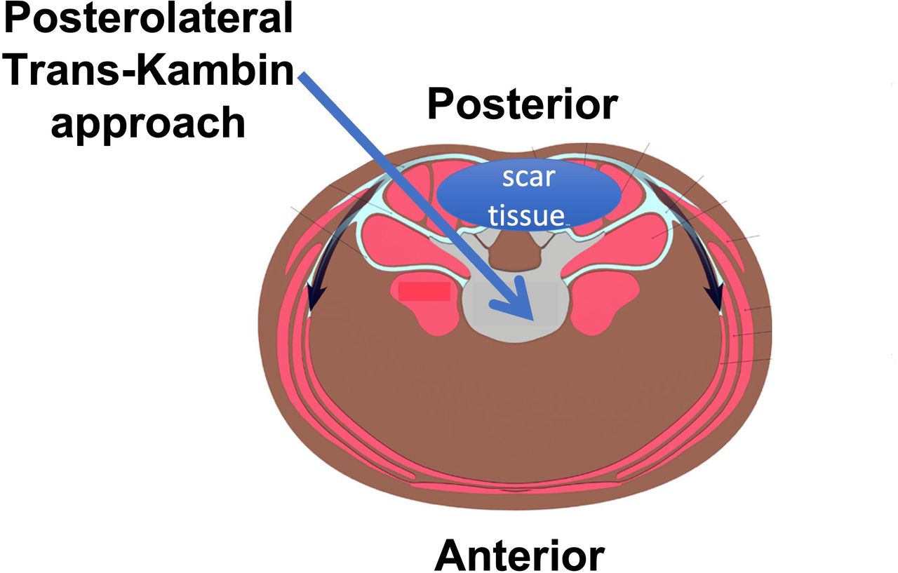
- Figure 1
An axial view of the lumbar spine and abdomen. In revision surgery cases after open transforaminal lumbar interbody fusion surgery, scar tissue can be usually found at the posterior part of the spine. The trans-Kambin approach to the disc allows bypassing of posterior scar tissue (arrow).
- Figure 2
(A) X-ray images of the lumbar spine in anterior/posterior (A/P) (left side) and lateral (right side) views for case 1, 3 mo before he presented to our clinic. Notice the undersized interbody cage at level L3-L4. (B) X-ray images of the lumbar spine in A/P (left side) and lateral (right side) views for case 1 at the time when he presented to our clinic. Also notice the migrated interbody cage into the right neuroforamen at level L3-L4 (marked with a red oval).
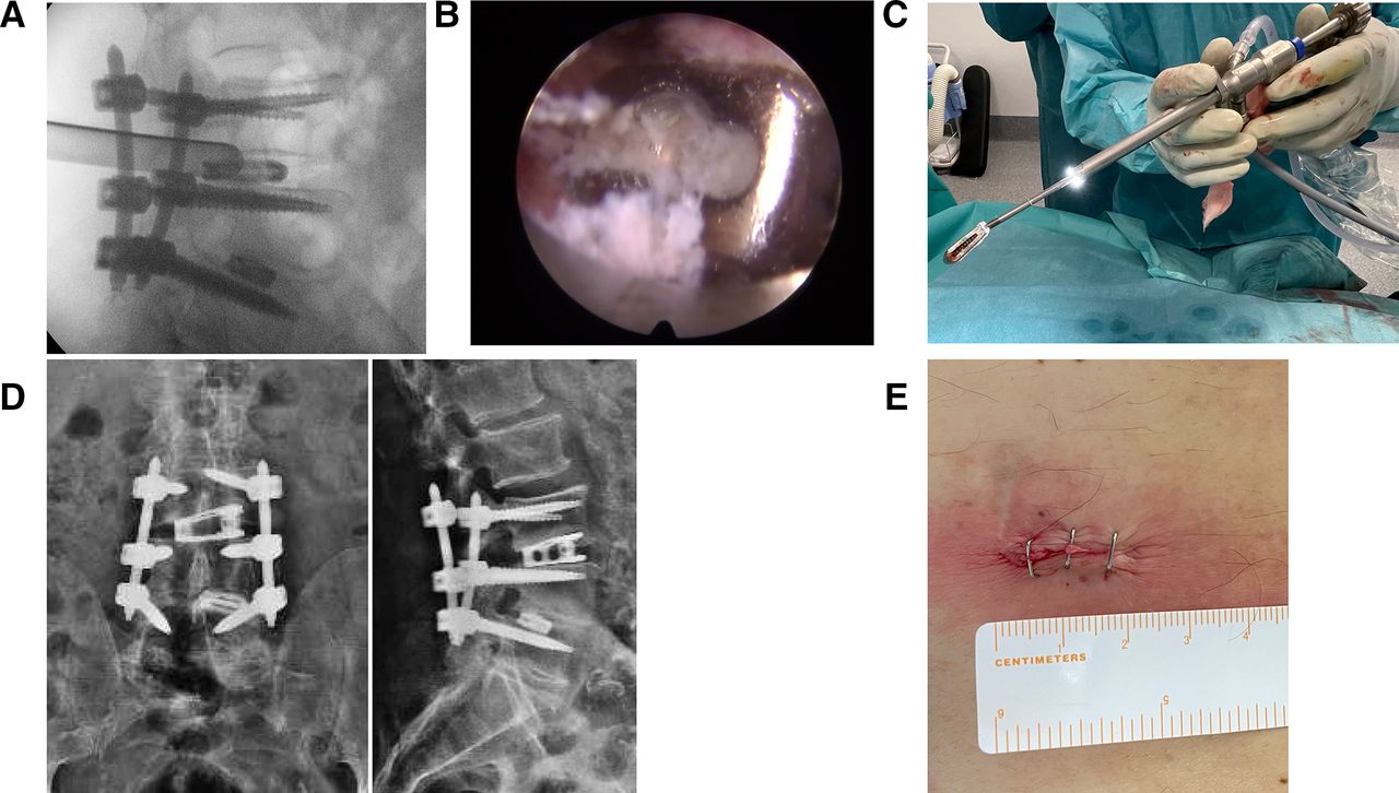
- Figure 3
(A) Lateral intraoperative fluoroscopic image of the specialized fusion dilator sleeve positioned into the disc close to the migrated cage at L3-L4. The sleeve protects the right L3 exiting nerve root. (B) Endoscopic view of the migrated titanium interbody cage. (C) Photo of the removed interbody cage attached to the cage handle. The cage handle can be seen looking out the working channel of the endoscope. (D) Postoperative x-ray control images of the lumbar spine in anterior/posterior (left side) and lateral (right side) views. Notice the large footprint, expandable titanium cage that was placed as a substitute at L3-L4. (E) Skin incision of 2-cm length through which the migrated interbody cage was removed, and the substitute large footprint expandable cage was placed into L3-L4 with the trans-Kambin approach.
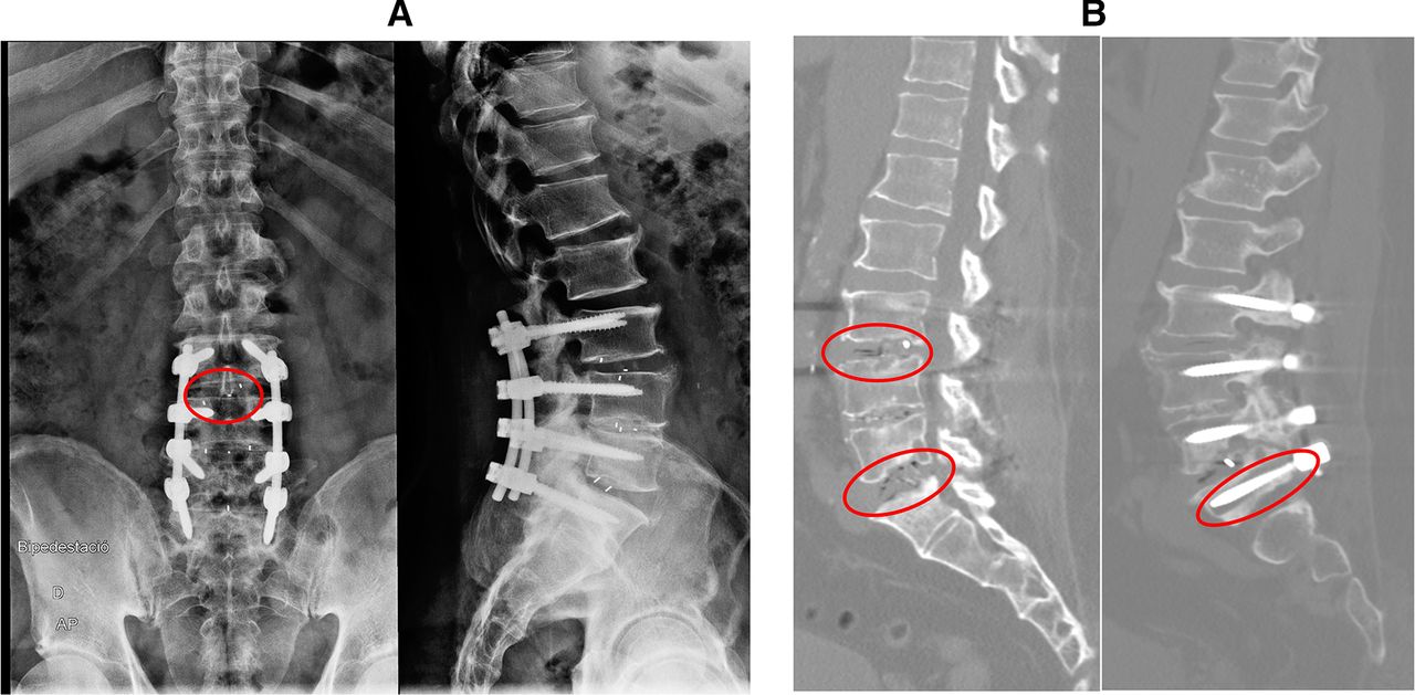
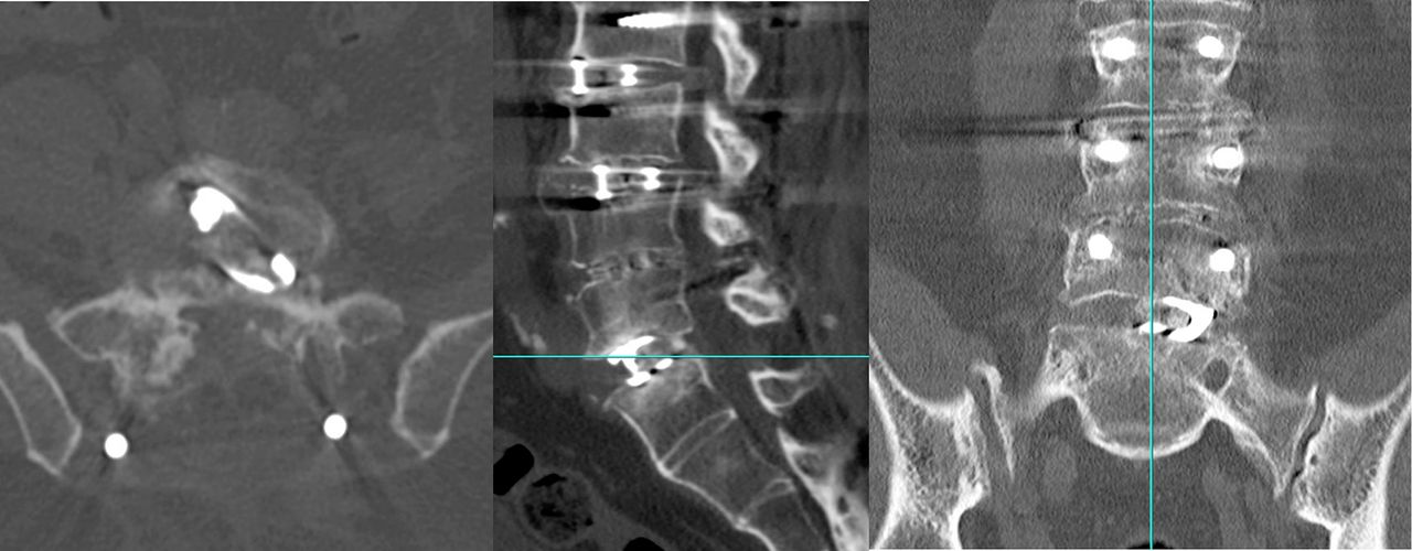
- Figure 4
(A) Preoperative x-ray control images of the lumbar spine in anterior/posterior (A/P) (left side) and lateral (right side) views for case 2. Notice the impacted interbody cage into the upper endplate of L4 at L3-L4 (marked with red circle). (B) Preoperative computed tomography image of the lumbar spine. Notice the vacuum sign suggestive of pseudarthrosis in L3-L4 and L5-S1 in the A/P view (marked with red circles), as well as the osteolysis due to screw loosening at S1 in the lateral view (marked with a red circle).
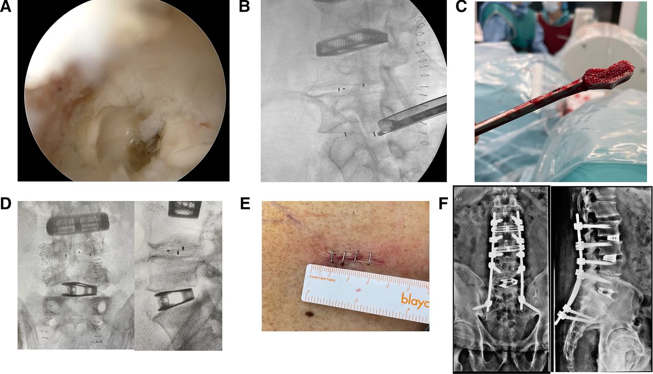
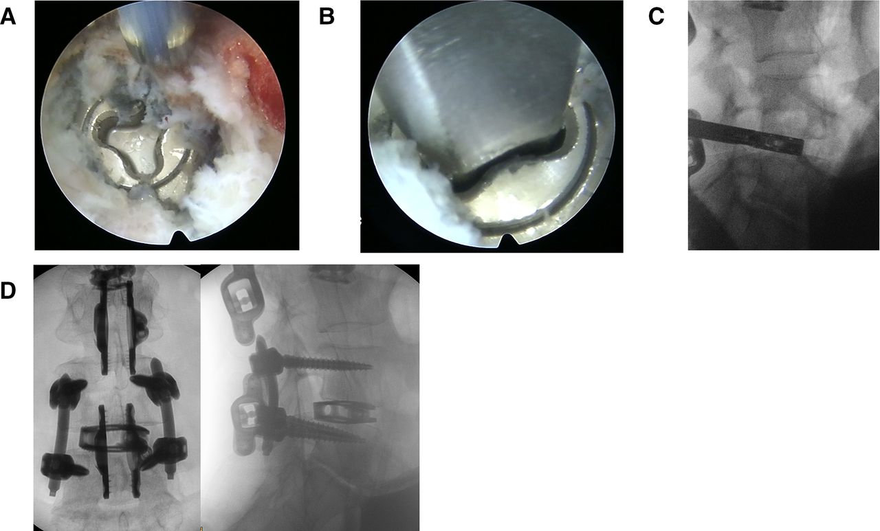
- Figure 5
(A) An endoscopic view of the pseudarthrotic PEEK interbody cage. (B) Fluoroscopic lateral intraoperative view of a forceps grasping the interbody cage. A protective sleeve keeps the exiting nerve root protected. (C) Photograph of the removed interbody PEEK cage attached to the forceps. (D) Intraoperative fluoroscopic images of the lumbar spine in anterior/posterior (A/P) (left side) and lateral (right side) views. Notice the large footprint, expandable titanium cage that was placed as a substitute at L5-S1. (E) Skin incision of 2-cm length through which the PEEK interbody cage was removed, and the substitute large footprint expandable cage was placed into L5-S1 with the trans-Kambin approach. (F) Postoperative x-ray control images of the lumbar spine in A/P (left side) and lateral (right side) views.
- Figure 6
(A) Image depicting a protective sleeve that has been placed through Kambin’s triangle into the disc to protect the exiting nerve root, while a foraminoplasty is performed with manual reamers with progressively increasing diameters. (B) Removed disc material after careful disc preparation of level L5-S1 using the trans-Kambin approach. (C) Removed disc material after careful disc preparation for level L3-L4 using the anterior-to-psoas open lateral approach. (D) Image depicting a protective sleeve that has been placed to protect the exiting nerve root while it allows inserting a large footprint interbody cage into the disc.
- Figure 7
Postoperative computed tomography image at 1-year follow-up showing spinal fusion at all operated levels.
- Figure 8
(A) Preoperative x-ray control images of the lumbar spine in anterior/posterior (left side) and lateral (right side) views for case 3. Notice the migrated interbody cage into the right neuroforamen of L4-L5 (marked with red circle). (B) Preoperative computed tomography image of the lumbar spine with lateral (above) and axial (below) views of L4-L5 showing a migrated interbody cage into the right neuroforamen of L4-L5.
- Figure 9
(A) An endoscopic view of a migrated titanium expandable cage. (B) An endoscopic view of the screwdriver docked into the mechanism of the expandable cage used to collapse the cage and reduce its footprint. (C) Fluoroscopic lateral intraoperative view of a forceps grasping the collapsed expandable cage. (D) Postoperative x-ray control images of the lumbar spine in anterior/posterior (left side) and lateral (right side) views with a large footprint expandable cage placed into L4-L5 and a posterior fixation.


Tables
- Table
Pre- and postoperative VAS for back and leg scores and ODI scores for all 3 reported full-endoscopic revision surgery cases.
Case VAS Back/VAS Leg/ODI Scores P Value Follow-up Preoperative Scores Postoperative Scores at Hospital Discharge Postoperative Scores at Latest Follow-up Case 1 1/6/36 2/0/26 1/1/14 <0.05a 12 mo Case 2 6/10/31 6/7/33 5/4/25 <0.05a 12 mo Case 3 7/4/29 4/2/25 0/3/16 <0.05a 6 mo Abbreviations: ODI, Oswestry Disability Index; VAS, visual analog scale.
↵a Significant difference (paired Student t test).
online supplemental video 1.
online supplemental video 2.
In this issue


Jump to section
Related Articles
Cited By...
- No citing articles found.