


Article Figures & Data
Figures
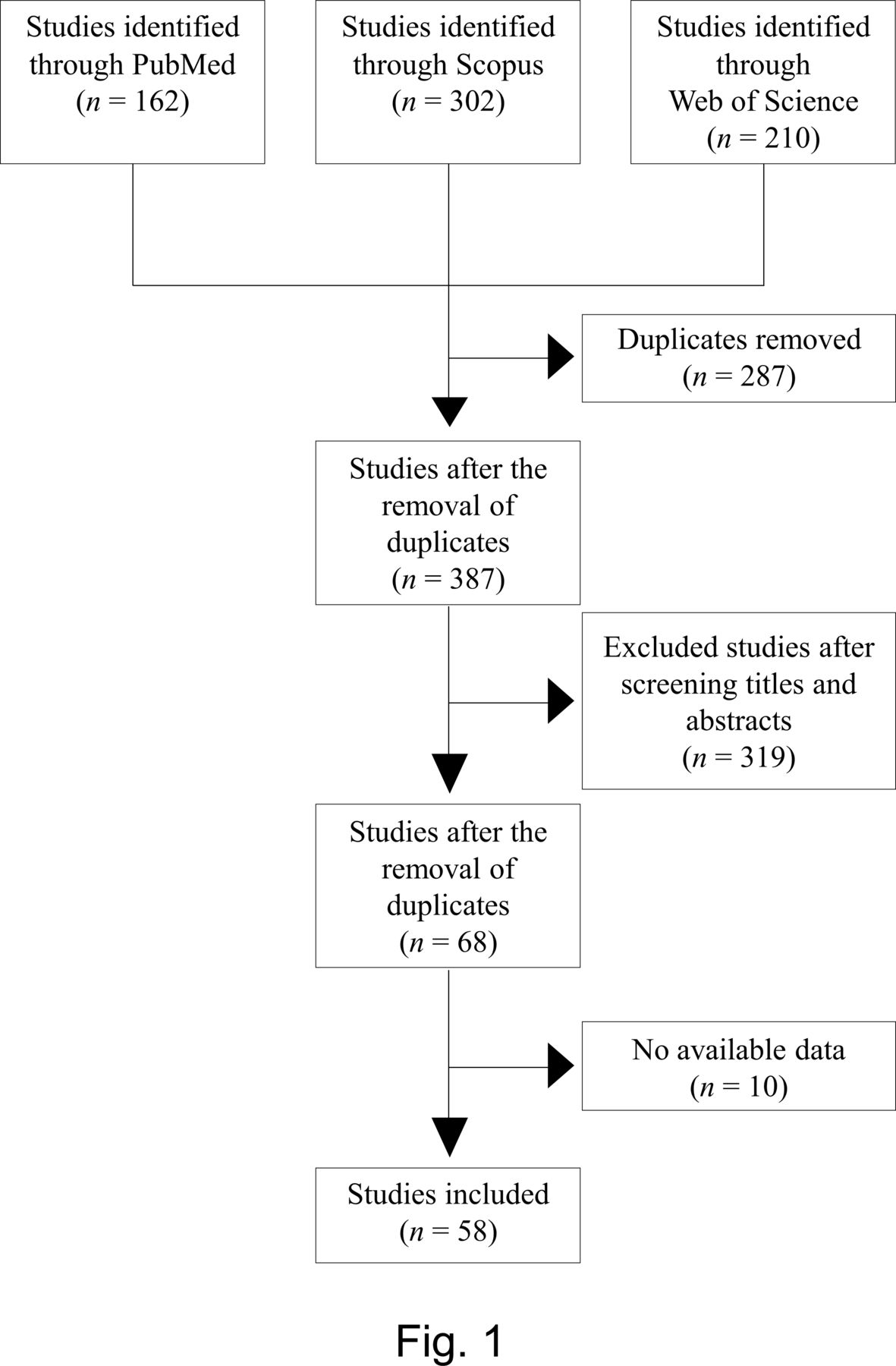
- Figure 1
Flow diagram of the study search and selection process.
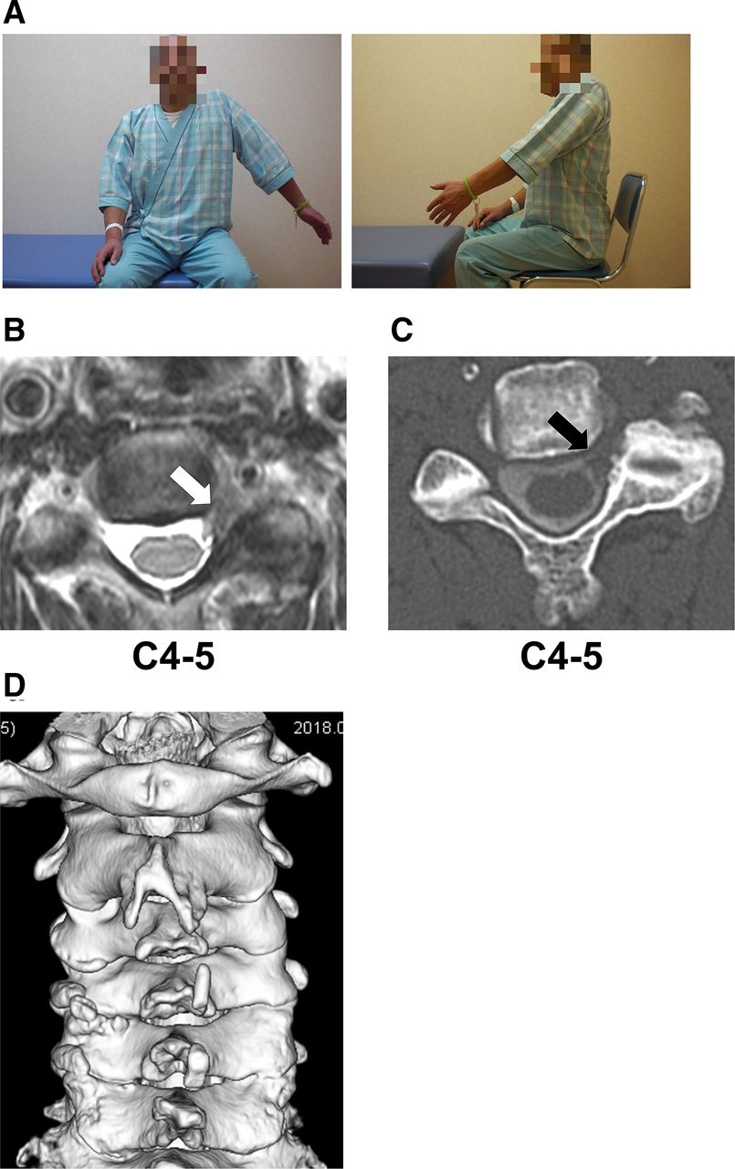
- Figure 2
Snapshots of the patient, magnetic resonance imaging (MRI) and computed tomography (CT) images prior to the surgery in Case 1. (A) The patient was unable to raise his left upper extremity. (B) T2-weighted axial MRI image in C4-5. The white arrow points to the lesion in the left C4-5 foramen. (C) C4-5 axial CT myelogram image. The black arrow points to the loss of contrast as a result of the lesion. (D) Preoperative 3D CT image of the cervical spine. It shows marked osteophyte formation at the left C4-5 and C5-6 facet joints.
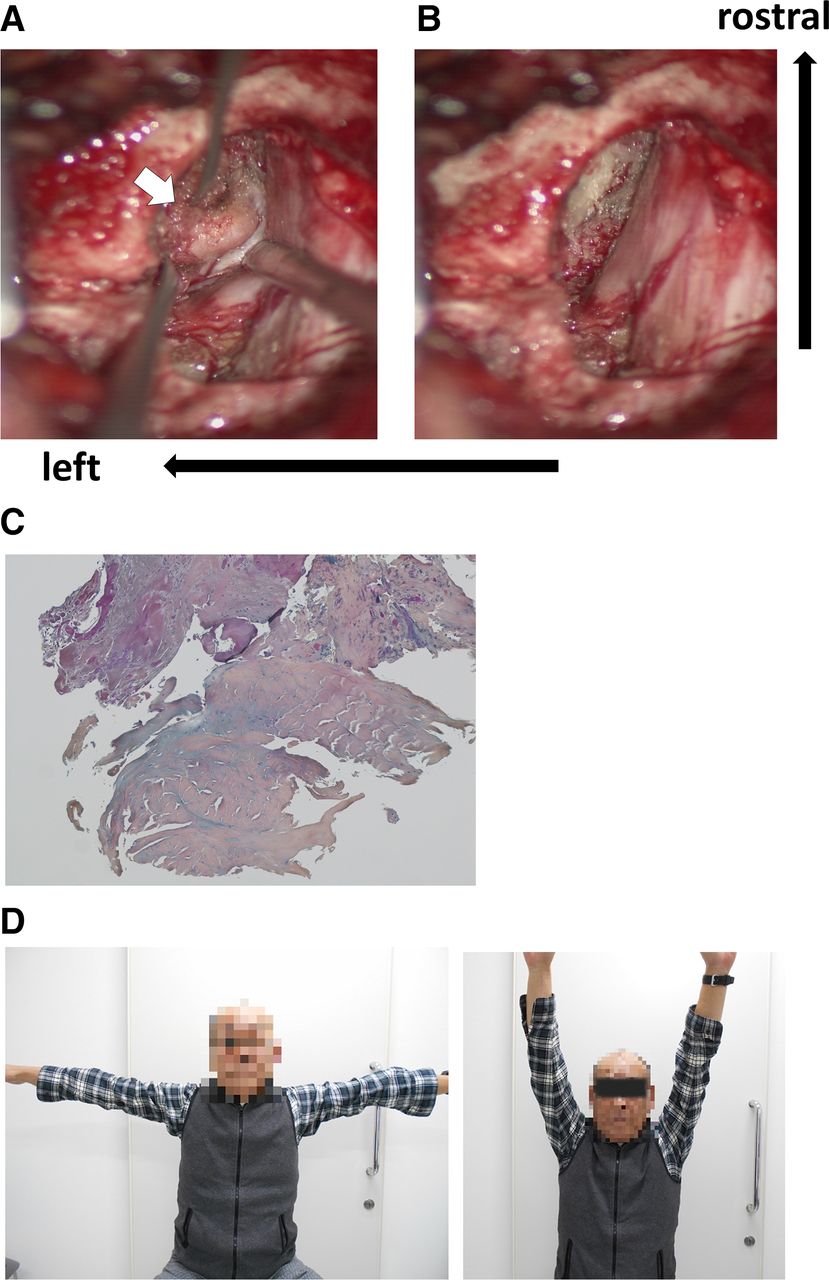
- Figure 3
Intraoperative photographs, histopathological images and postoperative photographs of the patient. (A) A photograph following microcervical foraminotomy before cyst removal. The white arrow points to the cyst. (B) A photograph following cyst removal. The left C5 nerve root is well decompressed. (C) A pathological image of resected cyst. Increased collagen fibers, fibroblasts and acidic mucus with positive Alcian blue staining point to the diagnosis of a ganglion cyst. (D) Six months following the surgery, the patient was able to raise his left upper extremity.
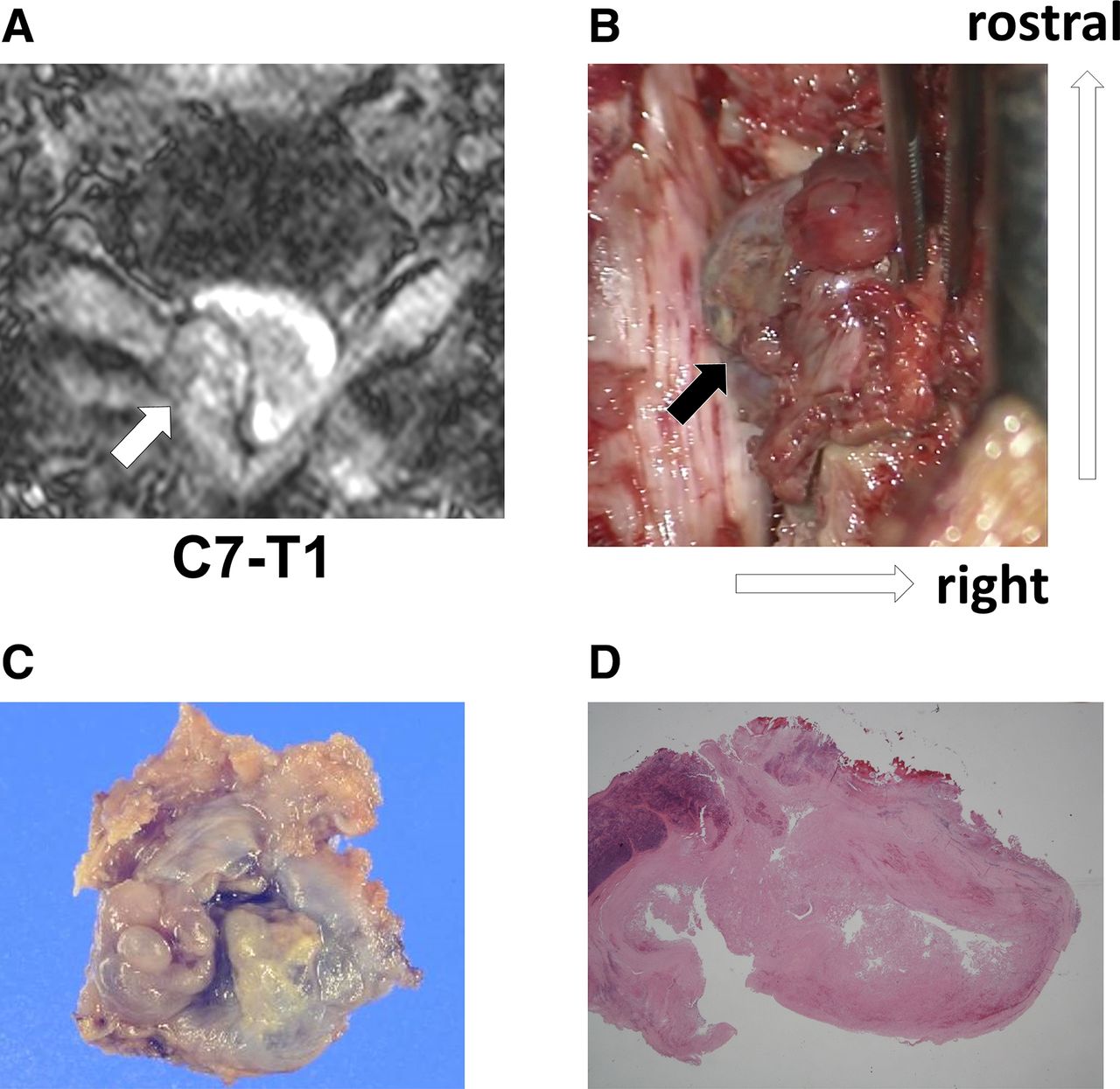
- Figure 4
A magnetic resonance image (MRI), an intraoperative photograph, a macrograph, and a pathological image of the resected cyst in Case 2. (A) T2-weighted axial MRI image in C7-T1. The white arrow points to the cyst. (B) An intraoperative photograph. The black arrow points to the cyst in ligamentum flavum. (C) A macrograph of the resected cyst. The color is dark red due to bleeding into the cyst. (D) A pathological image of the resected cyst. A ganglion cyst containing red blood cells and fibrin.
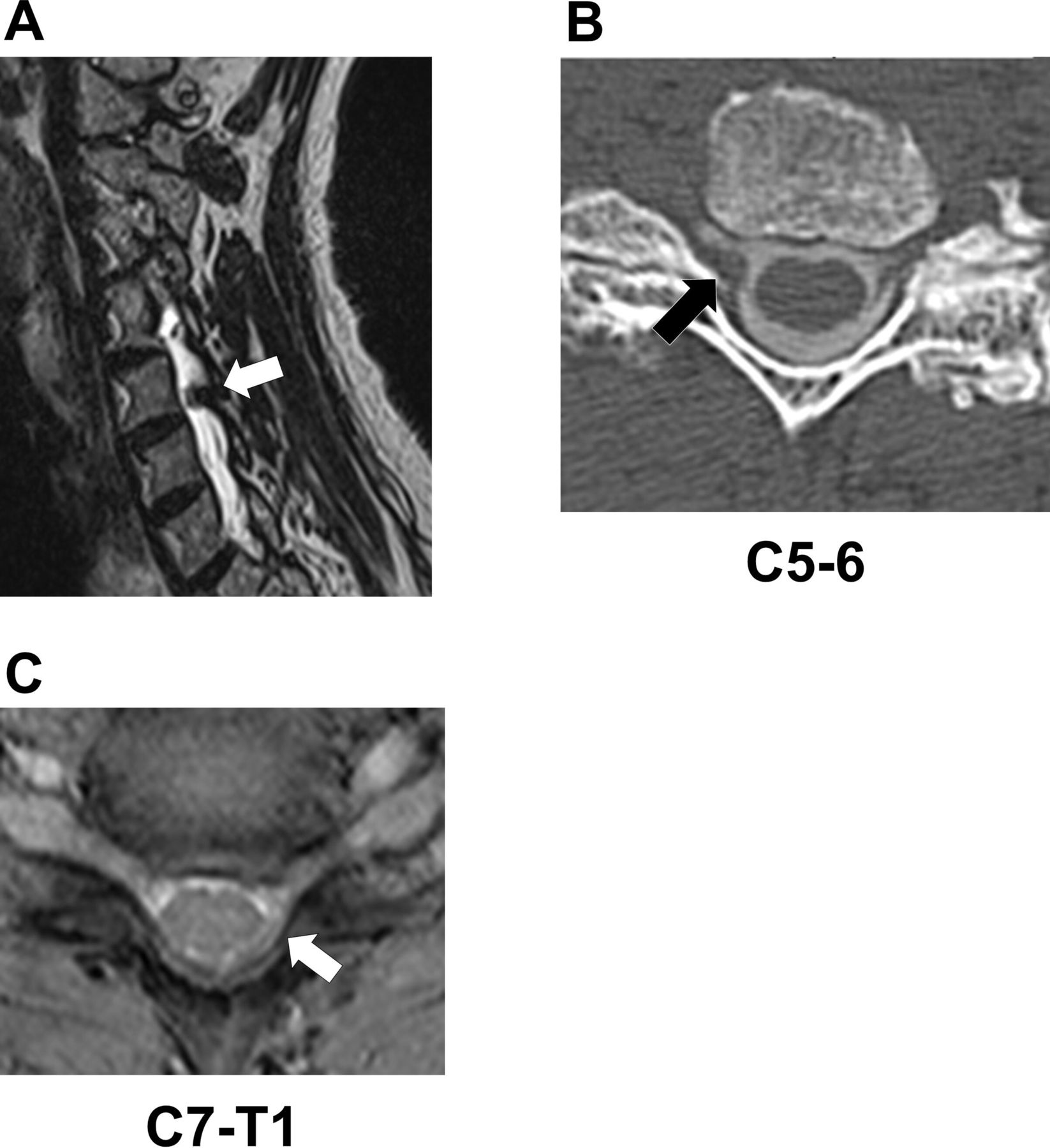
- Figure 5
Magnetic resonance imaging (MRI) and computed tomography (CT) images prior to the surgery in Case 3 and 4. (A) T2-weighted sagittal MRI image of Case 3. The white arrow points to the lesion at C5-C6. (B) C5-6 axial CT myelogram image of Case 3. The black arrow points to the loss of contrast as a result of the lesion at the border between the intervertebral foramen and spinal canal on the right side. (C) T2-weighted axial MRI image of Case 4. The white arrow points to the lesion from the spinal canal to the intervertebral foramen at the C7-T1 on the left side.


Tables
Age Sex Location Symptom Muscle Weakness Surgery Pathology Manual Muscle Test
(Preoperative/6 mo)69 Man C4-C5 Radiculopathy
(C5)Deltoid, biceps MCF Ganglion 1/5 72 Man C7-T1 Radiculopathy
(C8)Wrist flexor LP + MCF Ganglion 3/4 73 Woman C5-C6 Radiculopathy
(C6)Biceps MCF Ganglion 4/5 50 Man C7-T1 Radiculopathy
(C8)Finger extensor LP + MCF Synovial 4/5 Abbreviations: LP, laminoplasty; MCF, microcervical foraminotomy.
Characteristic n Gender, n Men 92 Women 51 NA 1 Age, y, mean (range) 65.2 (40–86) Cyst location C2–3 4 C3–4 17 C4–5 18 C5–6 10 C6–7 15 C7–T1 80 Symptoms Myelopathy 53 Myeloradiculopathy 15 Radiculopathy 74 NA 2 Motor weakness Yes 84 No 17 NA 43 Surgery Hemilaminectomy 59 Laminectomy 43 Posterior fusion 28 Laminoplasty 3 Endoscopic hemilaminectomy 7 Endoscopic foraminotomy 2 Needle aspiration 1 NA 1 Pathologic findings Synovial cyst 96 Ganglion cyst 32 NA 16 Abbreviation: NA, not available.
Note: Data presented as n except where otherwise noted.
Location Age, y, Mean (Range) Sex,
Men/WomenSymptom Motor Weakness,
With/WithoutPathology C2-C3 72.0
(58–79)4/0 Myelopathy: 2
Myeloradiculopathy: 1
Radiculopathy: 12/0 (100%)
(NA: 2)Synovial: 4
Ganglion: 0C3-C4 70.7
(60–81)14/3 Myelopathy: 12
Myeloradiculopathy: 4
Radiculopathy: 111/0 (100%)
(NA: 6)Synovial: 11
Ganglion: 5
NA: 6C4-C5 65.6
(42–86)15/3 Myelopathy: 5
Myeloradiculopathy: 2
Radiculopathy: 117/3 (70.0%)
(N/A: 8)Synovial: 12
Ganglion: 5
NA: 1C5-C6 62.5
(40–74)9/1 Myelopathy 3
Myeloradiculopathy 2
Radiculopathy 57/0 (100%)
(NA: 3)Synovial: 7
Ganglion: 2
NA: 1C6-C7 62.4
(48–83)9/6 Myelopathy: 4
Myeloradiculopathy: 2
Radiculopathy: 913/1 (92.9%)
(NA: 1)Synovial: 7
Ganglion: 4
NA: 4C7-T1 64.5
(41–84)42/37
(NA: 1)Myelopathy: 27
Myeloradiculopathy: 4
Radiculopathy: 47
NA: 245/13 (77.6%)
(NA: 22)Synovial: 55
Ganglion: 16
NA: 9Abbreviation: NA, not available.
Symptom Age, y, Mean (Range) Sex,
Men/WomenMotor Weakness,
With/WithoutSurgery Pathology Myelopathy 65.4
(41–86)41/11 (NA: 1) 35/3 (92.1%)
(NA: 15)Hemilaminectomy: 13
Laminectomy: 31
Fusion: 3
Laminoplasty: 1
Endoscopic hemilaminectomy: 5Synovial: 34
Ganglion: 15
NA: 4Myeloradiculopathy 66.1
(56–82)11/4 11/1 (91.7%)
(NA: 3)Laminectomy: 8
Fusion: 5
Laminoplasty: 2Synovial: 3
Ganglion: 8
NA: 4Radiculopathy 64.7
(40–84)39/35 38/13 (74.5%)
(NA: 23)Hemilaminectomy: 44
Laminectomy: 4
Fusion: 20
Endoscopic hemilaminectomy: 2
Endoscopic foraminotomy: 2
Needle aspiration: 1
NA: 1Synovial: 58
Ganglion: 8
NA: 8Abbreviation: NA, not available.
In this issue


Jump to section
Related Articles
Cited By...
- No citing articles found.