
Main menu

  • Home
  • Content
    • Current Issue
    • Advance Online Publication
    • Archive
  • About Us
    • About ISASS
    • About the Journal
    • Author Instructions
    • Editorial Board
    • Reviewer Guidelines & Publication Criteria
  • More
    • Advertise
    • Subscribe
    • Alerts
    • Feedback
  • Join Us
  • Reprints & Permissions
  • Sponsored Content
  • Other Publications
    • ijss

User menu

  • My alerts

Search

  • Advanced search
International Journal of Spine Surgery
  • My alerts
International Journal of Spine Surgery

Advanced Search

  • Home
  • Content
    • Current Issue
    • Advance Online Publication
    • Archive
  • About Us
    • About ISASS
    • About the Journal
    • Author Instructions
    • Editorial Board
    • Reviewer Guidelines & Publication Criteria
  • More
    • Advertise
    • Subscribe
    • Alerts
    • Feedback
  • Join Us
  • Reprints & Permissions
  • Sponsored Content
  • Follow ijss on Twitter
  • Visit ijss on Facebook
Research ArticleLumbar Spine

Minimally Invasive Surgery Transpedicular Intrabody Cage Technique for the Management of Kummell Disease

Junseok Bae, Chay-you Ang, Ifthekar Syed, Seong Kyun Jeong, Sang Ha Shin and Sang-Ho Lee
International Journal of Spine Surgery February 2024, 18 (1) 73-80; DOI: https://doi.org/10.14444/8570
Junseok Bae
1 Department of Neurosurgery, Wooridul Spine Hospital, Seoul, South Korea
MD
  • Find this author on Google Scholar
  • Find this author on PubMed
  • Search for this author on this site
Chay-you Ang
2 Division of Musculoskeletal Sciences, Singapore General Hospital, Singapore, Singapore
MBBS, FRCS
  • Find this author on Google Scholar
  • Find this author on PubMed
  • Search for this author on this site
  • For correspondence: angchayyou@msn.com
Ifthekar Syed
3 Sri Siddhartha Medical College, Tumakuru, India
MBBS
  • Find this author on Google Scholar
  • Find this author on PubMed
  • Search for this author on this site
Seong Kyun Jeong
1 Department of Neurosurgery, Wooridul Spine Hospital, Seoul, South Korea
MD
  • Find this author on Google Scholar
  • Find this author on PubMed
  • Search for this author on this site
Sang Ha Shin
1 Department of Neurosurgery, Wooridul Spine Hospital, Seoul, South Korea
MD
  • Find this author on Google Scholar
  • Find this author on PubMed
  • Search for this author on this site
Sang-Ho Lee
1 Department of Neurosurgery, Wooridul Spine Hospital, Seoul, South Korea
MD
  • Find this author on Google Scholar
  • Find this author on PubMed
  • Search for this author on this site
  • Article
  • Figures & Data
  • Info & Metrics
  • PDF
Loading

Article Figures & Data

Figures

  • Tables
  • Figure 1
    • Download figure
    • Open in new tab
    • Download powerpoint
    Figure 1

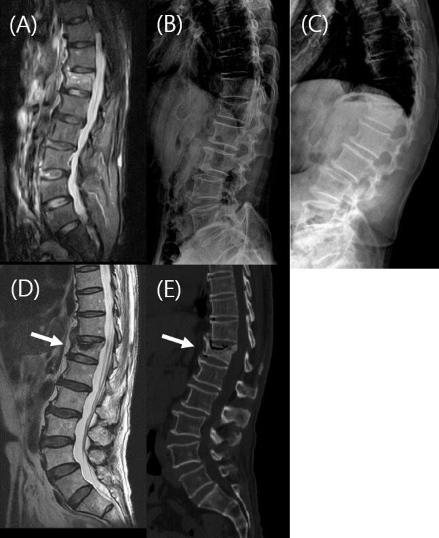
    Initial magnetic resonance imaging (MRI) showing an acute T12 fracture (A). Initial lateral radiograph showing T12 fracture with superior endplate depression (B). Lateral radiograph after 4 months showing kyphotic deformity (C). MRI after 4 months showing intervertebral cleft sign (arrow) and kyphotic deformity (D). Computed tomography image after 4 months showing intervertebral cleft (arrow) and nonunion of T12 fracture (E).

  • Figure 2
    • Download figure
    • Open in new tab
    • Download powerpoint
    Figure 2

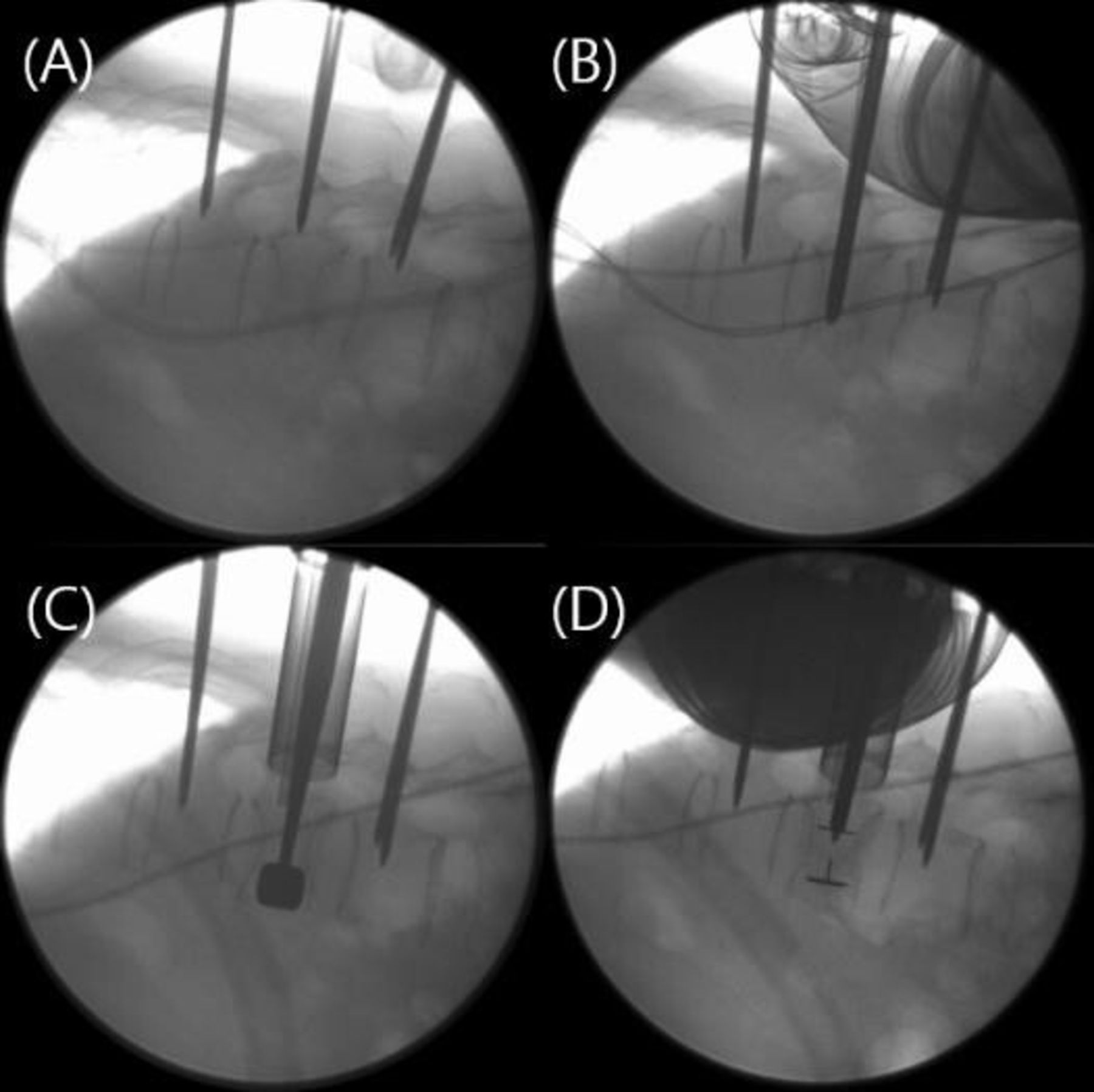
    Intraoperative lateral radiograph after insertion of Jamshidi needles (A). Insertion of the METRx dilator through one of the pedicles at the fractured vertebrae. (B). Insertion of cage trial into the fractured vertebrae to restore the vertebral height (C). Postinsertion of crescent cage (Medtronic) demonstrates satisfactory vertebral height restoration (D).

  • Figure 3
    • Download figure
    • Open in new tab
    • Download powerpoint
    Figure 3

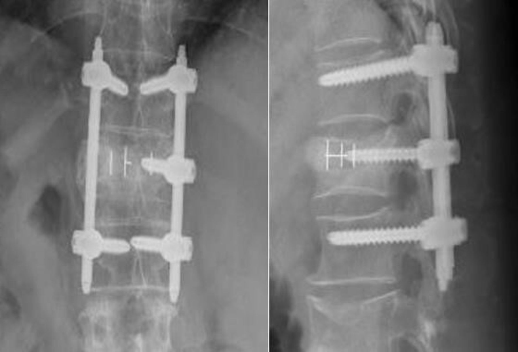
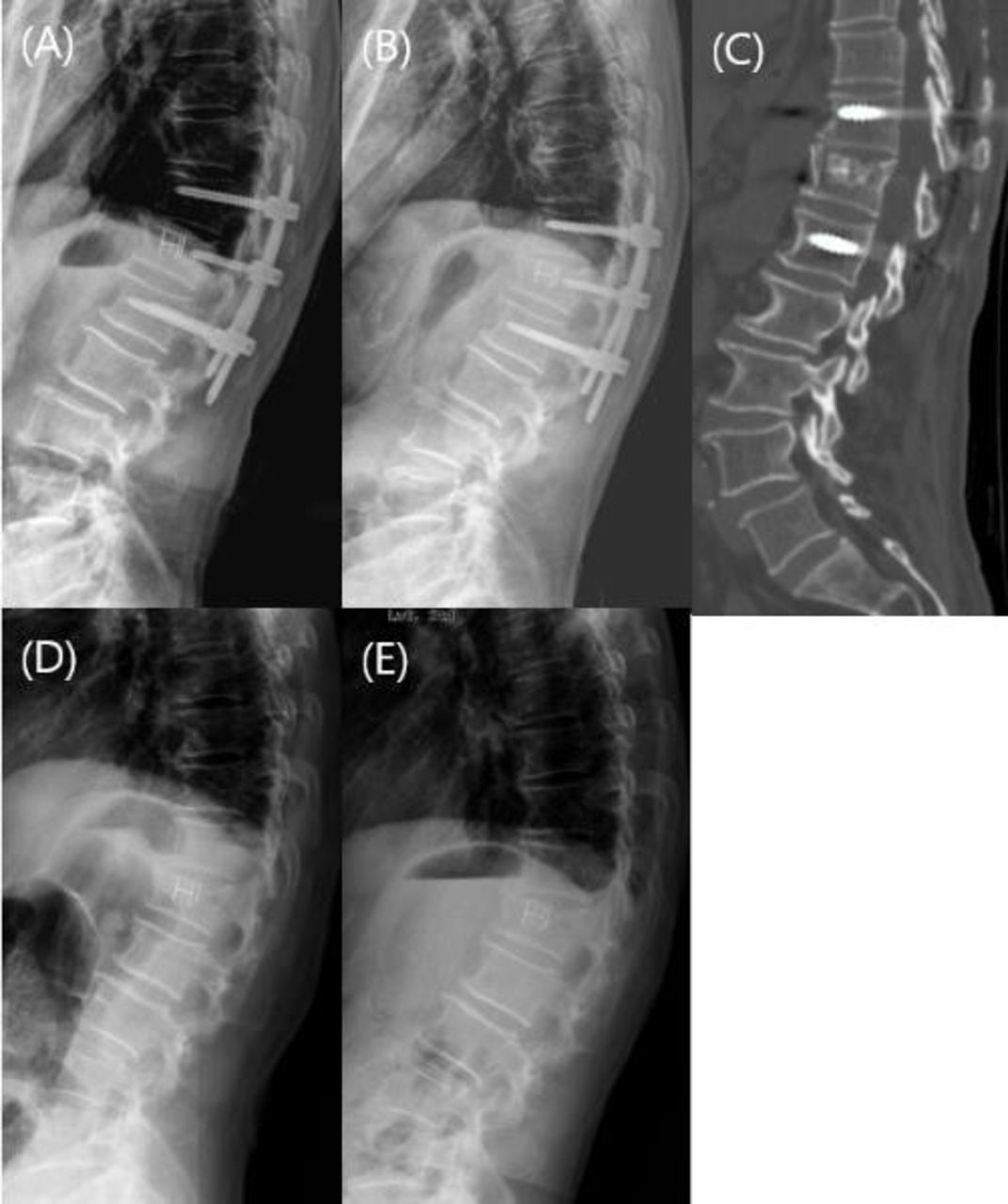
    Immediate postoperative radiograph demonstrating correction of the kyphosis (A). Lateral radiograph at 12 months postoperation (B).The computed tomography at 12 months postsurgery demonstrating the union of the fracture (C). Radiograph immediately postremoval of implants (D). One year postremoval of implants (E).

  • Figure 4
    • Download figure
    • Open in new tab
    • Download powerpoint
    Figure 4

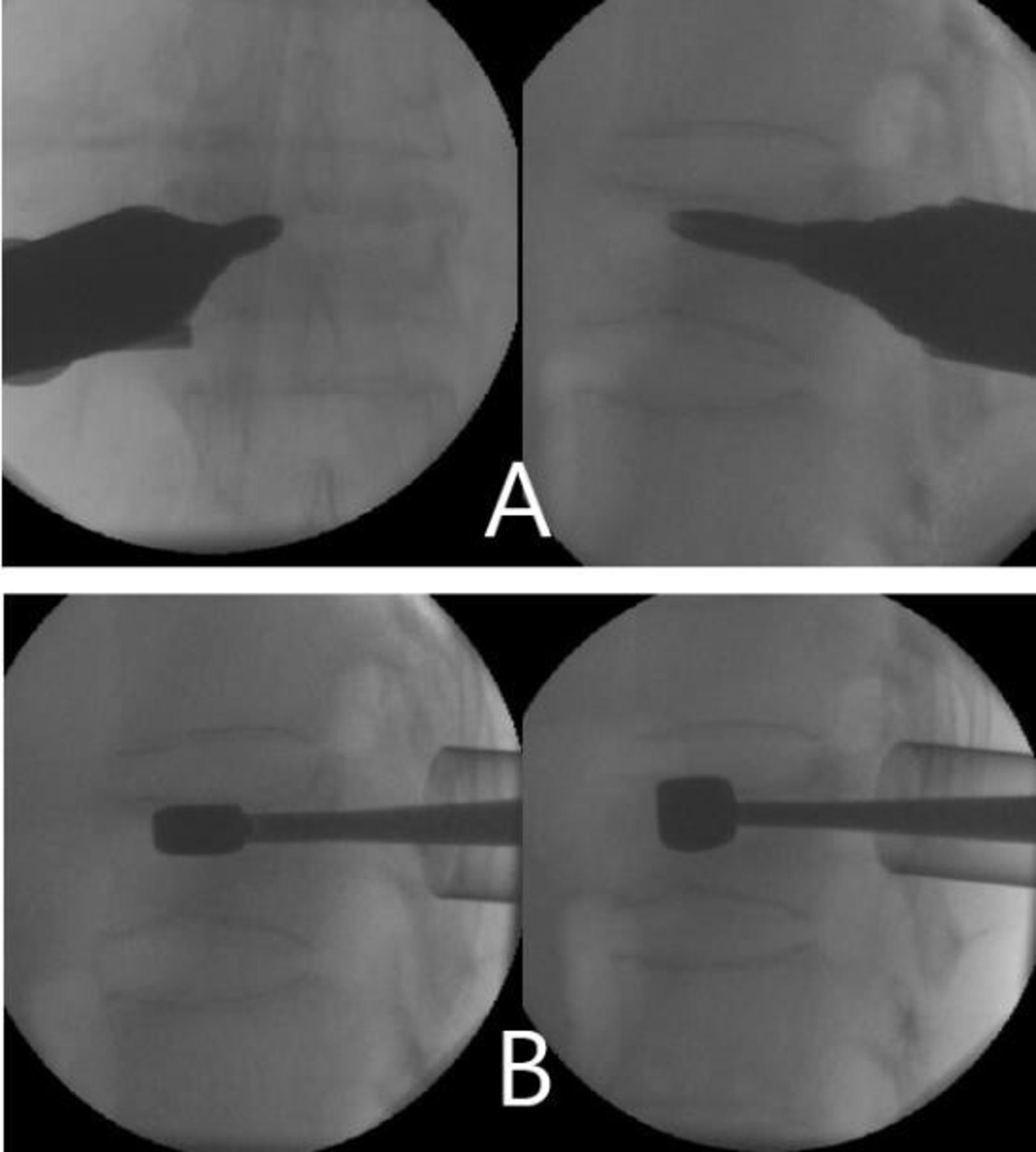
    A tubular retractor is placed after serial dilatations via the guidewire (A). Serial cage trials were introduced into the anterior column of the vertebral body to restore the vertebral body height (B).

  • Figure 5
    • Download figure
    • Open in new tab
    • Download powerpoint
    Figure 5

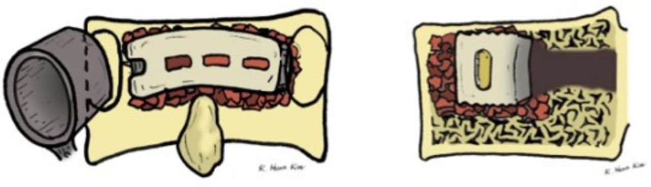
    A banana-shaped polyetheretherketone cage filled with demineralized bone matrix and allograft was placed into the verterbae, restoring the height of the vertebral body.

  • Figure 6
    • Download figure
    • Open in new tab
    • Download powerpoint
    Figure 6

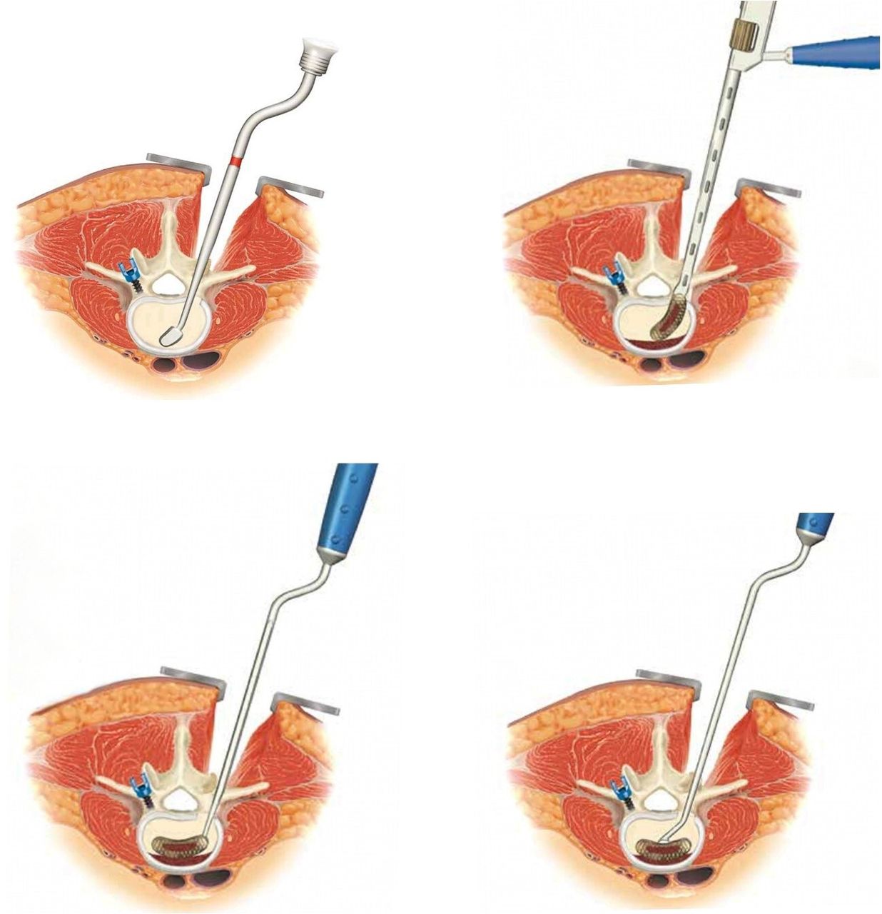
    Progressive dilatation was first performed using sequential trials (A). The cage was inserted obliquely into the vertebral body using a cage holder (B). Using a ball-tipped tamp, the cage was rotated into place through the gentle use of the mallet (C). The position of the cage was ascertained under fluoroscopy, and an anterior pusher can be used to push the cage anteriorly if necessary (D).

  • Figure 7
    • Download figure
    • Open in new tab
    • Download powerpoint
    Figure 7

    A percutaneous pedicle screw was inserted into each pedicle at the cranial and caudal vertebrae. A short pedicle screw was inserted into the contralateral pedicle at the index level for further stabilization.

Tables

  • Figures
    • View popup
    Table 1

    Demographic data of patients.

    Demographics N = 20
    Age, y, mean ± SD73.1 ± 6.1
    Sex, M/F, n 10/10
    History of trauma (mo before surgery), mean ± SD5.1 ± 3.9
    Follow-up, mo, mean ± SD18.4 ± 5.1
    Bone mineral density, mean ± SD−3.0 ± 1.2
    Level of surgery, n (%)
     T111 (5%)
     T126 (30%)
     L18 (40%)
     L24 (20%)
     L31 (5%)
    Operation time, min, mean ± SD118 ± 36.2
    Estimated blood loss, mL, mean ± SD216 ± 66.8
    • View popup
    Table 2

    Radiological data.

    DataPreoperativePostoperative P a 12-mo Follow-up P a
    Segmental angle, °15.2 ± 8.71.2 ± 5.2<0.013.6 ± 6.7<0.01
    Anterior body height, mm13.3 ± 14.622.6 ± 12.2<0.0121.9 ± 12.6<0.01
    Mean vertebral body height, mm18.5 ± 6.825.6 ± 7.6<0.0123.6 ± 4.4<0.01
    • ↵a P value in comparison with preoperative state.

    • View popup
    Table 3

    Visual analog scale and Oswestry Disability Index scores.

    Outcome MeasurePreoperative12-mo Follow-up P
    Visual analog scale8.0 ± 0.92.3 ± 0.5<0.01
    Oswestry disability index43 ± 13.822.9 ± 15.2<0.05
PreviousNext
Back to top

In this issue

International Journal of Spine Surgery
Vol. 18, Issue 1
1 Feb 2024
  • Table of Contents
  • Index by author

Print
Download PDF
Article Alerts
Sign In to Email Alerts with your Email Address
Email Article

Thank you for your interest in spreading the word on International Journal of Spine Surgery.

NOTE: We only request your email address so that the person you are recommending the page to knows that you wanted them to see it, and that it is not junk mail. We do not capture any email address.

Enter multiple addresses on separate lines or separate them with commas.
Minimally Invasive Surgery Transpedicular Intrabody Cage Technique for the Management of Kummell Disease
(Your Name) has sent you a message from International Journal of Spine Surgery
(Your Name) thought you would like to see the International Journal of Spine Surgery web site.
CAPTCHA
This question is for testing whether or not you are a human visitor and to prevent automated spam submissions.
Citation Tools
Minimally Invasive Surgery Transpedicular Intrabody Cage Technique for the Management of Kummell Disease
Junseok Bae, Chay-you Ang, Ifthekar Syed, Seong Kyun Jeong, Sang Ha Shin, Sang-Ho Lee
International Journal of Spine Surgery Feb 2024, 18 (1) 73-80; DOI: 10.14444/8570

Citation Manager Formats

  • BibTeX
  • Bookends
  • EasyBib
  • EndNote (tagged)
  • EndNote 8 (xml)
  • Medlars
  • Mendeley
  • Papers
  • RefWorks Tagged
  • Ref Manager
  • RIS
  • Zotero
Share
Minimally Invasive Surgery Transpedicular Intrabody Cage Technique for the Management of Kummell Disease
Junseok Bae, Chay-you Ang, Ifthekar Syed, Seong Kyun Jeong, Sang Ha Shin, Sang-Ho Lee
International Journal of Spine Surgery Feb 2024, 18 (1) 73-80; DOI: 10.14444/8570
Twitter logo Facebook logo Mendeley logo
  • Tweet Widget
  • Facebook Like
  • Google Plus One

Jump to section

  • Article
    • Abstract
    • Introduction
    • Case Presentation
    • Materials and Methods
    • Results
    • Discussion
    • Conclusion
    • Footnotes
    • References
  • Figures & Data
  • Info & Metrics
  • PDF

Related Articles

  • No related articles found.
  • PubMed
  • Google Scholar

Cited By...

  • No citing articles found.
  • Google Scholar

More in this TOC Section

Lumbar Spine

  • Comparison of Stand-Alone Anterior Lumbar Interbody Fusion, 360° Anterior Lumbar Interbody Fusion, and Arthroplasty for Recurrent Lumbar Disc Herniation: Focus on Nerve Decompression and Painful Spinal Instability Resolution
  • Recovery Trajectories After Lumbar Fusion Stratified by Baseline Patient-Reported Outcomes Measurement Information System Physical Function Disability Levels
  • Association Between Nonsteroidal Anti-inflammatory Drugs Use and Surgical Outcomes Following Posterior Lumbar Fusion: A Medical Claims Database Analysis
Show more Lumbar Spine

Minimally Invasive Surgery

  • Comparative Review of Lateral and Oblique Lumbar Interbody Fusion: Technique, Outcomes, and Complications
  • Key Considerations in Surgical Decision-Making on the Side of Approach for Lumbar Lateral Transpsoas Interbody Fusion Techniques
  • A Spine Surgeon’s Learning Curve With the Minimally Invasive L5 to S1 Lateral ALIF Surgical Approach: Perioperative Outcomes and Technical Considerations
Show more Minimally Invasive Surgery

Similar Articles

Keywords

  • Kummell disease
  • avascular necrosis
  • minimally invasive surgery
  • MISTIC
  • intrabody cage

Content

  • Current Issue
  • Latest Content
  • Archive

More Information

  • About IJSS
  • About ISASS
  • Privacy Policy

More

  • Subscribe
  • Alerts
  • Feedback

Other Services

  • Author Instructions
  • Join ISASS
  • Reprints & Permissions

© 2025 International Journal of Spine Surgery

International Journal of Spine Surgery Online ISSN: 2211-4599

Powered by HighWire