
Main menu

  • Home
  • Content
    • Current Issue
    • Advance Online Publication
    • Archive
  • About Us
    • About ISASS
    • About the Journal
    • Author Instructions
    • Editorial Board
    • Reviewer Guidelines & Publication Criteria
  • More
    • Advertise
    • Subscribe
    • Alerts
    • Feedback
  • Join Us
  • Reprints & Permissions
  • Sponsored Content
  • Other Publications
    • ijss

User menu

  • My alerts

Search

  • Advanced search
International Journal of Spine Surgery
  • My alerts
International Journal of Spine Surgery

Advanced Search

  • Home
  • Content
    • Current Issue
    • Advance Online Publication
    • Archive
  • About Us
    • About ISASS
    • About the Journal
    • Author Instructions
    • Editorial Board
    • Reviewer Guidelines & Publication Criteria
  • More
    • Advertise
    • Subscribe
    • Alerts
    • Feedback
  • Join Us
  • Reprints & Permissions
  • Sponsored Content
  • Follow ijss on Twitter
  • Visit ijss on Facebook
Research ArticleArticle
Open Access

Endoscopic Foraminal Decompression for Failed Back Surgery Syndrome under local Anesthesia

Anthony Yeung and Satishchandra Gore
International Journal of Spine Surgery January 2014, 8 22; DOI: https://doi.org/10.14444/1022
Anthony Yeung
1Desert Institute for Spine Care, Phoenix, AZ, USA
MD
  • Find this author on Google Scholar
  • Find this author on PubMed
  • Search for this author on this site
Satishchandra Gore
2Prime Surgical Centre, Pune, India
MD
  • Find this author on Google Scholar
  • Find this author on PubMed
  • Search for this author on this site
  • Article
  • Figures & Data
  • Info & Metrics
  • PDF
Loading

Article Figures & Data

Figures

  • Fig. 1
    • Download figure
    • Open in new tab
    • Download powerpoint
    Fig. 1

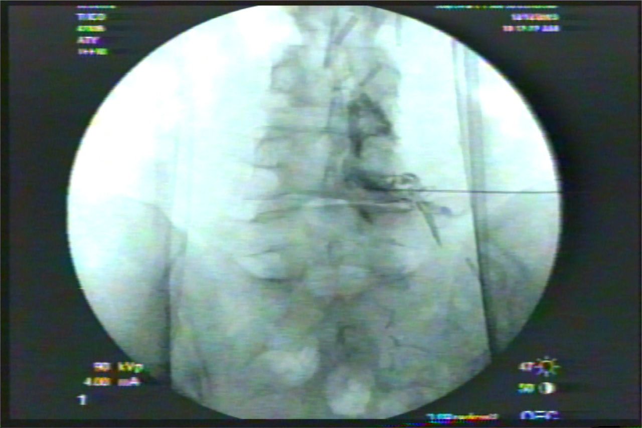
    A diagnostic and therapeutic epidural gram may help identify unrecognized lateral recess stenosis underestimated by MRI. An excellent result from a therapeutic block lends excellent prognosis for a more lasting and “permanent” result from transforaminal endoscopic lateral recess decompression.

  • Fig. 2
    • Download figure
    • Open in new tab
    • Download powerpoint
    Fig. 2

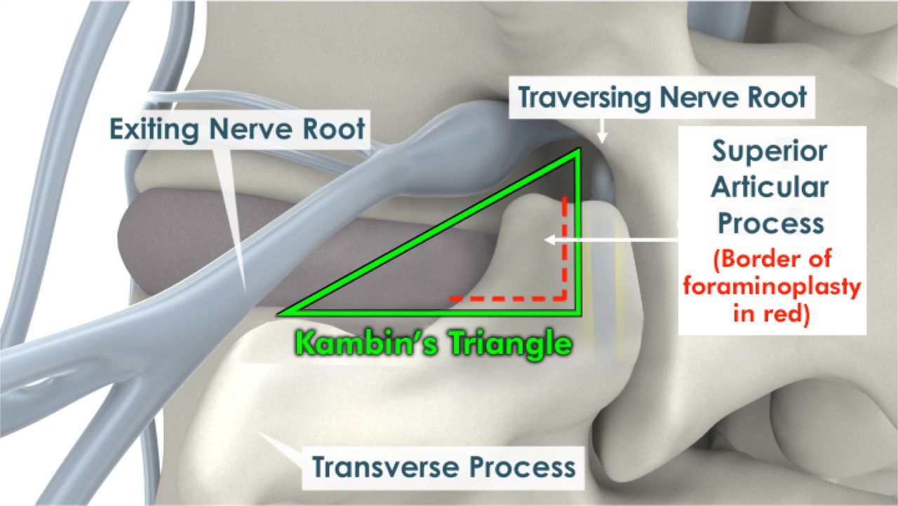
    Kambin's Triangle provides access to the “hidden zone” of Macnab by foraminoplasty. The foramen and lateral recess is decompressed by removing the ventral aspect and tip of the superior articular process to gain access to the axilla between the traversing and exiting nerve. FBSS contains patho-anatomy in the axilla between the traversing and exiting nerve that hides the pain generators of FBSS.

  • Fig. 3
    • Download figure
    • Open in new tab
    • Download powerpoint
    Fig. 3

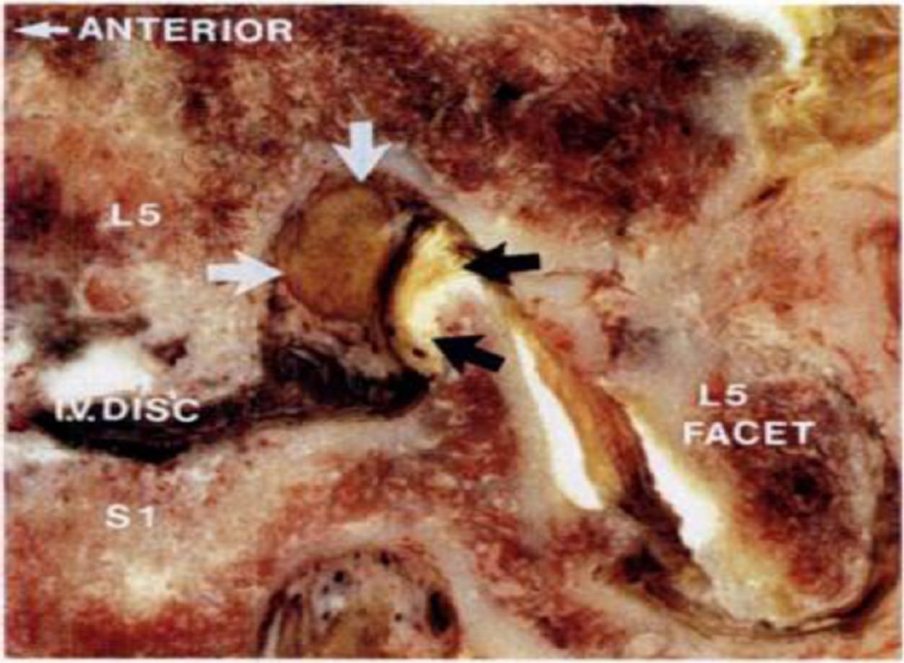
    Cadaver Illustration of Foraminal Stenosis (courtesy of Wolfgang Rauschning). As the disc narrows, the superior articular process impinges on the exiting nerve and DRG, creating lateral recess stenosis, lumbar spondylosis, and facet arthrosis.

  • Fig. 4
    • Download figure
    • Open in new tab
    • Download powerpoint
    Fig. 4

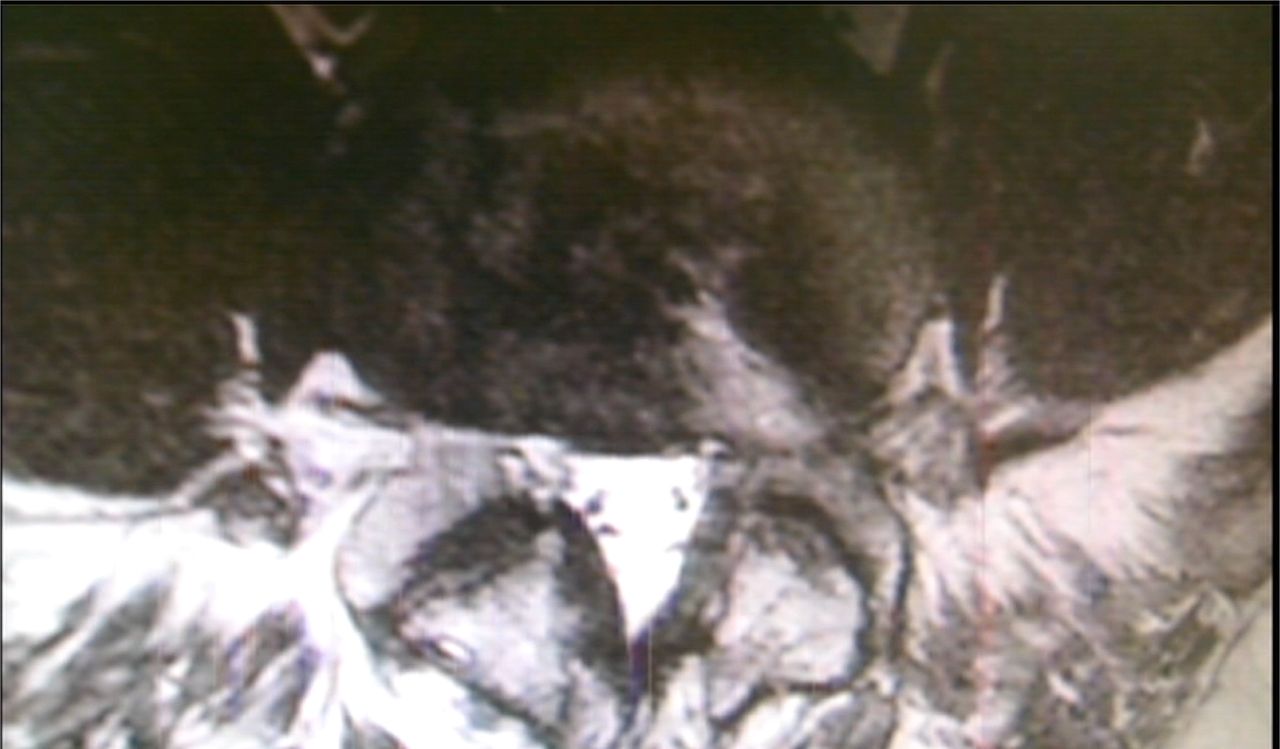
    MRI of FBSS following two open decompressions from midline and Wiltse approach. Patient had residual symptoms of numbness from persistent lateral stenosis.

  • Fig. 5
    • Download figure
    • Open in new tab
    • Download powerpoint
    Fig. 5

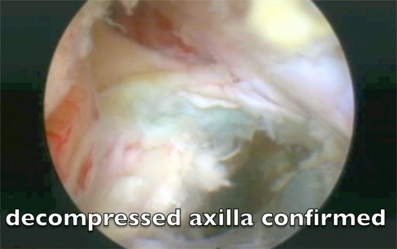
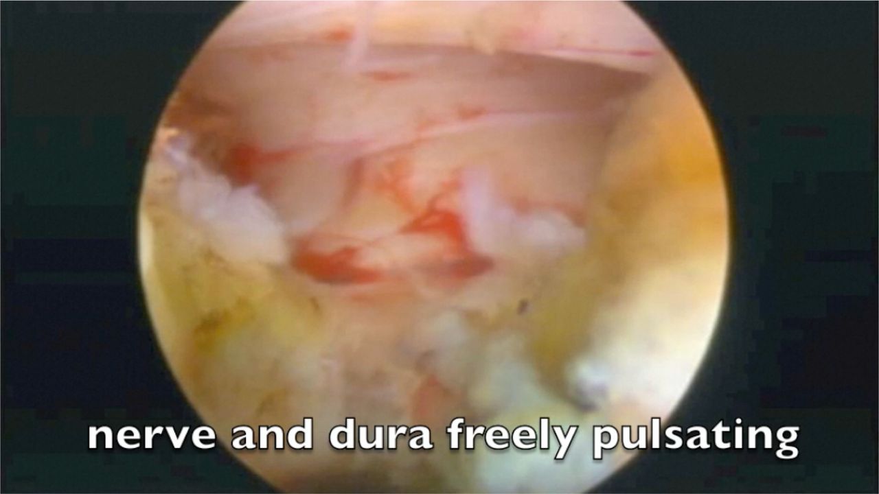
    The axilla between the traversing and exiting nerve is the location of missed patho-anatomy in FBSS. Image of a completely decompressed axilla will allow the surgeon to see that both exiting and traversing nerve are decompressed, and FBSS from patho-anatomy in this zone known as the “hidden zone,” when decompressed, will resolve FBSS from residual/recurrent HNP and lateral recess stenosis.

  • Fig. 6
    • Download figure
    • Open in new tab
    • Download powerpoint
    Fig. 6

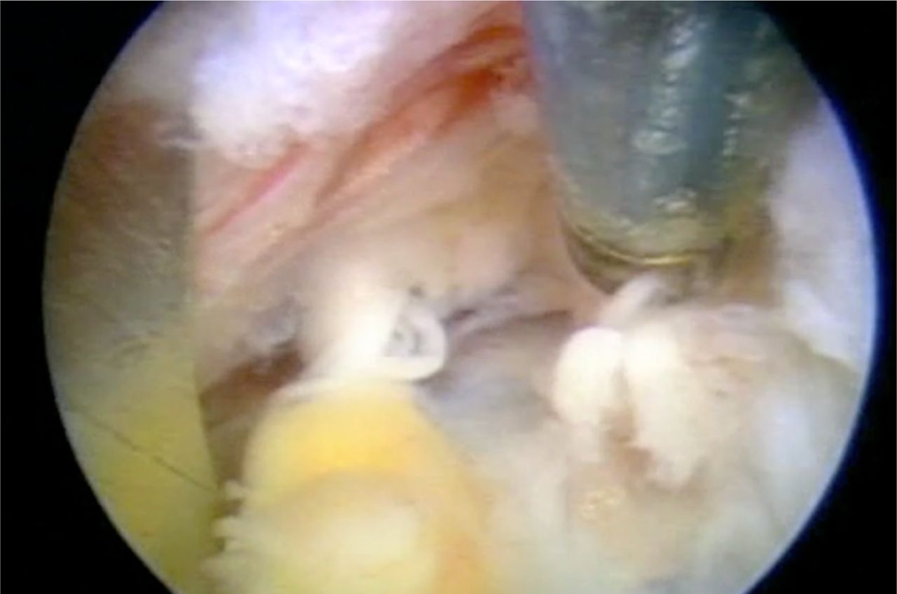
    View after extensive decompressing the ventral SAP after FBSS. Although medial translaminar decompression provides good access to the medial epidural space and the traversing nerve, further decompression through a transforaminal approach is sometimes, needed. Here transforaminal decompression confirms complete decompression of the nerve and the dura above, in a patient with residual sciatica following traditional decompression, but improving the patient further with by decompressing the lateral recess further to expose the lateral aspect of the foramen. A calcified annulus was also found. The medial capsule and ligamentum flavum usually protects the lateral edge of the traversing nerve and dura. Here, it was lifted up to visualize the nerve in the epidural space.

  • Fig. 7
    • Download figure
    • Open in new tab
    • Download powerpoint
    Fig. 7

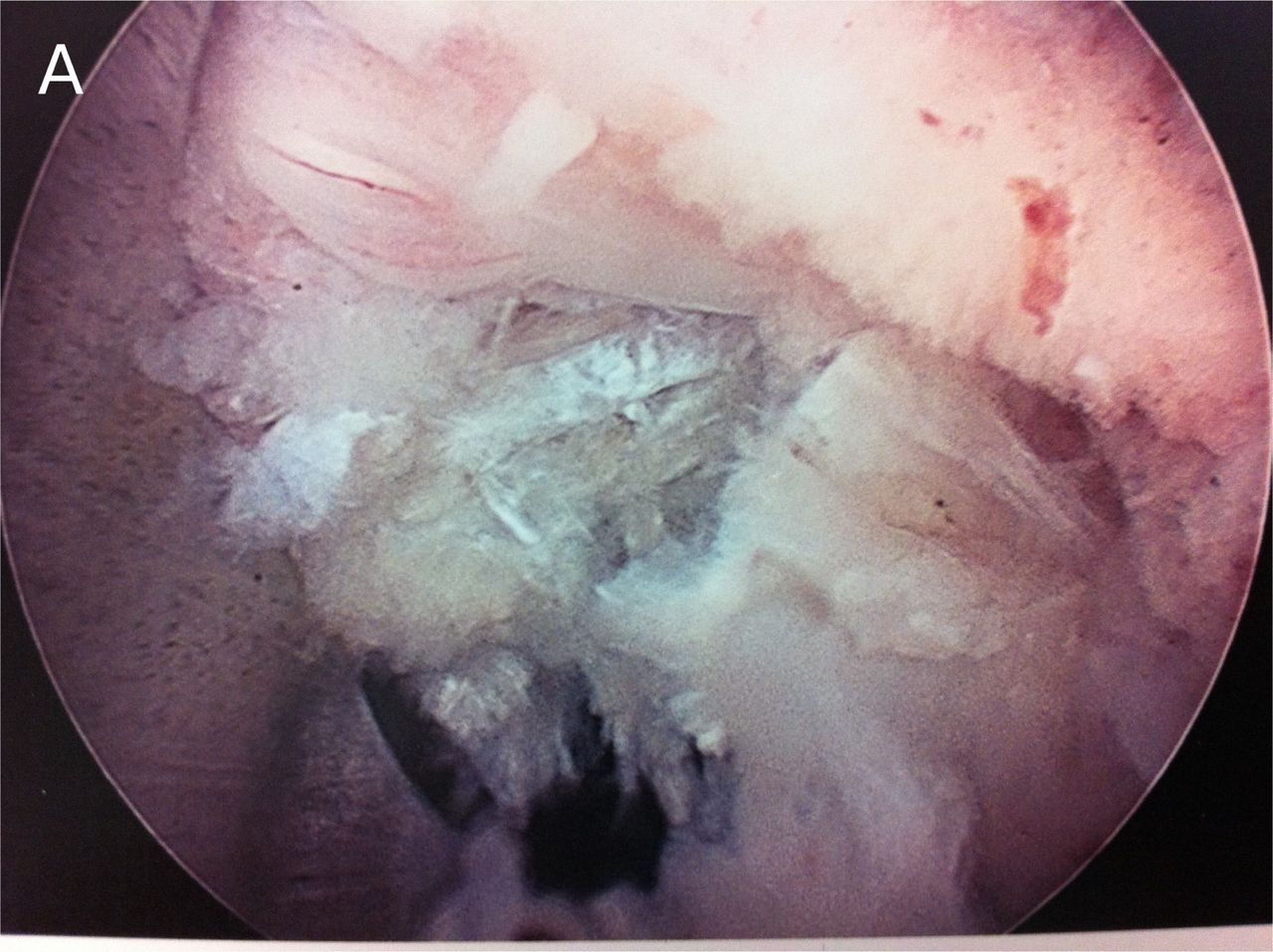
    The axilla contains hidden patho-anatomy such as foraminal disc protrusions, synovial cysts and in this example a rare neuroma of the dorsal ramus is visualized in the in the axilla of the foramen at the L4-5 spinal level. The dorsal ramus is easily mistaken for a furcal nerve.

  • Fig. 8
    • Download figure
    • Open in new tab
    • Download powerpoint
    Fig. 8

    Cadaver dissection of the Dorsal Ramus and its branches ventral to the intertransverse ligament. One medial branch crosses the transverse process, as does the intermediate and lateral branch. Other branches are demonstrated to reach the facet from the foramen ventral to the intertransverse ligament. Location of the branches is most consistent as described by Bogduk, but various locations and configurations are common.
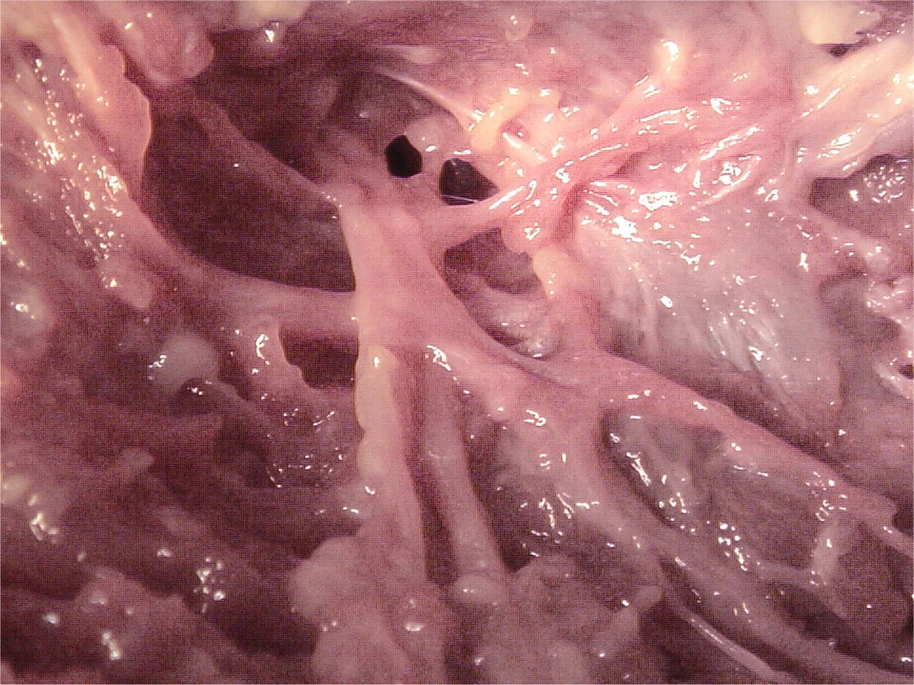
  • Fig. 9a
    • Download figure
    • Open in new tab
    • Download powerpoint
    Fig. 9a

    Medial branch of the dorsal ramus visualized in the foramen before it exits the foramen to innervate the facet. This is easily mistaken for a furcal nerve. Complete transection or ablation will not produce dysesthesia as it is a pure sensory nerve. Injury or partial ablation of a furcal nerve will produce temporary dysesthesia.
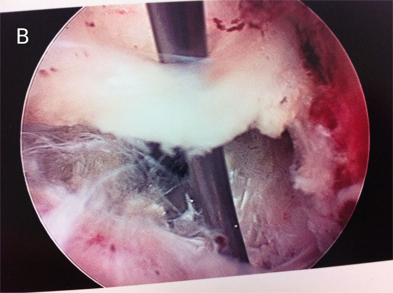
  • Fig. 9b
    • Download figure
    • Open in new tab
    • Download powerpoint
    Fig. 9b

    Medial branch of dorsal ramus in the foramen vs furcal nerve. It is not always possible to differentiate. If the nerve can be traced branching from a spinal nerve ablating it may cause dysesthesia. Usually very small nerves can be cut. It is better to completely transect that to partially ablate the nerve. The exiting nerve is partially visualized at 7 o'clock.
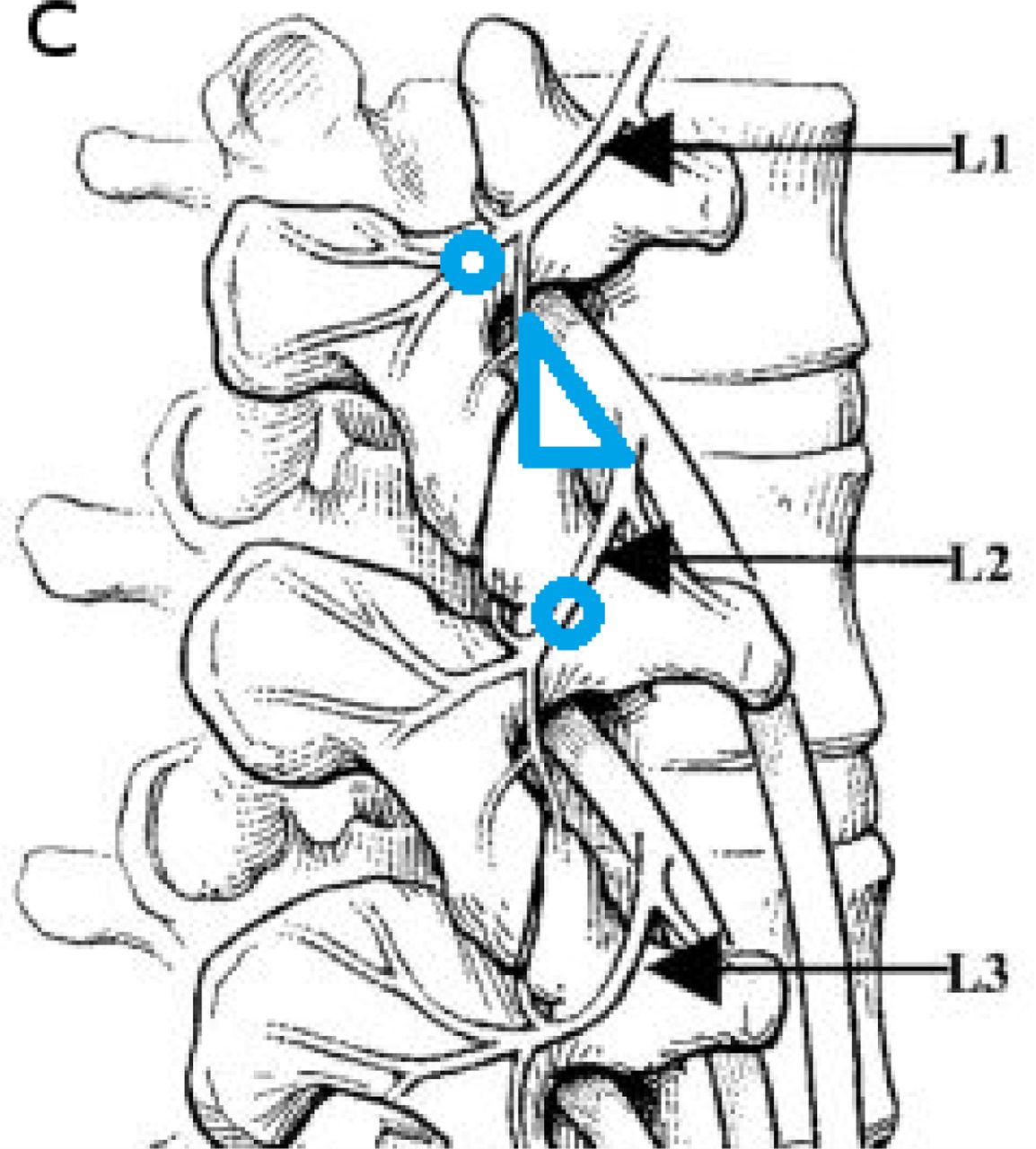
  • Fig. 9c
    • Download figure
    • Open in new tab
    • Download powerpoint
    Fig. 9c

    Locations and variations of the branches of the dorsal ramus. Position of the medial branch dorsal ramus in Kambin's triangle (in blue). The target for medial branch ablation on the transverse process is highlighted by the blue circles. The medial branch can be transected or ablated in the foramen as it traverses along the foraminal ligament on the ventral facet before it exits to innervate the joint. The nerve, if transected and provide axial back pain relief during foraminoplasty. These nerves can be mistaken for furcal nerves, that, if less than 1-2 mm, can be ablated without causing dysesthesia. If the nerve causes pain when ablated with bipolar RF, It would be advisable to avoid injuring it, if possible.

PreviousNext
Back to top

In this issue

International Journal of Spine Surgery
Vol. 8
1 Jan 2014
  • Table of Contents
  • Index by author

Print
Download PDF
Article Alerts
Sign In to Email Alerts with your Email Address
Email Article

Thank you for your interest in spreading the word on International Journal of Spine Surgery.

NOTE: We only request your email address so that the person you are recommending the page to knows that you wanted them to see it, and that it is not junk mail. We do not capture any email address.

Enter multiple addresses on separate lines or separate them with commas.
Endoscopic Foraminal Decompression for Failed Back Surgery Syndrome under local Anesthesia
(Your Name) has sent you a message from International Journal of Spine Surgery
(Your Name) thought you would like to see the International Journal of Spine Surgery web site.
CAPTCHA
This question is for testing whether or not you are a human visitor and to prevent automated spam submissions.
Citation Tools
Endoscopic Foraminal Decompression for Failed Back Surgery Syndrome under local Anesthesia
Anthony Yeung, Satishchandra Gore
International Journal of Spine Surgery Jan 2014, 8 22; DOI: 10.14444/1022

Citation Manager Formats

  • BibTeX
  • Bookends
  • EasyBib
  • EndNote (tagged)
  • EndNote 8 (xml)
  • Medlars
  • Mendeley
  • Papers
  • RefWorks Tagged
  • Ref Manager
  • RIS
  • Zotero
Share
Endoscopic Foraminal Decompression for Failed Back Surgery Syndrome under local Anesthesia
Anthony Yeung, Satishchandra Gore
International Journal of Spine Surgery Jan 2014, 8 22; DOI: 10.14444/1022
Twitter logo Facebook logo Mendeley logo
  • Tweet Widget
  • Facebook Like
  • Google Plus One

Jump to section

  • Article
    • Abstract
    • Introduction
    • Materials and Methods
    • Results
    • Discussion
    • Complications: Avoidance of irritation of the DRG
    • Conclusion
    • Disclosures
    • References
  • Figures & Data
  • Info & Metrics
  • PDF

Related Articles

  • No related articles found.
  • Google Scholar

Cited By...

  • Long-term Clinical Outcomes Following Endoscopic Foraminoplasty for Patients With Single-Level Foraminal Stenosis of the Lumbar Spine
  • Percutaneous Endoscopic Lumbar Discectomy Versus Microdiscectomy for the Treatment of Lumbar Disc Herniation: Pain, Disability, and Complication Rate--A Randomized Clinical Trial
  • Google Scholar

More in this TOC Section

  • Management of Spinal Langerhans Cell Histiocytosis in Children: A Systematic Review
  • Meta-Analysis Investigating Optimal Timing of Chemoprophylaxis for Venous Thromboembolism in Operatively Managed Blunt Spinal Injuries
  • Accuracy of Pedicle Screw Placement Using the ExcelsiusGPS Robotic Navigation Platform: An Analysis of 728 Screws
Show more Article

Similar Articles

Keywords

  • Failed Back Surgery Syndrome(FBSS)
  • Hidden zone
  • Foraminal decompression
  • Recurrent herniation
  • Lateral stenosis
  • Foraminal osteophyte

Content

  • Current Issue
  • Latest Content
  • Archive

More Information

  • About IJSS
  • About ISASS
  • Privacy Policy

More

  • Subscribe
  • Alerts
  • Feedback

Other Services

  • Author Instructions
  • Join ISASS
  • Reprints & Permissions

© 2025 International Journal of Spine Surgery

International Journal of Spine Surgery Online ISSN: 2211-4599

Powered by HighWire