
Main menu

  • Home
  • Content
    • Current Issue
    • Advance Online Publication
    • Archive
  • About Us
    • About ISASS
    • About the Journal
    • Author Instructions
    • Editorial Board
    • Reviewer Guidelines & Publication Criteria
  • More
    • Advertise
    • Subscribe
    • Alerts
    • Feedback
  • Join Us
  • Reprints & Permissions
  • Sponsored Content
  • Other Publications
    • ijss

User menu

  • My alerts

Search

  • Advanced search
International Journal of Spine Surgery
  • My alerts
International Journal of Spine Surgery

Advanced Search

  • Home
  • Content
    • Current Issue
    • Advance Online Publication
    • Archive
  • About Us
    • About ISASS
    • About the Journal
    • Author Instructions
    • Editorial Board
    • Reviewer Guidelines & Publication Criteria
  • More
    • Advertise
    • Subscribe
    • Alerts
    • Feedback
  • Join Us
  • Reprints & Permissions
  • Sponsored Content
  • Follow ijss on Twitter
  • Visit ijss on Facebook
Research ArticleArticle
Open Access

“Outside-in” Technique, Clinical Results, and Indications with Transforaminal Lumbar Endoscopic Surgery: a Retrospective Study on 220 Patients on Applied Radiographic Classification of Foraminal Spinal Stenosis

Kai-Uwe Lewandrowski
International Journal of Spine Surgery January 2014, 8 26; DOI: https://doi.org/10.14444/1026
Kai-Uwe Lewandrowski
Center for Advanced Spinal Surgery of Southern Arizona, Tucson, AZ
MD
  • Find this author on Google Scholar
  • Find this author on PubMed
  • Search for this author on this site
  • Article
  • Figures & Data
  • Info & Metrics
  • PDF
Loading

Article Figures & Data

Figures

  • Tables
  • Fig. 1
    • Download figure
    • Open in new tab
    • Download powerpoint
    Fig. 1

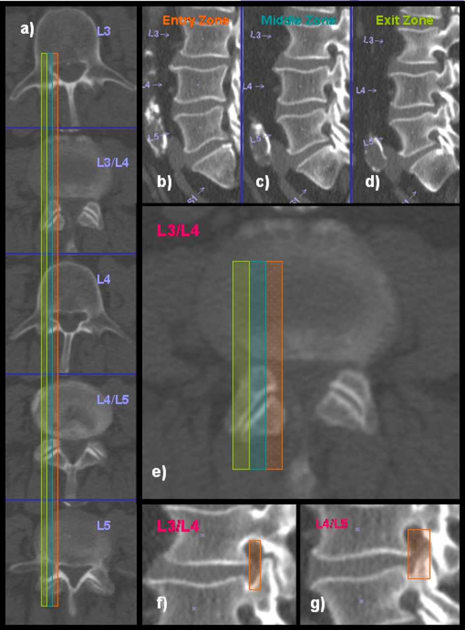
    Preoperative CT scans of a 70 year old male: a) panel on the left shows axial CT cuts from L3 to L5, b-d) panel shows sagittal CT cuts through the entry (shaded orange), middle (shaded turquoise), and exit zone (shaded green) of the lumbar neuroforamina, e) axial CT cut through the L3-4 disc space showing the stenotic lesion in the middle zone at that level, f-g) sagittal CT cuts through the middle zone at L3-4, and the L4-5 level. The neuroforaminal height (orange shade area) is less than 5 mm. The neuroforaminal width is less than 2 mm. Both indicators are consistent with spinal stenosis.

  • Fig. 2
    • Download figure
    • Open in new tab
    • Download powerpoint
    Fig. 2

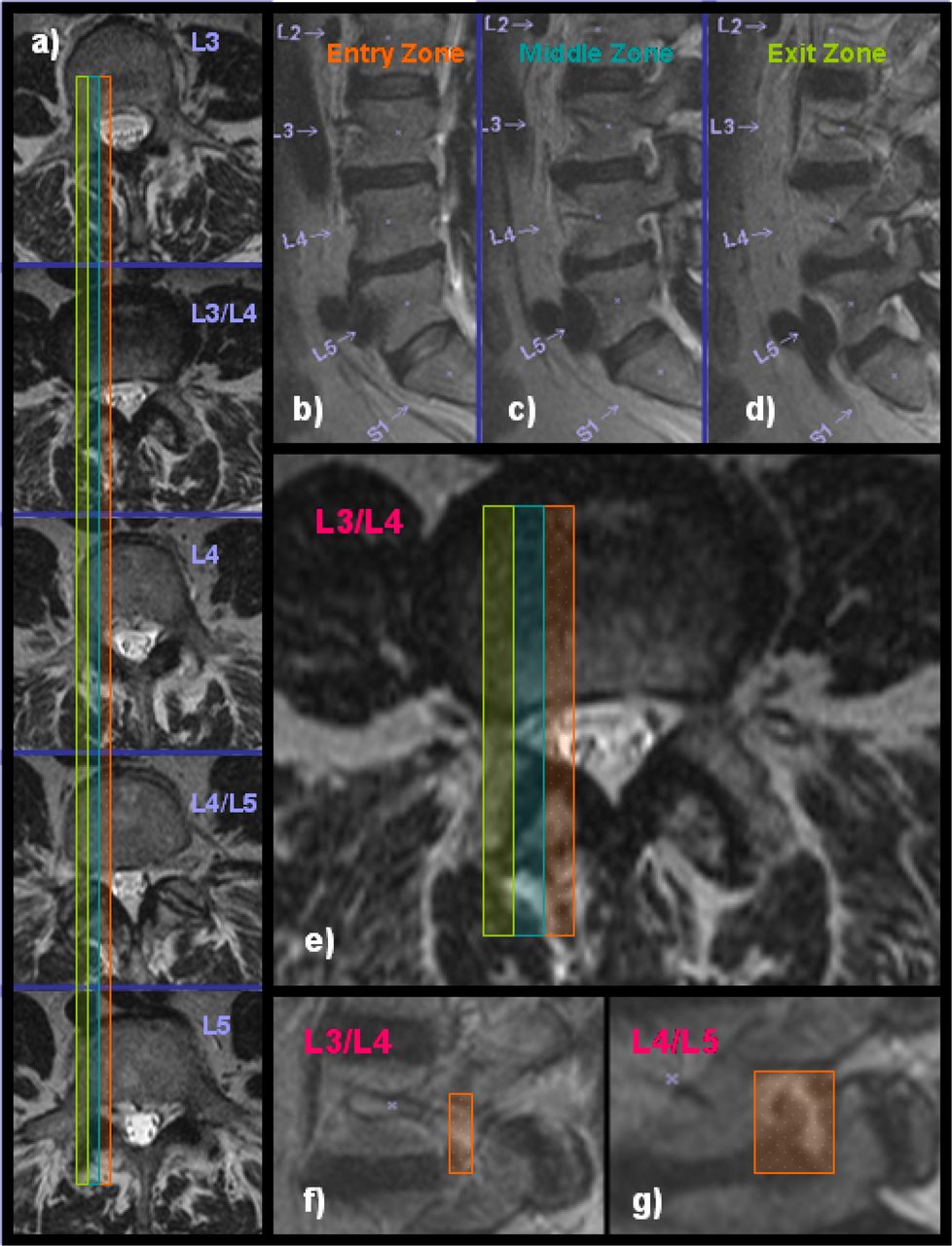
    Preoperative MRI scans of a 64 year old female: a) panel on the left shows axial MRI cuts from L3 to L5, b-d) panel shows sagittal MRI cuts through the entry (shaded orange), middle (shaded turquoise), and exit zone (shaded green) of the lumbar neuroforamina, e) axial MRI cut through the L3-4 disc space showing the stenotic lesion in the exit zone at that level, f-g) sagittal MRI cuts through the exit zone at L3-4, and the L4-5 level. The neuroforaminal height (orange shade area) is less than 3 mm and hence consistent with spinal stenosis. At L4-5, the neuroforaminal height is 5 mm (orange shade area).

  • Fig. 3
    • Download figure
    • Open in new tab
    • Download powerpoint
    Fig. 3

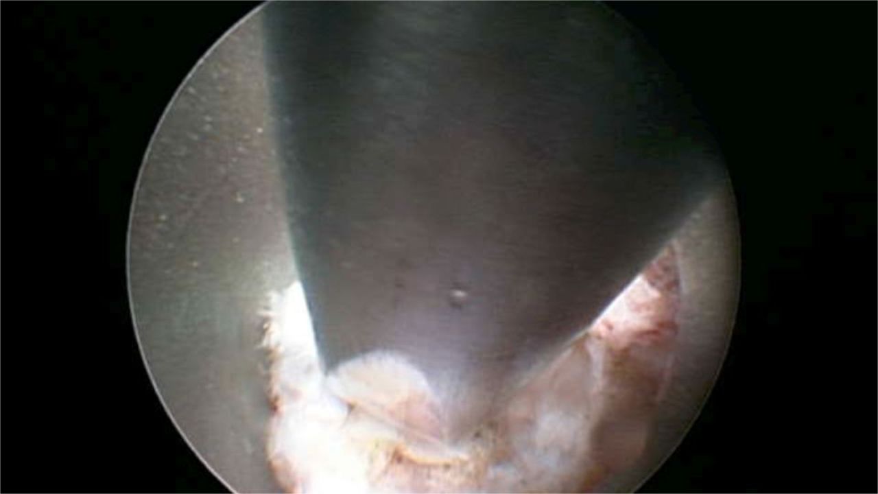
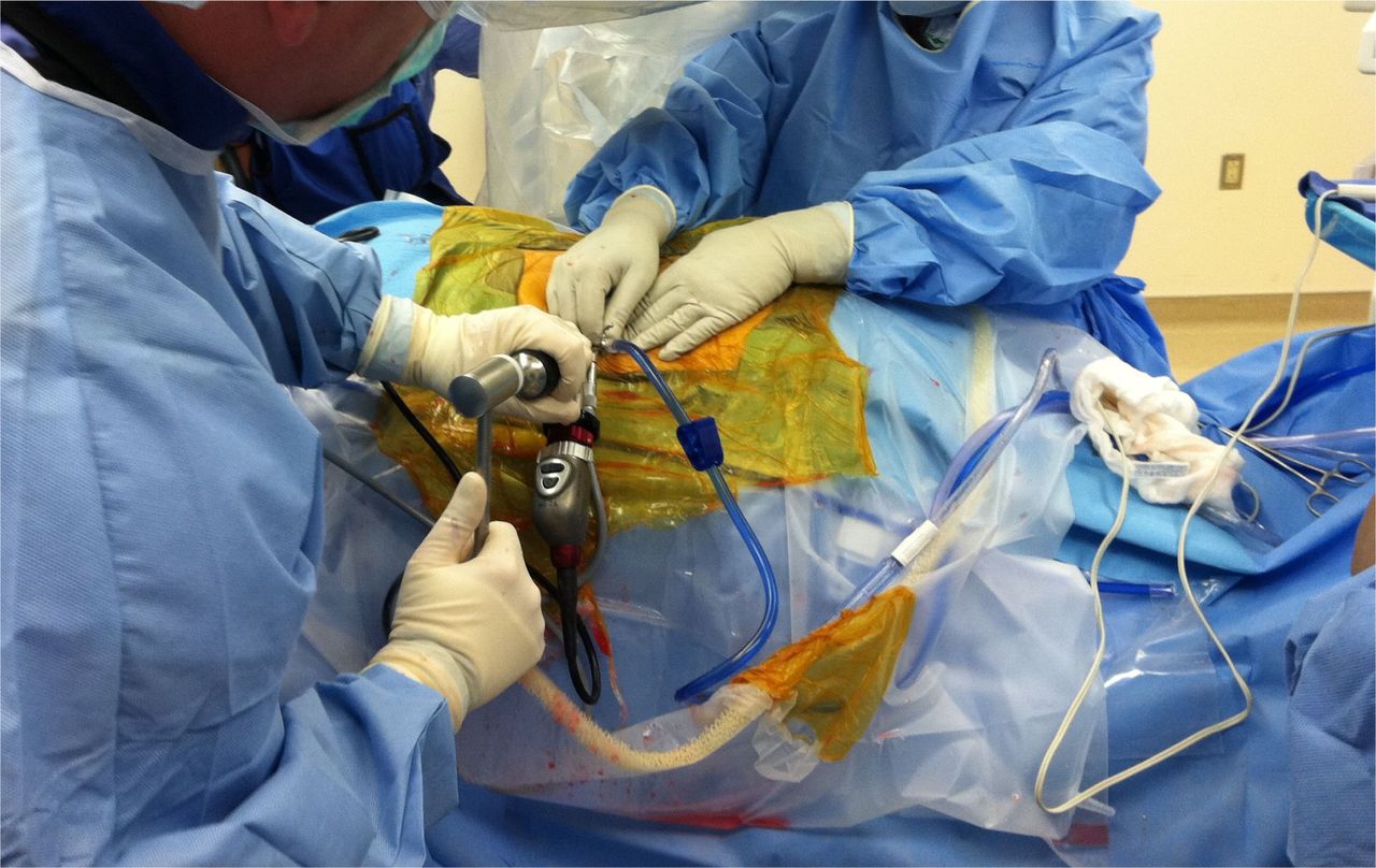
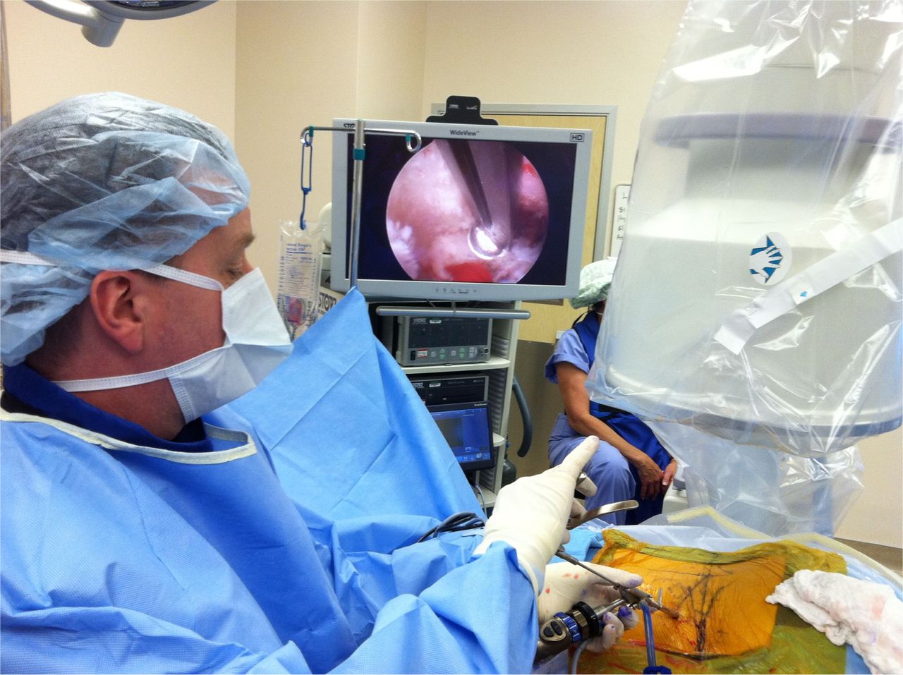
    Endoscopic view of the chisel used to perform a foraminoplasty. The cannula is docked at the lateral superior aspect of the facet joint. The chisel is introduced through the working channel of the endoscope starting in an upward direction and then chiseling in a downward direction by rotating the chisel 180 degrees to remove bone from the facet joint.

  • Fig. 4
    • Download figure
    • Open in new tab
    • Download powerpoint
    Fig. 4

    The chisel is advanced through the central working channel of the endoscope. A mallet may be used to advance the chisel for the foraminoplasty. Typically, a direct lateral approach to the foramen by dropping one's hand is more advantageous. The foraminoplasty can be facilitaed by chiseling in an upward diretion, then by rotating the chisel by 180 degrees followed by downward chiseling.

  • Fig. 5
    • Download figure
    • Open in new tab
    • Download powerpoint
    Fig. 5

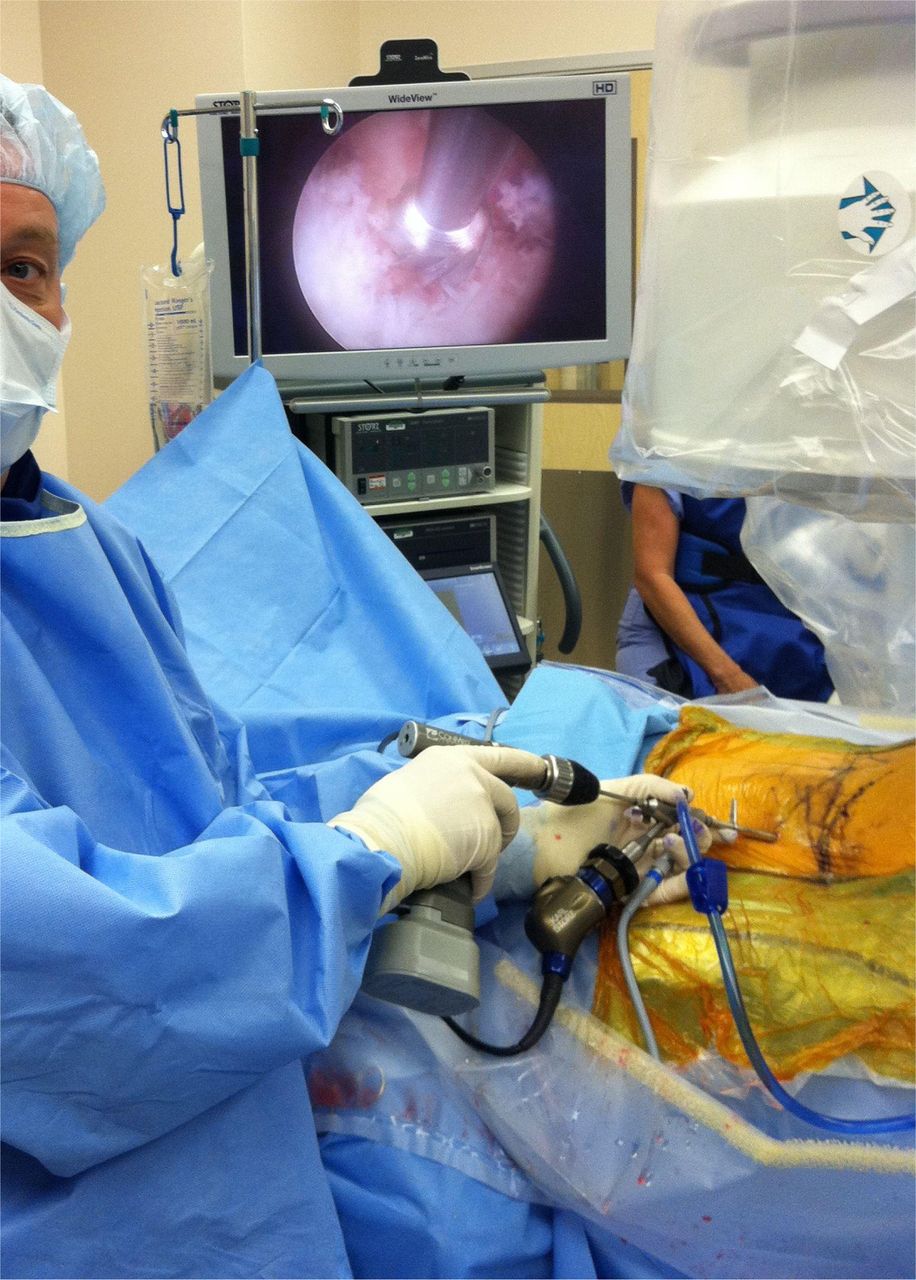
    An endoscopic Kerrison rongeur can be used to finalize the foraminoplasty. It is most suitable for decompressing the most medial portion of the lateral recess by using a Kerrison with a 135 degree footplate. The Kerrison is directed past the leading edge of the facet joint, then rotated 180 degrees to remove bone by dropping one's hand.

  • Fig. 6
    • Download figure
    • Open in new tab
    • Download powerpoint
    Fig. 6

    A foraminal drill can be advanced directly to the inner working channel. The drill is attached to a power driver and can be used in forward and reverse. It is most suitable for expansile foraminoplasty around the inferior pedicle.

Tables

  • Figures
    • View popup
    Table 1

    Patient population and type of lesion.

    220 Study Patients
    Extruded Disc 24 Patients
    Contained Disc 82 Patients
    Disc Bulge 33 Patients
    Foraminal Stenosis 114 Patients
    • View popup
    Table 2

    Foraminoplasty instruments.

    Embedded ImageEmbedded ImageThe transforaminal gold 7 and 9 mm mm reamer is intended for the initial foraminoplasty to remove bony overhang from arthritic facet joints. It is cannulated and fits through the working cannula to minimize tissue trauma and to reduce pain during surgery. It has a mostly side cutting beveled tip which is less aggressive when advancing thus minimizing risk for dural injury. The outer diameter of the beveled tip ranges from 4 – 7 mm. It comes with a multidirectional T-handle.
    Embedded ImageEmbedded ImageThe 4 mm chisel is intended to perform a foraminoplasty using the outside-in technique. The instrument is semi sharp to obviate the need for sharpening. It has a handle with a metal cap for tapping with a surgical mallet.
    Embedded ImageEmbedded ImageThe 4 mm round drill is also intended for a foraminoplasty. It can be used on power for a more controlled removal of bony tissue. This drill bit is less likely to cause dural injury. It is used best for the final steps of the foraminoplasty. For example, it is very useful to drill of bone spurs of the superior articular process that compress the traversing nerve root.
    Embedded ImageEmbedded ImageThe 3.5 mm kerrison rongeur is designed to perform a foraminoplasty. This is done by hooking the instrument underneath the structure to be removed and cutting it by squeezing the handle.
    Embedded ImageEmbedded ImageThe 3-7 mm trephine cutters are intended to enlarge the foraminoplasty. It can be placed at the pedicle or facet joint. It is cannulated and fits over the 1mm and 1.65 mm steel or nitinol guide wire. The end of the trephine has serrations for improved grip and fine motor control. The handle attaches to the opposite end of the trephine and is cannulated as well to accommodate for the long guide wire.
PreviousNext
Back to top

In this issue

International Journal of Spine Surgery
Vol. 8
1 Jan 2014
  • Table of Contents
  • Index by author

Print
Download PDF
Article Alerts
Sign In to Email Alerts with your Email Address
Email Article

Thank you for your interest in spreading the word on International Journal of Spine Surgery.

NOTE: We only request your email address so that the person you are recommending the page to knows that you wanted them to see it, and that it is not junk mail. We do not capture any email address.

Enter multiple addresses on separate lines or separate them with commas.
“Outside-in” Technique, Clinical Results, and Indications with Transforaminal Lumbar Endoscopic Surgery: a Retrospective Study on 220 Patients on Applied Radiographic Classification of Foraminal Spinal Stenosis
(Your Name) has sent you a message from International Journal of Spine Surgery
(Your Name) thought you would like to see the International Journal of Spine Surgery web site.
CAPTCHA
This question is for testing whether or not you are a human visitor and to prevent automated spam submissions.
Citation Tools
“Outside-in” Technique, Clinical Results, and Indications with Transforaminal Lumbar Endoscopic Surgery: a Retrospective Study on 220 Patients on Applied Radiographic Classification of Foraminal Spinal Stenosis
Kai-Uwe Lewandrowski
International Journal of Spine Surgery Jan 2014, 8 26; DOI: 10.14444/1026

Citation Manager Formats

  • BibTeX
  • Bookends
  • EasyBib
  • EndNote (tagged)
  • EndNote 8 (xml)
  • Medlars
  • Mendeley
  • Papers
  • RefWorks Tagged
  • Ref Manager
  • RIS
  • Zotero
Share
“Outside-in” Technique, Clinical Results, and Indications with Transforaminal Lumbar Endoscopic Surgery: a Retrospective Study on 220 Patients on Applied Radiographic Classification of Foraminal Spinal Stenosis
Kai-Uwe Lewandrowski
International Journal of Spine Surgery Jan 2014, 8 26; DOI: 10.14444/1026
Twitter logo Facebook logo Mendeley logo
  • Tweet Widget
  • Facebook Like
  • Google Plus One

Jump to section

  • Article
    • Abstract
    • Introduction
    • Materials and Methods
    • Results
    • Discussion
    • Conclusion
    • Disclosures
    • References
  • Figures & Data
  • Info & Metrics
  • PDF

Related Articles

  • No related articles found.
  • Google Scholar

Cited By...

  • Evolving Role of Lumbar Decompression: A Narrative Review
  • Difficulties, Challenges, and the Learning Curve of Avoiding Complications in Lumbar Endoscopic Spine Surgery
  • Artificial Intelligence Comparison of the Radiologist Report With Endoscopic Predictors of Successful Transforaminal Decompression for Painful Conditions of the Lumber Spine: Application of Deep Learning Algorithm Interpretation of Routine Lumbar Magnetic Resonance Imaging Scan
  • Reliability Analysis of Deep Learning Algorithms for Reporting of Routine Lumbar MRI Scans
  • Google Scholar

More in this TOC Section

  • Accuracy of Pedicle Screw Placement Using the ExcelsiusGPS Robotic Navigation Platform: An Analysis of 728 Screws
  • Management of Spinal Langerhans Cell Histiocytosis in Children: A Systematic Review
  • Meta-Analysis Investigating Optimal Timing of Chemoprophylaxis for Venous Thromboembolism in Operatively Managed Blunt Spinal Injuries
Show more Article

Similar Articles

Keywords

  • Preoperative Classification of Lumbar Foraminal Spinal Stenosis
  • Percutaneous endoscopic transforaminal lumbar foraminotomy
  • outside-in technique
  • microdiscectomy

Content

  • Current Issue
  • Latest Content
  • Archive

More Information

  • About IJSS
  • About ISASS
  • Privacy Policy

More

  • Subscribe
  • Alerts
  • Feedback

Other Services

  • Author Instructions
  • Join ISASS
  • Reprints & Permissions

© 2025 International Journal of Spine Surgery

International Journal of Spine Surgery Online ISSN: 2211-4599

Powered by HighWire