


Article Figures & Data
Figures
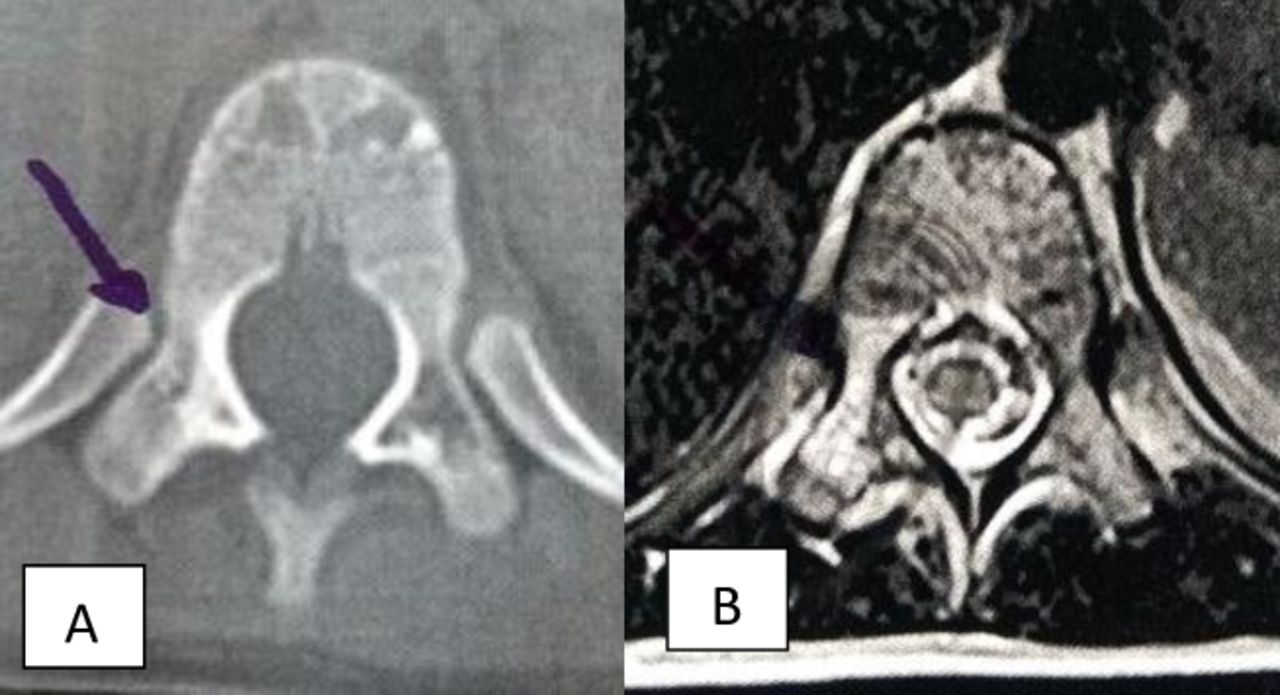
- Figure 1
(A) Axial computed tomography of the spine. The image shows absence of the right T11 pedicle, its space being replaced by cell attenuation coefficient material. (B) Axial T2-weighted magnetic resonance image. The image shows the right T11 pedicle with local hypersignal, indicating spondylolysis without signs of listesis.
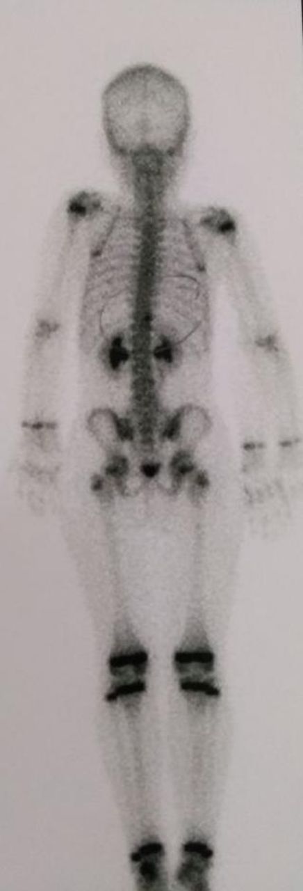
- Figure 2
Bone scintigraphy. The image shows a slight increase in osteoblastic activity on the right edge of the T11 vertebra.
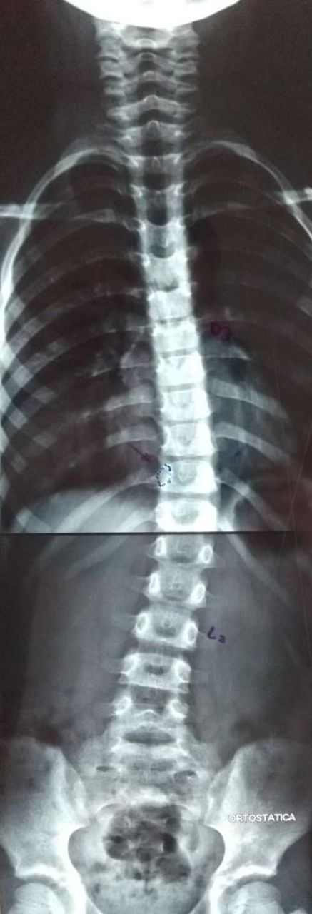
- Figure 3
Panoramic radiography. The image shows dextrochondral scoliosis with a 20° Cobb angle.
- Figure 4
Positioning the working cannula at the extraforaminal T10/T11 endoscopic approach.
- Figure 5
Intraoperative endoscopic images. (A) Initial image of the extraforaminal access, with view of the lateral border of the pedicle and the lower part of the foramen T10/T11 on the right. (B) Beginning of foraminoplasty and bone resection of the T11 upper right joint process using a foraminoplasty burr. (C) Macroscopic aspect of pathological bone tissue. (D) Lateral image of the central canal and the right T11 pedicle partially resected.
- Figure 6
The conclusion of bone resection. Keeping the inferior cortical bone of the right T11 pedicle intact, local hemostasis, and bipolar cauterization of all beds and residual trabecular tissue are reviewed.
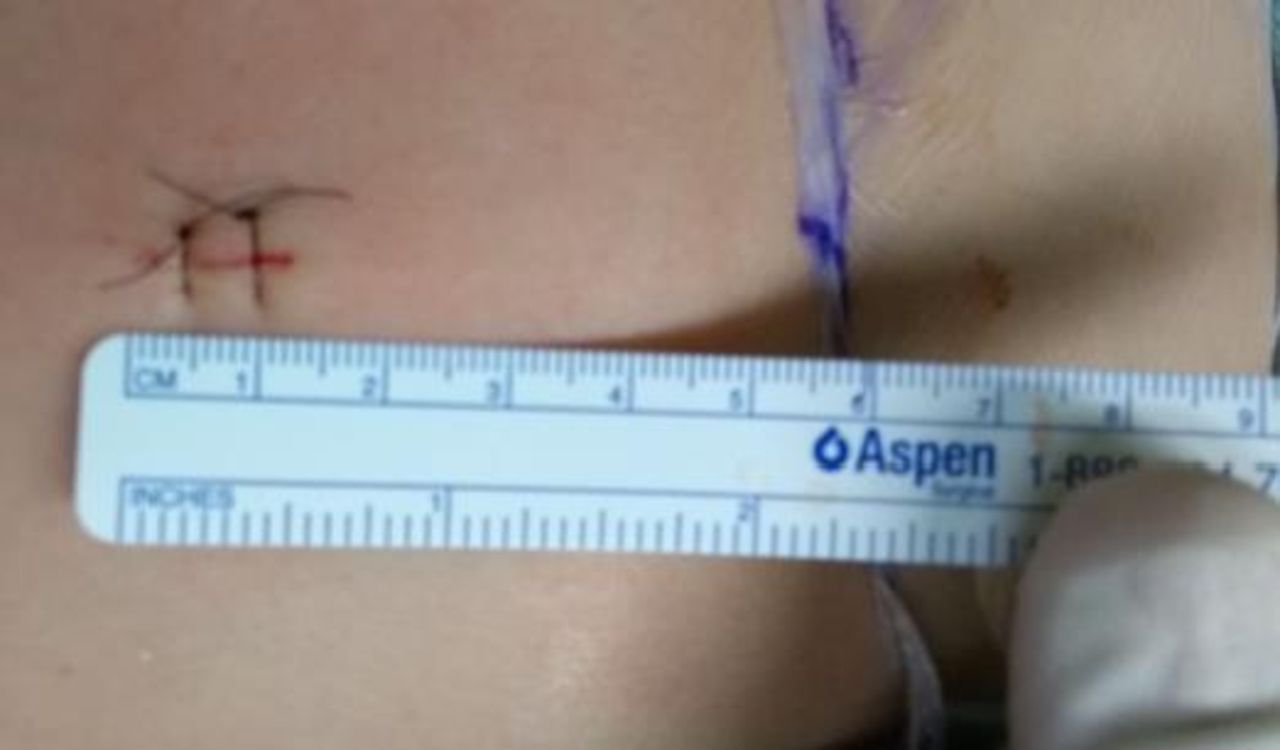
- Figure 7
An 8-mm skin incision is made ∼50 mm lateral to the midline at the height of the right T10/T11.
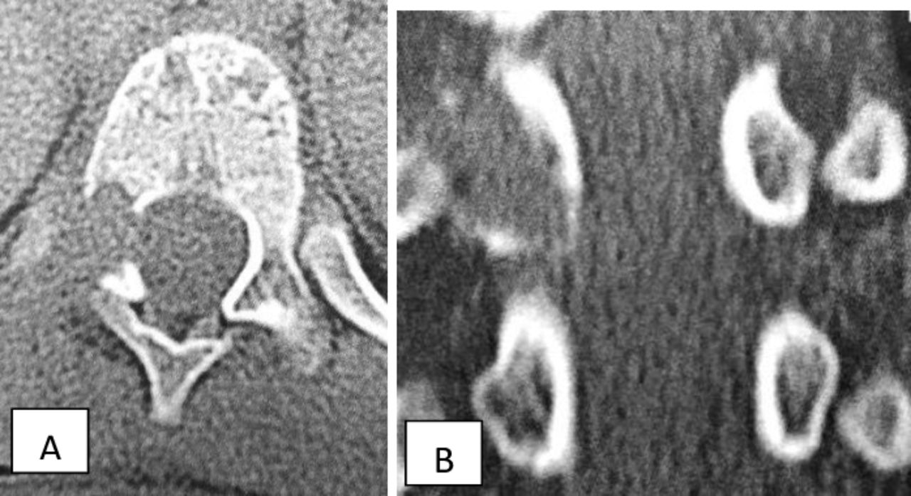
- Figure 8
Imaging at the first year. (A) Axial spine computed tomography and (B) coronal spine computed tomography, without radiological signs of lesion recurrence.


In this issue


