
Main menu

  • Home
  • Content
    • Current Issue
    • Advance Online Publication
    • Archive
  • About Us
    • About ISASS
    • About the Journal
    • Author Instructions
    • Editorial Board
    • Reviewer Guidelines & Publication Criteria
  • More
    • Advertise
    • Subscribe
    • Alerts
    • Feedback
  • Join Us
  • Reprints & Permissions
  • Sponsored Content
  • Other Publications
    • ijss

User menu

  • My alerts

Search

  • Advanced search
International Journal of Spine Surgery
  • My alerts
International Journal of Spine Surgery

Advanced Search

  • Home
  • Content
    • Current Issue
    • Advance Online Publication
    • Archive
  • About Us
    • About ISASS
    • About the Journal
    • Author Instructions
    • Editorial Board
    • Reviewer Guidelines & Publication Criteria
  • More
    • Advertise
    • Subscribe
    • Alerts
    • Feedback
  • Join Us
  • Reprints & Permissions
  • Sponsored Content
  • Follow ijss on Twitter
  • Visit ijss on Facebook
Research ArticleLumbar Spine

Restoring Segmental Spinal Alignment in Mini-Open Lateral Spinal Deformity Surgery—Determiners of Radiographic Outcome

Alexander Spiessberger, Nicholas Dietz, Basil E. Gruter, Justin Virojanapa and Peter Hollis
International Journal of Spine Surgery June 2022, 16 (3) 540-547; DOI: https://doi.org/10.14444/8246
Alexander Spiessberger
1 Department of Neurosurgery, North Shore University Hospital – Hofstra School of Medicine, Manhasset, NY, USA
MD
  • Find this author on Google Scholar
  • Find this author on PubMed
  • Search for this author on this site
Nicholas Dietz
2 Department of Neurosurgery, University of Kentucky – Louisville, Lexington, KY, USA
MD
  • Find this author on Google Scholar
  • Find this author on PubMed
  • Search for this author on this site
Basil E. Gruter
3 Department of Neurosurgery, University Zurich, Zurich, Switzerland
MD
  • Find this author on Google Scholar
  • Find this author on PubMed
  • Search for this author on this site
Justin Virojanapa
1 Department of Neurosurgery, North Shore University Hospital – Hofstra School of Medicine, Manhasset, NY, USA
DO
  • Find this author on Google Scholar
  • Find this author on PubMed
  • Search for this author on this site
Peter Hollis
1 Department of Neurosurgery, North Shore University Hospital – Hofstra School of Medicine, Manhasset, NY, USA
MD
  • Find this author on Google Scholar
  • Find this author on PubMed
  • Search for this author on this site
  • Article
  • Figures & Data
  • Info & Metrics
  • PDF
Loading

Article Figures & Data

Figures

  • Tables
  • Figure 1
    • Download figure
    • Open in new tab
    • Download powerpoint
    Figure 1

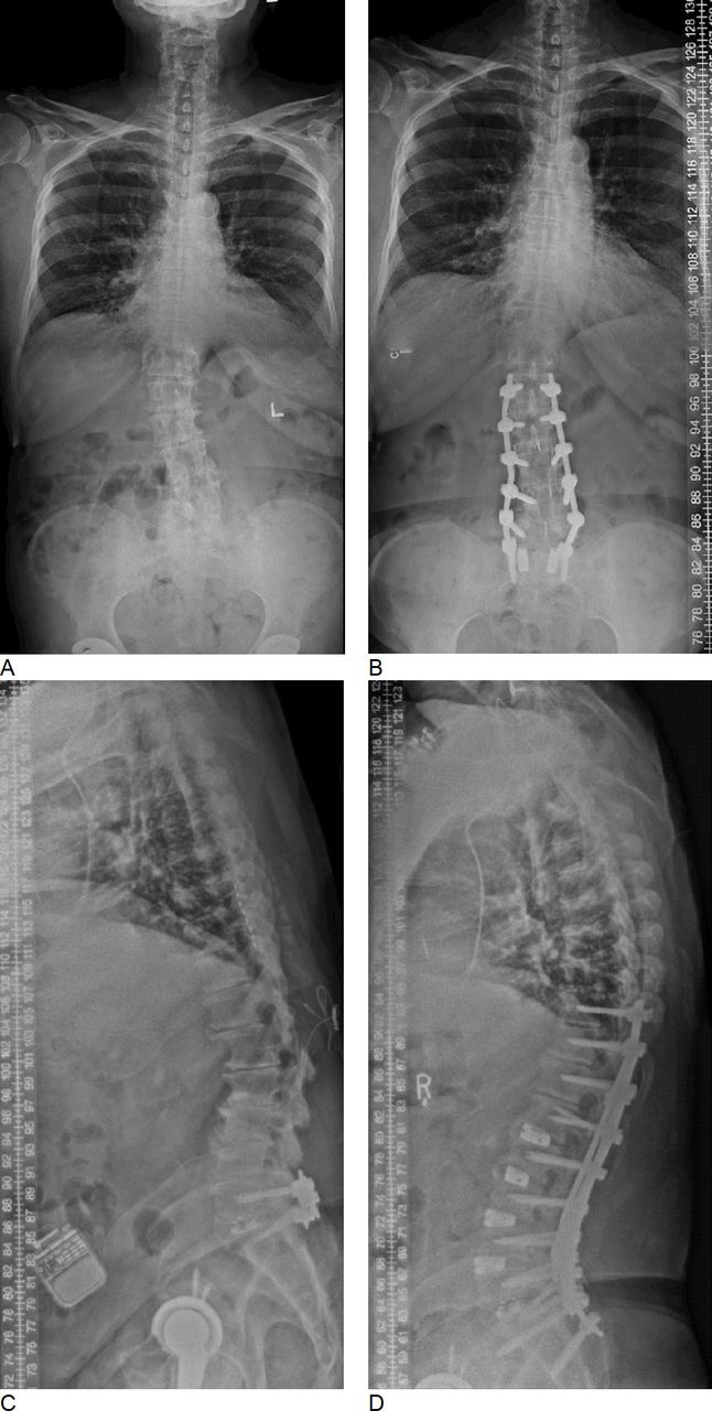
    (A and B) Pre- and postoperative whole spine x-ray images. The correction of coronal deformity can be appreciated. (C and D) Pre- and postoperative whole spine x-ray images in a patient with marked correction of sagittal alignment.

  • Figure 2
    • Download figure
    • Open in new tab
    • Download powerpoint
    Figure 2

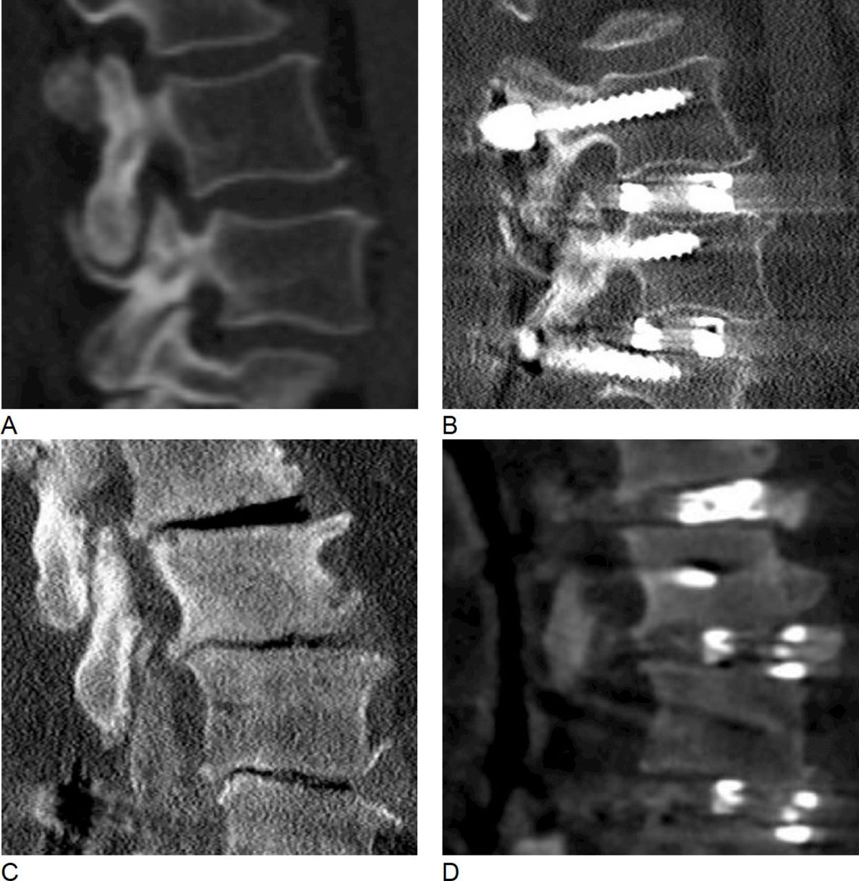
    Restoration of segmental sagittal profile. (A and B) Pre- and postoperative sagittal computed tomography (CT) images of the L3/4 disc space. Preoperatively, there was little loss of disc space height. The perioperative change of segmental sagittal Cobb angle was +3°. (C and D) Pre- and postoperative sagittal CT images of the L3/4 disc space in a patient with marked loss of disc space height. In this case, the perioperative gain in segmental lordosis was 5°.

  • Figure 3
    • Download figure
    • Open in new tab
    • Download powerpoint
    Figure 3

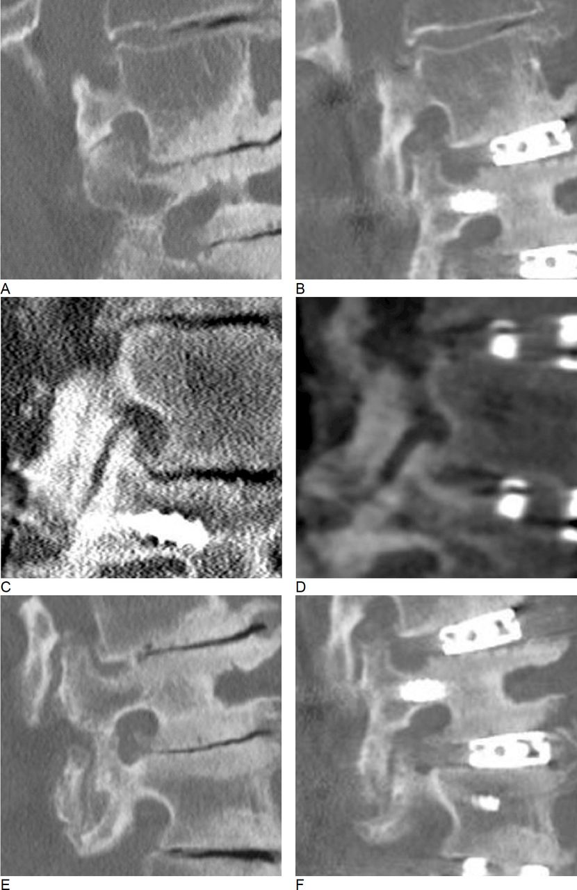
    Foraminal decompression. (A and B) Pre- and postoperative sagittal views of the L2/3 neuroforamen. The superior articulating process causes significant foraminal stenosis preoperatively. Postoperatively, the SAP is displaced caudally, which leads to foraminal decompression. (C and D) Similarly, Pre- and postoperative views of a left-sided L4/5 foramen. Again, postoperative displacement of the SAP decompresses the neuroforamen. (E and F) Failure of indirect decompression. Even though the craniocaudal dimension of the neuroforamen significantly increases postoperatively, an osteophyte causes persistent nerve root compression.

Tables

  • Figures
    • View popup
    Table 1

    Patient demographics and overview of surgical details (N = 49).

    DemographicData
    Age, y, mean (SD) (range)68.7 (8.6) (39.9–84.7)
    Sex, female/male63%/37%
    Fusion construct
     Distribution of upper instrumented vertebra
      T42%
      T106%
      T112%
      T122%
      L117%
      L260%
      L310%
     Distribution of lower instrumented vertebra
      L5a 60%
      S135%
      Pelvis15%
    No. of levels fused, mean (SD) (range)4.3 (2.3) (3–15)
    Operative complications
     Seromab 4%
     Wound dehiscence4%
     Transient dysesthesia18%
     Transient hip flexor weakness6%
    • ↵a 4% of L4/5 underwent transforaminal lumbar interbody fusion.

    • ↵b Occurring after open posterior stage of the surgery.

    • View popup
    Table 2

    Radiographic measurements on whole spine standing x-rays before and after surgery.

    MeasurementMean (SD) (Range) P value
    C7SVA, mm0.1
     Preoperative79.6 (63) (−43−254)
     Postoperative60 (51.8) (−26–217)
     ∆C7SVA−33.6 (57.9) (−166–60)
    TK, ° 0.01
     Preoperative36.1 (15.4) (5–77)
     Postoperative43.8 (15) (21−91)
     ∆4.2 (15.3) (−35–39)
    LL, ° 0.003
     Preoperative32.9 (15.9) (−14–78)
     Postoperative41.6 (12.4) (24–84)
     ∆9.3 (10.5) (−4–46)
    T10-L2 sag Cobb, °0.8
     Preoperative3 (12.7) (−24–30)
     Postoperative2.3 (11.2) (−30–32)
     ∆−1 (9.4) (-21–32)
    PI, °49 (10.7) (21–77)
    PT, °0.7
     Preoperative21.1 (9.6) (−4–54)
     Postoperative21.8 (7.3) (2–34)
     ∆0.1 (10) (−28−26)
    Degenerative scoliosisLeft convex 63%
    Scoliotic apex level
     L11%
     L225%
     L2/34%
     L3/46%
     L46%
    Coronal Cobb, ° 0.02
     Preoperative16.3 (12) (0−48)
     Postoperative10.8 (10.3) (0–45)
     ∆−4.8 (8.4) (−24–30)
    C7PL, mm0.6
     Preoperative14.1 (15.1) (0–54)
     Postoperative12.9 (9.1) (0–28)
     ∆−3.7 (15.3) (−45–26)
    • C7PL, C7 plumb line (coronal); C7SVA, C7 sagittal vertical axis; LL, lumbar lordosis; PI, pelvic incidence; PT, pelvic tilt; TK, thoracic kyphosis.

    • View popup
    Table 3

    Radiographic measurements before and after surgery for each anatomic level.

    Mean (SD) (Range)
    MeasurementL1/2L2/3L3/4L4/5
    Sagittal Cobb, °
     Preoperative−1.8 (5.6) (−11–13)2.5 (4) (−6-16)5.6 (4.9) (−5–21)9.5 (5.7) (1–21)
     Postoperative2.2 (4.9) (−5–17)5.2 (3.8) (0–18)7.3 (4.4) (0–14)11 (4.8) (3–24)
     ∆4 (3.2) (0–11)2.7 (3.4) (−5–10)1.8 (4.2) (−10–14)1.6 (4.6) (−7–17)
    Coronal Cobb, °
     Preoperative5.5 (5.1) (0–16)7.4 (5.8) (0–26)5.8 (5.6) (0–23)4.2 (3.8) (0–15)
     Postoperative4.2 (3.5) (0–11)3.4 (3.8) (0–17)3.3 (4) (0−20)2.7 (2.2) (0–9)
     ∆−1.3 (6) (−14−11)−4 (4.6) (−14–6)−2.4 (5.5) (−22–18)4.4 (2.5) (−1–11)
    Foraminal height, mm
     Preoperative15.2 (3.8) (11–23)16.5 (4) (8−26)15.7 (3.8) (8–24)15.5 (3.6) (6–24)
     Postoperative18 (5.1) (11–30)19.6 (4.4) (9–37)19.3 (4) (8-32)18.6 (3.5) (9–25)
     ∆2.8 (2.6) (−1–7)3.1 (3.2) (−2–16)3.6 (3.5) (−3–14)2.7 (4.7) (−16–13)
    Foraminal width, mm
     Preoperative7.1 (2) (3-10)8 (2) (5−13)8.3 (1.6) (5–12)8.5 (1.9) (5–14)
     Postoperative9.5 (3) (6−15)9.4 (2.1) (5–14)9.8 (1.9) (7–14)9.8 (1.9) (6–13)
     ∆2.4 (2.7) (−1–7)1.4 (2.2) (−2–8)1.4 (2.4) (−5–7)1 (2.5) (−6–7)
    Disc space height, mm
     Preoperative4 (3) (1−11)5 (2.6) (1–11)5.8 (2.9) (1–12)6.5 (3) (2−13)
     Postoperative9.2 (2.2) (7–16)9.8 (2) (7−15)10.6 (1.5) (8–14)10.9 (1.7) (8–16)
     ∆5.2 (2.3) (−1–9)4.8 (2.5) (−1–10)4.7 (2.9) (0–11)4.4 (2.5) (−1–11)
    • View popup
    Table 4

    Correlation between baseline radiographic measurements and perioperative change in segmental sagittal Cobb angle.

    MeasurementUnstandardized CoefficientsStandardized Coefficient P Value
    B Standard ErrorBeta t
    Preoperative segmental sagittal Cobb angle, °−0.40.1−0.5−5.70.01
    Preoperative disc space height, mm−0.20.1−0.2−1.70.09
    Preoperative osteophytes gradea −0.30.6−0.05−0.50.59
    Preoperative facet joint degeneration gradeb −0.10.3−0.02−0.20.81
    Anatomic level0.80.40.21.90.06
    Preoperative coronal Cobb angle, °0.10.10.22.3 0.02
    • ↵a Three grades: I, nonbridging disc space osteophytes; II, small bridging disc space osteophytes; III, ankylosed disc space.

    • ↵b Three grades: I, nonbridging osteophytes of the facet joint; II, small bridging osteophytes of the facet joint; III, ankylosed facet joint.

    • View popup
    Table 5

    Correlation between baseline radiographic measurements and perioperative change in neuroforaminal height.

     Unstandardized CoefficientsStandardized CoefficientSignificance
    B Standard ErrorBeta t
    Preoperative disc space height, mm−0.1.01−0.04−0.40.71
    Preoperative osteophytes gradea 0.70.60.11.30.18
    Preoperative foraminal height, mm−0.30.1−0.3−2.7 0.01
    Preoperative foraminal width, mm0.20.20.10.90.36
    Preoperative facet joint degeneration gradeb −0.10.3−0.02−0.20.81
    Preoperative coronal Cobb angle, °0.020.10.030.30.74
    Implant positionc −0.30.6-0.04−0.50.65
    Anatomic level0.20.40.040.40.68
    • ↵a Three grades: I, nonbridging disc space osteophytes; II, small bridging disc space osteophytes; III, ankylosed disc space.

    • ↵b Three grades: I, nonbridging osteophytes of the facet joint; II, small bridging osteophytes of the facet joint; III, ankylosed facet joint.

    • ↵c Location of the center of the interbody device in relation to 4 quadrants, which the disc space has been divided into on sagittal imaging (I anterior to IV posterior).

    • View popup
    Table 6

    Correlation between baseline radiographic measurements and perioperative change in neuroforaminal width.

     Unstandardized CoefficientsStandardized CoefficientSignificance
    B Standard ErrorBeta t
    Preoperative disc space height, mm−0.020.1−0.02−0.20.82
    Preoperative osteophytes gradea −0.60.3−0.2−1.80.08
    Preoperative foraminal width, mm−0.70.1−0.5−6.8 0.01
    Preoperative facet joint degenerationb 0.40.20.21.80.08
    Preoperative coronal Cobb angle, °−0.010.03−0.03−0.40.73
    Implant positionc −0.30.4−0.1−0.90.37
    Anatomic level−0.30.2−0.1−1.10.26
    • a Three grades: I, nonbridging disc space osteophytes; II, small bridging disc space osteophytes; III, ankylosed disc space.

    • ↵b Three grades: I, nonbridging osteophytes of the facet joint; II, small bridging osteophytes of the facet joint; III, ankylosed facet joint.

    • ↵c Location of the center of the interbody device in relation to 4 quadrants, which the disc space has been divided into on sagittal imaging (I anterior to IV posterior).

PreviousNext
Back to top

In this issue

International Journal of Spine Surgery
Vol. 16, Issue 3
1 Jun 2022
  • Table of Contents
  • Index by author

Print
Download PDF
Article Alerts
Sign In to Email Alerts with your Email Address
Email Article

Thank you for your interest in spreading the word on International Journal of Spine Surgery.

NOTE: We only request your email address so that the person you are recommending the page to knows that you wanted them to see it, and that it is not junk mail. We do not capture any email address.

Enter multiple addresses on separate lines or separate them with commas.
Restoring Segmental Spinal Alignment in Mini-Open Lateral Spinal Deformity Surgery—Determiners of Radiographic Outcome
(Your Name) has sent you a message from International Journal of Spine Surgery
(Your Name) thought you would like to see the International Journal of Spine Surgery web site.
CAPTCHA
This question is for testing whether or not you are a human visitor and to prevent automated spam submissions.
Citation Tools
Restoring Segmental Spinal Alignment in Mini-Open Lateral Spinal Deformity Surgery—Determiners of Radiographic Outcome
Alexander Spiessberger, Nicholas Dietz, Basil E. Gruter, Justin Virojanapa, Peter Hollis
International Journal of Spine Surgery Jun 2022, 16 (3) 540-547; DOI: 10.14444/8246

Citation Manager Formats

  • BibTeX
  • Bookends
  • EasyBib
  • EndNote (tagged)
  • EndNote 8 (xml)
  • Medlars
  • Mendeley
  • Papers
  • RefWorks Tagged
  • Ref Manager
  • RIS
  • Zotero
Share
Restoring Segmental Spinal Alignment in Mini-Open Lateral Spinal Deformity Surgery—Determiners of Radiographic Outcome
Alexander Spiessberger, Nicholas Dietz, Basil E. Gruter, Justin Virojanapa, Peter Hollis
International Journal of Spine Surgery Jun 2022, 16 (3) 540-547; DOI: 10.14444/8246
Twitter logo Facebook logo Mendeley logo
  • Tweet Widget
  • Facebook Like
  • Google Plus One

Jump to section

  • Article
    • Abstract
    • Introduction
    • Methods
    • Results
    • Discussion
    • Conclusion
    • Footnotes
    • References
  • Figures & Data
  • Info & Metrics
  • PDF

Related Articles

  • No related articles found.
  • PubMed
  • Google Scholar

Cited By...

  • No citing articles found.
  • Google Scholar

More in this TOC Section

  • Sexual Dysfunction and Urinary Incontinence in Female Patients Following Primary Anterior Lumbar Interbody Fusion: A Survey of 84 Patients
  • Comparison of Stand-Alone Anterior Lumbar Interbody Fusion, 360° Anterior Lumbar Interbody Fusion, and Arthroplasty for Recurrent Lumbar Disc Herniation: Focus on Nerve Decompression and Painful Spinal Instability Resolution
  • Safety and Viability of Anterior Lumbar Interbody Fusion in Complex Revision Lumbar Spine Surgeries: Insights From a Case Series of 135 Patients on Transforaminal Lumbar Interbody Fusion/Posterior Lumbar Interbody Fusion Cage Removal
Show more Lumbar Spine

Similar Articles

Keywords

  • mini-open lateral spine surgery
  • deformity surgery spine

Content

  • Current Issue
  • Latest Content
  • Archive

More Information

  • About IJSS
  • About ISASS
  • Privacy Policy

More

  • Subscribe
  • Alerts
  • Feedback

Other Services

  • Author Instructions
  • Join ISASS
  • Reprints & Permissions

© 2025 International Journal of Spine Surgery

International Journal of Spine Surgery Online ISSN: 2211-4599

Powered by HighWire