
Main menu

  • Home
  • Content
    • Current Issue
    • Advance Online Publication
    • Archive
  • About Us
    • About ISASS
    • About the Journal
    • Author Instructions
    • Editorial Board
    • Reviewer Guidelines & Publication Criteria
  • More
    • Advertise
    • Subscribe
    • Alerts
    • Feedback
  • Join Us
  • Reprints & Permissions
  • Sponsored Content
  • Other Publications
    • ijss

User menu

  • My alerts

Search

  • Advanced search
International Journal of Spine Surgery
  • My alerts
International Journal of Spine Surgery

Advanced Search

  • Home
  • Content
    • Current Issue
    • Advance Online Publication
    • Archive
  • About Us
    • About ISASS
    • About the Journal
    • Author Instructions
    • Editorial Board
    • Reviewer Guidelines & Publication Criteria
  • More
    • Advertise
    • Subscribe
    • Alerts
    • Feedback
  • Join Us
  • Reprints & Permissions
  • Sponsored Content
  • Follow ijss on Twitter
  • Visit ijss on Facebook
Research ArticleMinimally Invasive Surgery

Significant Reduction of Radiation Exposure Using Specific Settings of the O-Arm for Percutaneous Cementoplasty in Accordance With the ALARA Principle

Marc Prod’homme, Didier Grasset, Alain Akiki, Lionel Helfer, Gilles Dietrich, Jonas Pierre Müller, Mattia Tomasoni, Mahmoud Sellami and Duccio Boscherini
International Journal of Spine Surgery August 2023, 17 (4) 534-541; DOI: https://doi.org/10.14444/8477
Marc Prod’homme
1 Clinic La Source, Neuro Orthopedic Center, Lausanne, Switzerland
2 Orthopedic Surgery Department, Riviera-Chablais Hospital, Rennaz, Switzerland
MD
  • Find this author on Google Scholar
  • Find this author on PubMed
  • Search for this author on this site
  • For correspondence: marcprod86@gmail.com
Didier Grasset
1 Clinic La Source, Neuro Orthopedic Center, Lausanne, Switzerland
MD
  • Find this author on Google Scholar
  • Find this author on PubMed
  • Search for this author on this site
Alain Akiki
2 Orthopedic Surgery Department, Riviera-Chablais Hospital, Rennaz, Switzerland
MD
  • Find this author on Google Scholar
  • Find this author on PubMed
  • Search for this author on this site
Lionel Helfer
2 Orthopedic Surgery Department, Riviera-Chablais Hospital, Rennaz, Switzerland
MD
  • Find this author on Google Scholar
  • Find this author on PubMed
  • Search for this author on this site
Gilles Dietrich
2 Orthopedic Surgery Department, Riviera-Chablais Hospital, Rennaz, Switzerland
3 Orthopedic Surgery Department, Lausanne University Hospital, Lausanne, Switzerland
MD
  • Find this author on Google Scholar
  • Find this author on PubMed
  • Search for this author on this site
Jonas Pierre Müller
2 Orthopedic Surgery Department, Riviera-Chablais Hospital, Rennaz, Switzerland
MD
  • Find this author on Google Scholar
  • Find this author on PubMed
  • Search for this author on this site
Mattia Tomasoni
4 Ophtalmology Department, Jules-Gonin University Eye Hospital, University of Lausanne, Lausanne, Switzerland
PHD
  • Find this author on Google Scholar
  • Find this author on PubMed
  • Search for this author on this site
Mahmoud Sellami
4 Ophtalmology Department, Jules-Gonin University Eye Hospital, University of Lausanne, Lausanne, Switzerland
PHD
  • Find this author on Google Scholar
  • Find this author on PubMed
  • Search for this author on this site
Duccio Boscherini
1 Clinic La Source, Neuro Orthopedic Center, Lausanne, Switzerland
2 Orthopedic Surgery Department, Riviera-Chablais Hospital, Rennaz, Switzerland
MD, PHD
  • Find this author on Google Scholar
  • Find this author on PubMed
  • Search for this author on this site
  • Article
  • Figures & Data
  • Info & Metrics
  • PDF
Loading

Article Figures & Data

Figures

  • Tables
  • Figure 1
    • Download figure
    • Open in new tab
    • Download powerpoint
    Figure 1

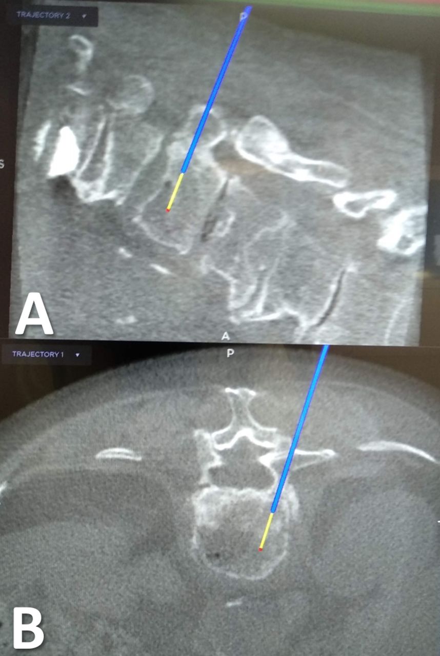
    Intraoperative views of the 3D navigation on O-arm with the trajectory of the navigated Jamshidi needle. (A) Sagittal plane. (B) Axial plane.

  • Figure 2
    • Download figure
    • Open in new tab
    • Download powerpoint
    Figure 2

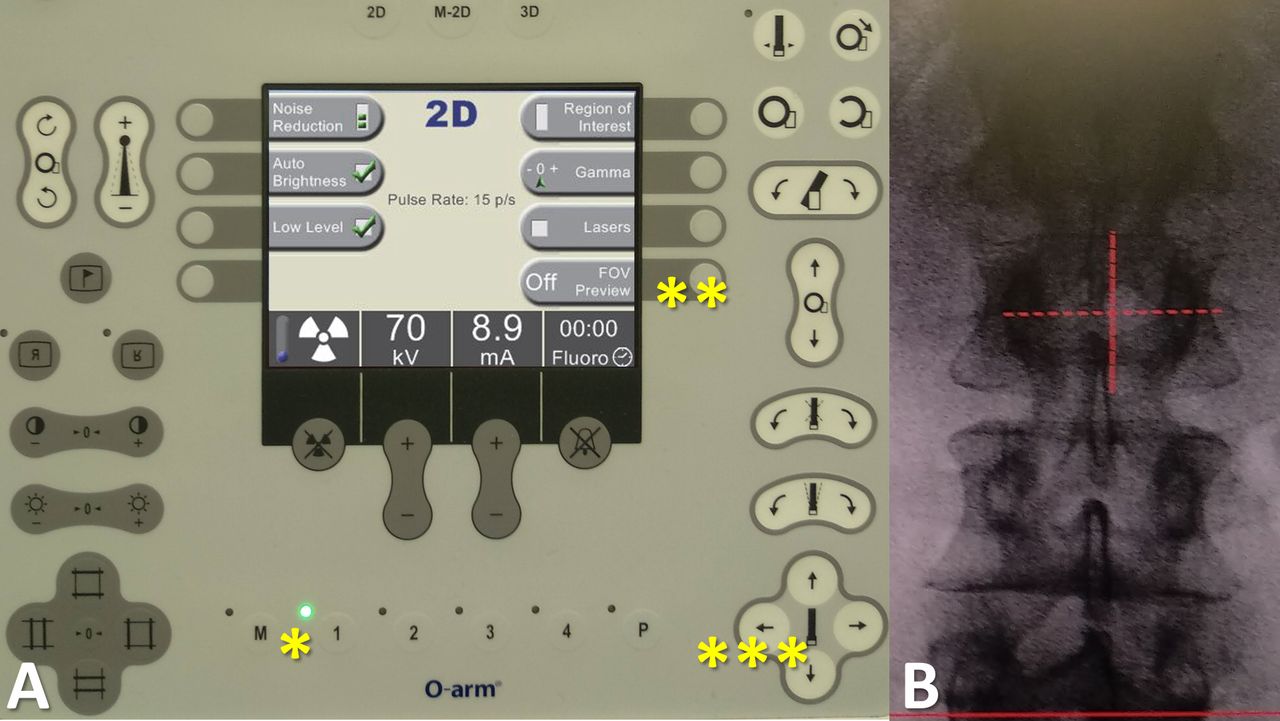
    View of the control panel for settings of imaging acquisition. (A) First, an anteroposterior view can be taken. Then, the image and the O-arm position (*) are recorded. Second, the radiological technician can activate the navigation on the images by pressing the "FOV (field of view) Preview" button (**). Third, the radiological technician can navigate on the screen, using the position arrows of the control panel (***), especially in order to center the view on the fractured vertebra. (B) Finally, the lateral view is obtained and the height of the region of interest can be reached without another acquisition. Thus, a 3-dimensional navigated scan can be performed.

  • Figure 3
    • Download figure
    • Open in new tab
    • Download powerpoint
    Figure 3

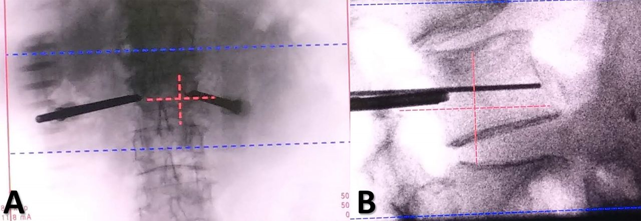
    Intraoperative views of a cementoplasty procedure using the field of view (FOV) preview. (A) Anteroposterior view after placement of navigated Jamshidi needles in the fractured vertebra. (B) Lateral view. The region of interest may be reached with the FOV principle, allowing the fractured vertebra to be at the center of the image. Collimation can then be applied and also a zoom on the screen, to follow the cementoplasty procedure, and to decrease radiation exposure.

Tables

  • Figures
    • View popup
    Table 1

    O-arm settings for 2D and 3D procedures.

    O-arm SettingGroup AGroup B
    2D Mode (pulses/s)Fluorography (30)Low dose (15)
    Navigation on the FOVNoYes
    CollimationNoYes
    3D ModeStandardStandard or low dose
    • Abbreviations: 2D, 2-dimensional; 3D, 3-dimensional; FOV, field of view.

    • View popup
    Table 2

    Patient demographics and surgery data.

    Patient DataGroup A (n = 43)Group B (n = 27) P Value
    Sex ratio, female/male25/1819/80.32a
    Age, y, mean ± SD (range)76 ± 11 (43–89)67 ± 16 (34–96)0.02b
    Body mass index, kg/m2, mean ± SD (range)25 ± 3 (19–30)24 ± 5 (17–33)0.46b
    Magerl classification, n (%)0.61a
     A150 (85)45 (90)
     A22 (3)2 (4)
     A37 (12)3 (6)
    Location of the fractures0.08a
     Thoracic spine, n (%)32 (54)18 (36)
     Lumbar spine, n (%)27 (46)32 (64)
    Operative time, min, mean ± SD (range)34.6 ± 14.3 (15–90)41.9 ± 19.0 (12–90)0.13b
    Operative time per level, min, mean ± SD (range)27.3 ± 10.9 (15–60)28.9 ± 13.4 (4.4–60)0.74b
    Cemented vertebrae, n 5950
     Vertebroplasties, n (%)39 (66)30 (60)0.55a
     Kyphoplasties, n (%)20 (34)20 (40)
    Unipedicular approaches, n (%)25 (42)26 (52)0.04a
    Surgeon experience, senior/junior16/276/210.29a
    Cement volume injected, cc, mean ± SD (range)6.8 ± 3.8 (2–18)8.2 ± 3.5 (4–18)0.13b
    Cement volume injected per level, cc, mean ± SD (range)4.9 ± 1.5 (2–7)5.3 ± 1.5 (2–8)0.30b
    • ↵a Fisher exact test.

    • ↵b Student t test.

    • View popup
    Table 3

    Garnier classification of vertebral filling and cement leakage.

    Garnier ClassificationGroup A (n = 43)Group B (n = 27) P Valuea
    Cement filling n (%)0.11
     Satisfactory (Y3, Z2, Z3)56 (95)42 (84)-
     Acceptable (X2, X3, Y2)2 (3)1 (2)-
     Poor (X1, Y1, Z1)1 (2)7 (14)-
    Cement leakage, n 0.62
     Spinal canal21-
     Intravascular21-
     Intradiscal36-
     Paravertebral64-
      Total, n (%)13 (22)12 (24)-
    • ↵a Fisher exact test.

    • View popup
    Table 4

    Radiation exposure data comparison in the 2 groups.

    Radiation Exposure, mean ± SD (range)Group A
    (n = 43)
    Group B(n = 27) P Valuea
    2D Imaging duration, s23.67 ± 15.32 (9.70–84.87)17.82 ± 10.01 (2.82–46.07)0.06
    2D Imaging duration/level, s18.56 ± 10.40 (4.41–62.61)12.93 ± 8.49 (1.95–42.30)0.02
    Number of 3D acquisitions/patient1.5 ± 1.0 (1–5)1.4 ± 0.9 (1–4)0.72
    Dose length product, mGy·cm534 ± 385 (175–1975)271 ± 165 (88–769)<0.01
    Dose area product, mGy·cm2 9173 ± 10,430 (1298–63,331)1614 ± 1656 (332–9070)<0.01
    Overall E, mSv9.94 ± 6.67 (2.76–31.48)4.34 ± 2.52 (1.41–11.33)<0.01
    E per level, mSv7.65 ± 4.82 (1.39–19.69)2.97 ± 1.42 (0.49–5.67)<0.01
    E2D, mSv2.12 ± 2.66 (0.25–16.47)0.37 ± 0.33 (0.06–1.72)<0.01
    E3D, mSv7.82 ± 5.67 (2.46–29.63)3.97 ± 2.34 (1.23–10.77)<0.01
    E per 3D acquisition, mSv5.60 ± 3.14 (2.46–12.56)2.90 ± 0.80 (1.23–4.12)<0.01
    Absolute risk of radiation-induced cancer equivalent to a whole-body exposure, %5.0 × 10⁻⁴ ± 3.3 × 10⁻⁴ (1.4 × 10⁻⁴–1.6 × 10⁻³)2.2 × 10⁻⁴ ± 1.3 × 10⁻⁴ (7.1 × 10⁻⁵–5.7 × 10⁻⁴)<0.01
    • Abbreviations: 2D, 2-dimensional; 3D, 3-dimensional; E, effective dose.

    • ↵a Student t test.

    • View popup
    Table 5

    Correlation study: E, E2D, and surgeon level with other outcomes.

    P Value
    CorrelationGroup A (n = 43)Group B (n = 27)Overall (n = 70)
    E and agea <0.010.130.31
    E and body mass indexa 0.460.240.20
    E and operative timea 0.280.010.43
    E and imaging durationa <0.010.06<0.01
    E2D and imaging durationa <0.01<0.01<0.01
    E and cement amounta 0.130.400.41
    E2D and cement amounta 0.230.450.55
    E and surgeon levelb 0.750.720.70
    E2D and surgeon levelb 0.710.140.45
    Surgeon level and operative timeb 0.740.610.41
    • Abbreviations: 2D, 2-dimensional; E, effective dose.

    • ↵a Student t test.

    • ↵b Analysis of variance.

    • View popup
    Table 6

    Subjective image quality assessment.

    Image QualityGroup A (n = 43)Group B (n = 27) P Valuea
    3 Dimensional>0.99
     Acceptable4327-
     Insufficient00-
    2 Dimensional>0.99
     Acceptable3623-
     Insufficient74-
    • ↵a Fisher exact test.

PreviousNext
Back to top

In this issue

International Journal of Spine Surgery
Vol. 17, Issue 4
1 Aug 2023
  • Table of Contents
  • Index by author

Print
Download PDF
Article Alerts
Sign In to Email Alerts with your Email Address
Email Article

Thank you for your interest in spreading the word on International Journal of Spine Surgery.

NOTE: We only request your email address so that the person you are recommending the page to knows that you wanted them to see it, and that it is not junk mail. We do not capture any email address.

Enter multiple addresses on separate lines or separate them with commas.
Significant Reduction of Radiation Exposure Using Specific Settings of the O-Arm for Percutaneous Cementoplasty in Accordance With the ALARA Principle
(Your Name) has sent you a message from International Journal of Spine Surgery
(Your Name) thought you would like to see the International Journal of Spine Surgery web site.
CAPTCHA
This question is for testing whether or not you are a human visitor and to prevent automated spam submissions.
Citation Tools
Significant Reduction of Radiation Exposure Using Specific Settings of the O-Arm for Percutaneous Cementoplasty in Accordance With the ALARA Principle
Marc Prod’homme, Didier Grasset, Alain Akiki, Lionel Helfer, Gilles Dietrich, Jonas Pierre Müller, Mattia Tomasoni, Mahmoud Sellami, Duccio Boscherini
International Journal of Spine Surgery Aug 2023, 17 (4) 534-541; DOI: 10.14444/8477

Citation Manager Formats

  • BibTeX
  • Bookends
  • EasyBib
  • EndNote (tagged)
  • EndNote 8 (xml)
  • Medlars
  • Mendeley
  • Papers
  • RefWorks Tagged
  • Ref Manager
  • RIS
  • Zotero
Share
Significant Reduction of Radiation Exposure Using Specific Settings of the O-Arm for Percutaneous Cementoplasty in Accordance With the ALARA Principle
Marc Prod’homme, Didier Grasset, Alain Akiki, Lionel Helfer, Gilles Dietrich, Jonas Pierre Müller, Mattia Tomasoni, Mahmoud Sellami, Duccio Boscherini
International Journal of Spine Surgery Aug 2023, 17 (4) 534-541; DOI: 10.14444/8477
Twitter logo Facebook logo Mendeley logo
  • Tweet Widget
  • Facebook Like
  • Google Plus One

Jump to section

  • Article
    • Abstract
    • Introduction
    • Methods
    • Results
    • Discussion
    • Conclusion
    • Footnotes
    • References
  • Figures & Data
  • Info & Metrics
  • PDF

Related Articles

  • No related articles found.
  • PubMed
  • Google Scholar

Cited By...

  • No citing articles found.
  • Google Scholar

More in this TOC Section

  • Comparative Review of Lateral and Oblique Lumbar Interbody Fusion: Technique, Outcomes, and Complications
  • Key Considerations in Surgical Decision-Making on the Side of Approach for Lumbar Lateral Transpsoas Interbody Fusion Techniques
  • A Spine Surgeon’s Learning Curve With the Minimally Invasive L5 to S1 Lateral ALIF Surgical Approach: Perioperative Outcomes and Technical Considerations
Show more Minimally Invasive Surgery

Similar Articles

Keywords

  • O-arm
  • vertebroplasty
  • kyphoplasty
  • radiation exposure
  • ALARA principle

Content

  • Current Issue
  • Latest Content
  • Archive

More Information

  • About IJSS
  • About ISASS
  • Privacy Policy

More

  • Subscribe
  • Alerts
  • Feedback

Other Services

  • Author Instructions
  • Join ISASS
  • Reprints & Permissions

© 2025 International Journal of Spine Surgery

International Journal of Spine Surgery Online ISSN: 2211-4599

Powered by HighWire