


Article Figures & Data
Figures
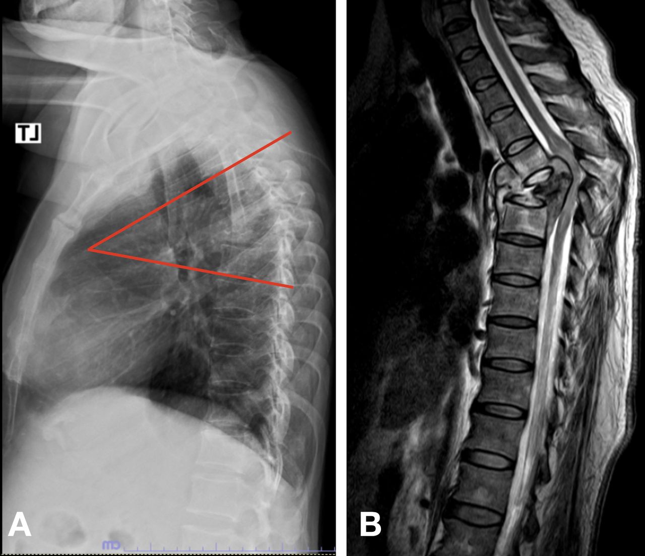
- Figure 1
(A) Measurement of kyphosis deformity using the Cobb method (ie, evaluation of the angle between the upper border of the upper normal vertebra and the lower border of the lower normal vertebra). (B) Hyperintense signal cord change (together with anterior and posterior spine destruction) at the area of the kyphotic deformity on the T2-weighted image.
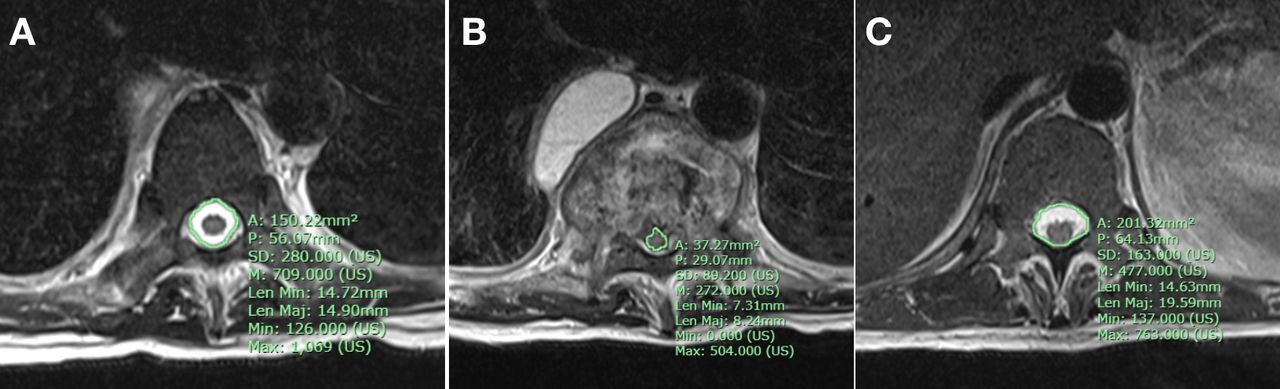
- Figure 2
Surgimap magnetic resonance imaging software was used to measure canal encroachment in an axial section. (A) The cross-sectional area of the canal level was above the site of maximum compression. (B) The area occupied by the spinal cord at maximum compression. (C) The cross-sectional area of the canal level was below the site of maximum compression. The average canal area was calculated by obtaining the average canal area of the vertebrae proximal and distal to the diseased segment. The percentage of canal encroachment area (CEA) reflected the maximum compression of the spinal cord area/average canal area ×100.
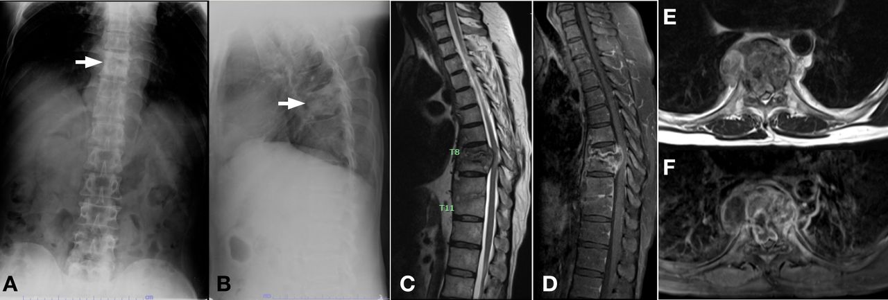
- Figure 3
A 46-year-old male patient diagnosed with spinal tuberculosis at the T8–T9 segment and a neurological deficit (Frankel grade B). Radiographs in the anteroposterior view (A) and lateral view (B) show blurred endplates of T8–T9 (arrow) with kyphosis of 20° measured using the Cobb method. Sagittal T2-weighted (T2W; C), sagittal T1-weighted + gadolinium (T1W+Gd; D), axial T2W (E), and axial T1W+Gd (F) magnetic resonance images show paradiscal involvement of the T8–T9 vertebral bodies with endplate destruction and changed marrow signal intensity, prevertebral and epidural collection, which caused loss of anterior and posterior CSF around the cord and cord compression.


Tables
- Table 1
Demographic data of the spinal thoracic tuberculosis patients at the initial visit.
Characteristic Neurodeficit Group
(n = 71)Control Group
(n = 44)P Value Age, y, mean ± SD 56.69 ± 16.95 58.00 ± 14.80 0.674 Sex 0.339 Male 38 (53.5%) 19 (43.2%) Female 33 (46.5%) 25 (56.8%) BMI, mean ± SD 21.53 ± 4.89 20.39 ± 3.42 0.182 Level of thoracic involvement Proximal thoracic (T1-T4) 9 (12.7%) 1 (2.3%) 0.037 Middle thoracic (T5-T8) 23 (32.4%) 9 (20.4%) Distal thoracic (T9-T12/L1) 38 (53.5%) 34 (77.3%) Extensive lesion 1 (1.4%) 0 (0%) Onset, d, mean ± SD 113.60 ± 179.8 100.02 ± 122.09 0.660 <30 18 (25.4%) 7 (15.9%) 0.398 30–90 32 (45.0%) 25 (56.8%) >90 21 (29.6%) 12 (27.3%) Peak ESR, mean ± SD 64.84 ± 27.83 62.37 ± 28.62 0.652 Peak CRP, mean ± SD 66.55 ± 63.73 47.88 ± 62.88 0.133 Type of involvement Paradiscal 49 (69%) 35 (79.5%) 0.050 Central 6 (8.5%) 7 (15.9%) Panvertebral 13 (18.3%) 2 (4.6%) Anterior 0 (0%) 0 (0%) Posterior 3 (4.2%) 0 (0%) Compression Abscess 53 (74.6%) 30 (68.2%) 0.281 Granulation tissue 7 (9.9%) 6 (13.6%) Disc 0 (0%) 1 (2.3%) Vertebral body bulging 9 (12.7%) 5 (11.4%) None 0 (0%) 2 (4.5%) Combined 2 (2.8%) 0 (0%) X-ray imaging Number of vertebral involvement 1.73 ± 0.64 1.68 ± 0.67 0.709 AVH loss >50% 36 (51.4%) 23 (54.8%) 0.845 <50% 35 (48.6%) 21 (45.2%) Kyphosis >30๐ 14 (20%) 7 (16.7%) 0.804 <30๐ 57 (80%) 37 (83.3%) MRI Loss of anterior CSF 69 (97.2%) 41 (93.2%) 0.369 Loss of posterior CSF 62 (87.3%) 21 (47.7%) <0.001 Cord signal change 56 (78.9%) 8 (18.2%) <0.001 Canal encroachment, %, mean [SD] 32.92 ± 15.64 56.79 ± 27.42 <0.001 Canal encroachment >50% 66 (92.9%) 21 (47.7%) <0.001 Abbreviations: AVH, anterior vertebral height; CRP, C-reactive protein; CSF, cerebrospinal fluid; ESR, erythrocyte sedimentation rate; MRI, magnetic resonance imaging.
Note: Data presented as n (%) unless otherwise noted. Bold values represent statistically significant comparisons (P < 0.05).
Possible Risk Factors OR 95% CI P Value Age 0.782 0.37–1.67 0.523 Sex Female 1.367 0.66–2.85 0.404 BMI >25 3.975 1.09–14.56 0.037 Location Proximal thoracic (T1-T4) Ref Middle thoracic (T5-T8) 0.283 0.03–2.58 0.263 Distal thoracic (T9-T12/L1) 0.124 0.01–1.03 0.053 Multilevel High ESR >80 1.636 0.72–3.71 0.238 High CRP >20 2.19 1.10–5.95 0.029 Onset <30 Ref 30–90 0.498 0.18–1.38 0.179 >90 0.681 0.22–2.10 0.221 Type of involvement Paradiscal Ref Central 0.612 0.19–1.98 0.413 Panvertebral 4.642 0.98–21.89 0.049 Anterior - - Posterior - - X-ray imaging AVH loss >50% 0.874 0.41–1.88 0.732 Kyphosis >30๐ 1.25 0.46–3.40 0.662 MRI (n [%]) Loss of anterior CSF 2.524 0.40–15.74 0.321 Loss of posterior CSF 7.545 3.02–18.85 <0.001 Cord signal change 16.800 6.47–43.65 <0.001 Canal encroachment >50% 18.07 5.61–58.22 <0.001 Abbreviations: AVH, anterior vertebral height; BMI, body mass index; CRP, C-reactive protein; CSF, cerebrospinal fluid; ESR, erythrocyte sedimentation rate; MRI, magnetic resonance imaging; Ref, reference.
Note: Bold values represent statistically significant comparisons (P < 0.05).
Possible Risk Factors Adjusted OR 95% CI P Value BMI >25 16.18 1.60–163.64 0.018 CRP >20 2.56 0.53–12.35 0.241 Onset, d <30 Ref - - 30–90 0.09 0.01–0.95 0.046 >90 0.43 0.04–4.04 0.467 Location Proximal thoracic (T1-T4) Ref - - Middle thoracic (T5-T8) 0.20 0.00–40.23 0.55 Distal thoracic (T9-T12/L1) 0.06 0.00–11.81 0.30 Panvertebral involvement 5.64 0.58–54.75 0.136 Loss posterior CSF 1.11 0.20–6.18 0.899 Cord signal change 7.42 1.85–29.74 0.005 Canal encroachment >50% 51.86 5.53–486.24 0.001 Abbreviations: BMI, body mass index; CRP, C-reactive protein; CSF, cerebrospinal fluid; Ref, reference.
Note: Bold values represent statistically significant comparisons (P < 0.05).
In this issue


Jump to section
Related Articles
Cited By...
- No citing articles found.