
Main menu

  • Home
  • Content
    • Current Issue
    • Advance Online Publication
    • Archive
  • About Us
    • About ISASS
    • About the Journal
    • Author Instructions
    • Editorial Board
    • Reviewer Guidelines & Publication Criteria
  • More
    • Advertise
    • Subscribe
    • Alerts
    • Feedback
  • Join Us
  • Reprints & Permissions
  • Sponsored Content
  • Other Publications
    • ijss

User menu

  • My alerts

Search

  • Advanced search
International Journal of Spine Surgery
  • My alerts
International Journal of Spine Surgery

Advanced Search

  • Home
  • Content
    • Current Issue
    • Advance Online Publication
    • Archive
  • About Us
    • About ISASS
    • About the Journal
    • Author Instructions
    • Editorial Board
    • Reviewer Guidelines & Publication Criteria
  • More
    • Advertise
    • Subscribe
    • Alerts
    • Feedback
  • Join Us
  • Reprints & Permissions
  • Sponsored Content
  • Follow ijss on Twitter
  • Visit ijss on Facebook
Research ArticleOther and Special Categories

Prone Position for Preoperative Planning in Lumbar Endoscopic and Minimally Invasive Fusion Procedures: Insights From a Magnetic Resonance Imaging Study

Miguel Relvas-Silva, José Maria Matos Sousa, Daniel Dias, Bernardo Sousa Pinto, António Sousa, José Fonseca, Miguel Loureiro, André Rodrigues Pinho, Vitorino Veludo, António Serdoura, Maria Dulce Madeira and Pedro Alberto Pereira
International Journal of Spine Surgery April 2025, 19 (2) 179-187; DOI: https://doi.org/10.14444/8731
Miguel Relvas-Silva
1 Department of Orthopedics and Traumatology, ULS São João, Porto, Portugal
2 Department of Surgery and Physiology, Faculty of Medicine, University of Porto, Alameda Professor Hernâni Monteiro, Porto, Portugal
3 Unit of Anatomy, Department of Biomedicine, Faculty of Medicine, University of Porto, Alameda Professor Hernâni Monteiro, Porto, Portugal
4 NeuroGen Research Group, Center for Health Technology and Services Research (CINTESIS), Rua Dr. Plácido da Costa, Porto, Portugal
MD
  • Find this author on Google Scholar
  • Find this author on PubMed
  • Search for this author on this site
  • ORCID record for Miguel Relvas-Silva
  • For correspondence: mrelvas.silva@gmail.com
José Maria Matos Sousa
5 Department of Neuroradiology, ULS São João, Porto, Portugal
MD
  • Find this author on Google Scholar
  • Find this author on PubMed
  • Search for this author on this site
  • ORCID record for José Maria Matos Sousa
Daniel Dias
1 Department of Orthopedics and Traumatology, ULS São João, Porto, Portugal
MD
  • Find this author on Google Scholar
  • Find this author on PubMed
  • Search for this author on this site
  • ORCID record for Daniel Dias
Bernardo Sousa Pinto
6 MEDCIDS—Department of Community Medicine, Information and Health Decision Sciences; Faculty of Medicine, University of Porto, Porto, Portugal
7 CINTESIS@RISE—Health Research Network, MEDCIDS, Faculty of Medicine, University of Porto, Porto, Portugal
MD, PhD
  • Find this author on Google Scholar
  • Find this author on PubMed
  • Search for this author on this site
  • ORCID record for Bernardo Sousa Pinto
António Sousa
1 Department of Orthopedics and Traumatology, ULS São João, Porto, Portugal
MD, PhD
  • Find this author on Google Scholar
  • Find this author on PubMed
  • Search for this author on this site
  • ORCID record for António Sousa
José Fonseca
5 Department of Neuroradiology, ULS São João, Porto, Portugal
MD
  • Find this author on Google Scholar
  • Find this author on PubMed
  • Search for this author on this site
Miguel Loureiro
1 Department of Orthopedics and Traumatology, ULS São João, Porto, Portugal
8 Hospital das Forças Armadas, Porto, Portugal
MD
  • Find this author on Google Scholar
  • Find this author on PubMed
  • Search for this author on this site
  • ORCID record for Miguel Loureiro
André Rodrigues Pinho
1 Department of Orthopedics and Traumatology, ULS São João, Porto, Portugal
3 Unit of Anatomy, Department of Biomedicine, Faculty of Medicine, University of Porto, Alameda Professor Hernâni Monteiro, Porto, Portugal
4 NeuroGen Research Group, Center for Health Technology and Services Research (CINTESIS), Rua Dr. Plácido da Costa, Porto, Portugal
9 CINTESIS@RISE, Faculty of Medicine, University of Porto, Alameda Professor Hernâni Monteiro, Porto, Portugal
MD, PhD
  • Find this author on Google Scholar
  • Find this author on PubMed
  • Search for this author on this site
  • ORCID record for André Rodrigues Pinho
Vitorino Veludo
1 Department of Orthopedics and Traumatology, ULS São João, Porto, Portugal
MD
  • Find this author on Google Scholar
  • Find this author on PubMed
  • Search for this author on this site
  • ORCID record for Vitorino Veludo
António Serdoura
1 Department of Orthopedics and Traumatology, ULS São João, Porto, Portugal
MD
  • Find this author on Google Scholar
  • Find this author on PubMed
  • Search for this author on this site
Maria Dulce Madeira
3 Unit of Anatomy, Department of Biomedicine, Faculty of Medicine, University of Porto, Alameda Professor Hernâni Monteiro, Porto, Portugal
4 NeuroGen Research Group, Center for Health Technology and Services Research (CINTESIS), Rua Dr. Plácido da Costa, Porto, Portugal
9 CINTESIS@RISE, Faculty of Medicine, University of Porto, Alameda Professor Hernâni Monteiro, Porto, Portugal
MD, PhD
  • Find this author on Google Scholar
  • Find this author on PubMed
  • Search for this author on this site
  • ORCID record for Maria Dulce Madeira
Pedro Alberto Pereira
3 Unit of Anatomy, Department of Biomedicine, Faculty of Medicine, University of Porto, Alameda Professor Hernâni Monteiro, Porto, Portugal
4 NeuroGen Research Group, Center for Health Technology and Services Research (CINTESIS), Rua Dr. Plácido da Costa, Porto, Portugal
9 CINTESIS@RISE, Faculty of Medicine, University of Porto, Alameda Professor Hernâni Monteiro, Porto, Portugal
PhD
  • Find this author on Google Scholar
  • Find this author on PubMed
  • Search for this author on this site
  • ORCID record for Pedro Alberto Pereira
  • Article
  • Figures & Data
  • Info & Metrics
  • PDF
Loading

Article Figures & Data

Figures

  • Tables
  • Figure 1
    • Download figure
    • Open in new tab
    • Download powerpoint
    Figure 1

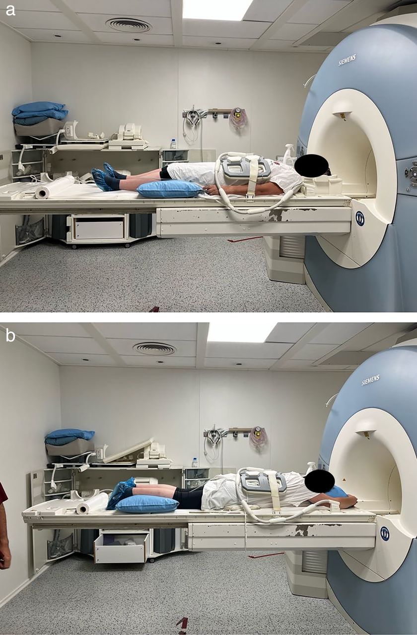
    Patient positioning for supine (a) and prone (b) magnetic resonance imaging.

  • Figure 2
    • Download figure
    • Open in new tab
    • Download powerpoint
    Figure 2

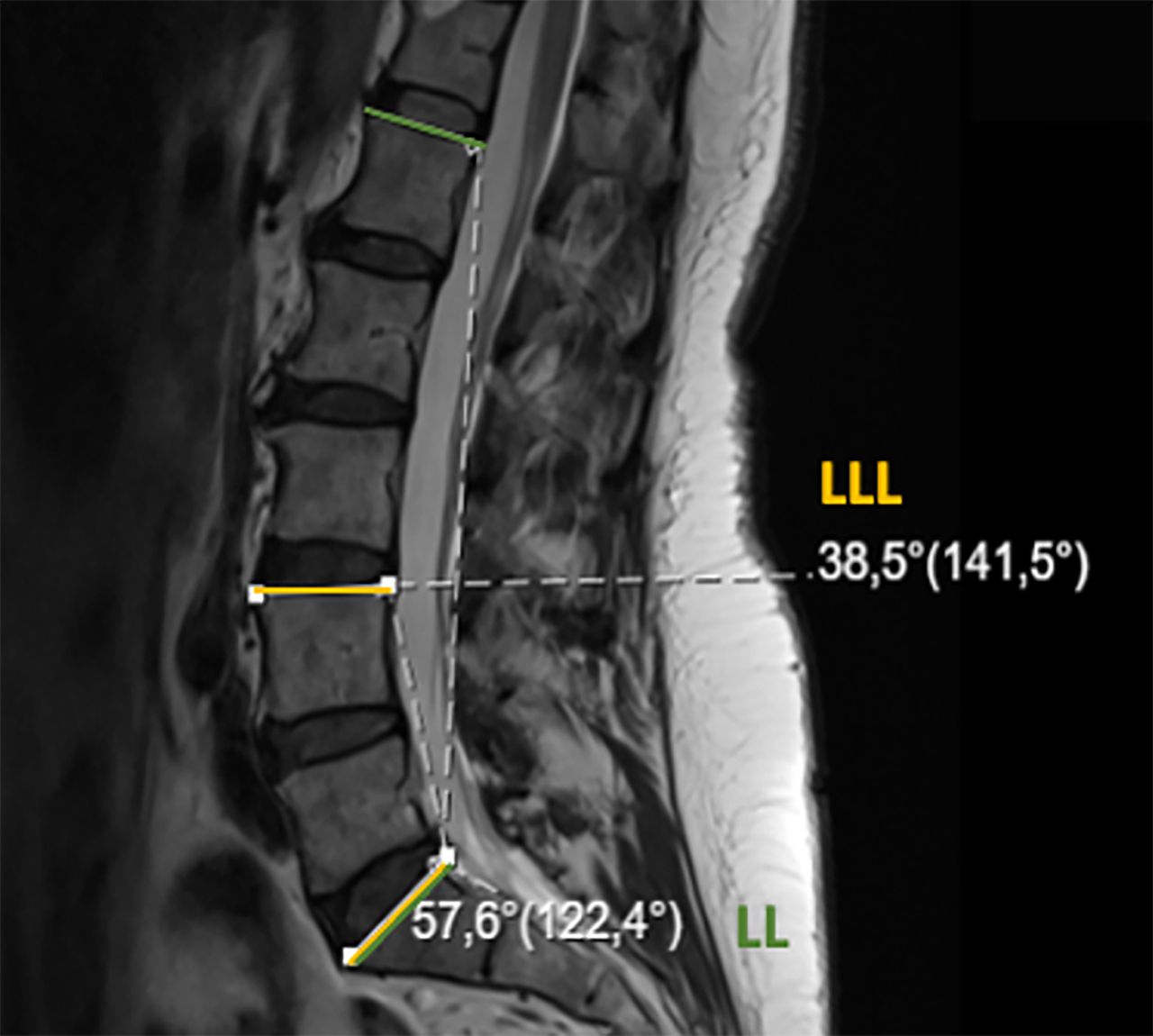
    Lumbar lordosis (LL; green) and lower lumbar lordosis (LLL; yellow).

  • Figure 3
    • Download figure
    • Open in new tab
    • Download powerpoint
    Figure 3

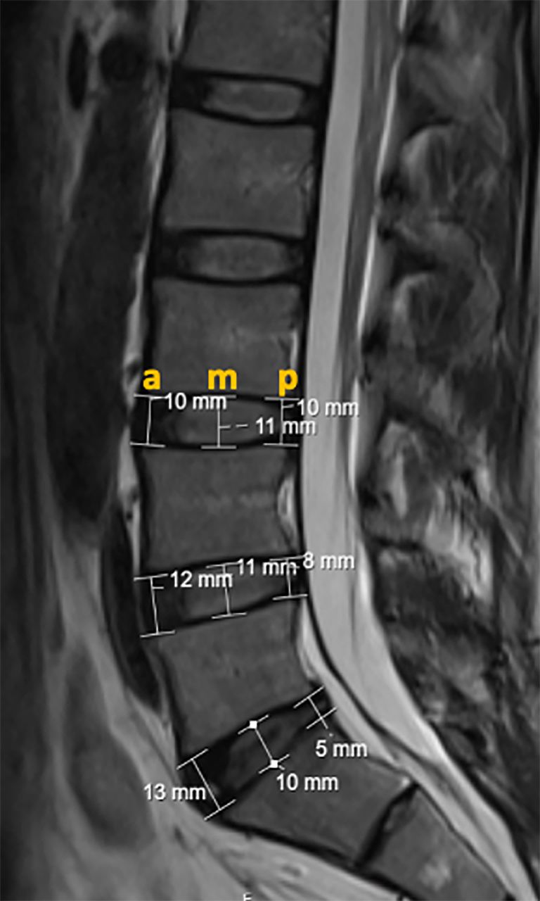
    Anterior (a), middle (m), and posterior (p) intervertebral disc height.

  • Figure 4
    • Download figure
    • Open in new tab
    • Download powerpoint
    Figure 4

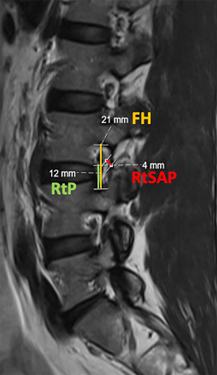
    Foraminal height (FH), root to pedicle (RtP), and root to superior articular process (RtSAP) distances.
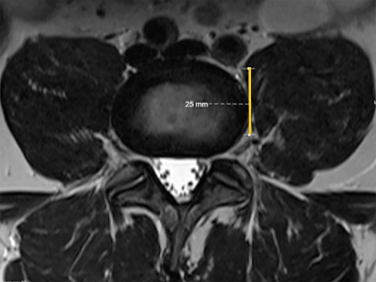
  • Figure 5
    • Download figure
    • Open in new tab
    • Download powerpoint
    Figure 5

    Left lateral safe corridor (yellow line) for lateral lumbar interbody fusion approach.
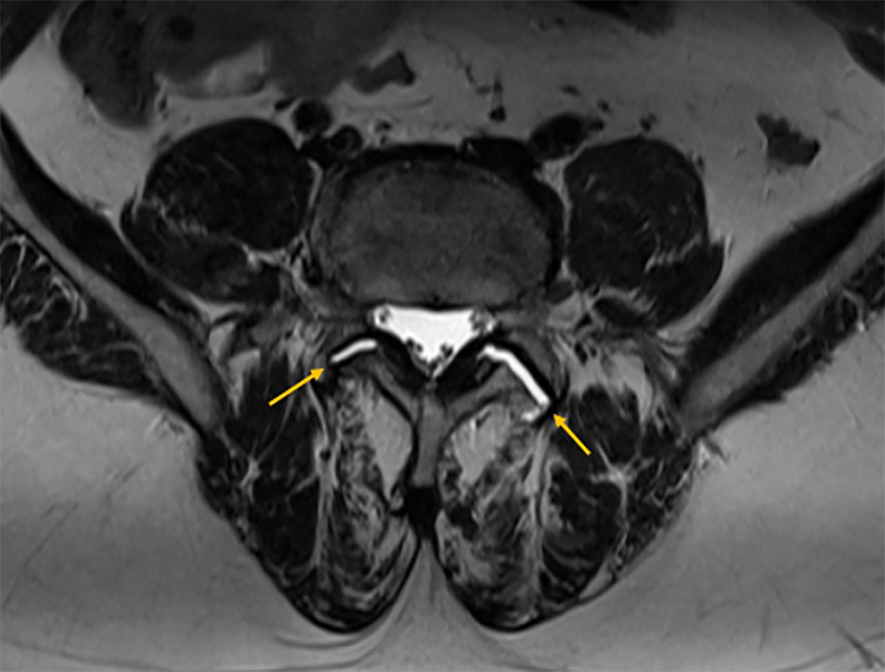
  • Figure 6
    • Download figure
    • Open in new tab
    • Download powerpoint
    Figure 6

    Disperse facet joint fluid signal changes (arrow), suggestive of dynamic lumbar spine instability.

Tables

  • Figures
    • View popup
    Table 1

    Lumbar lordosis and IVDh comparison from supine to prone.

    VariableSupine Position, Mean (SD)Prone Position, Mean (SD)Paired Difference (95% CI) P
    Lumbar lordosis (°)49.3 (9.3)52.1 (10.0)−2.8 (−4.7, −0.9)0.005
    Lower lumbar lordosis (°)36.9 (6.0)36.4 (6.5)0.5 (−0.7, 1.7)0.404
    L3−L4 anterior IVDh (mm)9.3 (1.6)9.7 (1.5)−0.3 (−0.6, −0.1)0.015
    L3−L4 middle IVDh (mm)10.2 (2.1)10.4 (1.9)−0.2 (−0.5, 0.0)0.101
    L3−L4 posterior IVDh (mm)6.5 (1.5)6.5 (1.5)0.0 (−0.3, 0.2)0.800
    L4−L5 anterior IVDh (mm)10.2 (2.2)10.4 (2.0)−0.2 (−0.5, 0.1)0.142
    L4−L5 middle IVDh (mm)10.2 (2.3)10.2 (2.1)0.0 (−0.2, 0.0)0.914
    L4−L5 posterior IVDh (mm)6.0 (1.5)6.3 (1.5)−0.3 (−0.6, −0.1]0.353
    L5−S1 anterior IVDh (mm)11.7 (2.9)11.7 (2.7)0.0 (−0.4, 0.4)1.000
    L5−S1 middle IVDh (mm)9.2 (2.7)9.2 (2.9)0.0 (−0.3, 0.3)0.876
    L5−S1 posterior IVDh (mm)5.1 (1.3)5.4 (1.7)−0.3 (−0.6, 0.0)0.032
    • Abbreviations: CI, confidence interval; IVDh, intervertebral disc height.

    • View popup
    Table 2

    Foraminal height, root-to-pedicle, and RtSAP distances comparison from supine to prone.

    VariableSupine Position, Mean (SD)Prone Position, Mean (SD)Paired Difference (95% CI) P
    Left Foraminal Height
     L3−L419.7 (2.2)19.4 (2.0)0.3 (−0.1, 0.7)0.172
     L4−L518.2 (1.7)18.2 (1.7)0.0 (−0.4, 0.3)0.859
     L5−S115.6 (2.0)16.4 (1.7)−0.8 (−1.3, −0.3)0.001
    Right Foraminal Height
     L3−L419.5 (1.9)19.2 (2.1)0.4 (−0.1, 0.8)0.086
     L4−L518.3 (1.6)17.8 (2.0)0.4 (0.0, 0.9)0.039
     L5−S116.9 (2.0)16.5 (2.1)0.4 (−0.1, 0.8)0.093
    Left Root-to-Pedicle
     L3−L48.2 (1.8)8.6 (2.1)−0.3 (−0.8, 0.1)0.166
     L4−L58.3 (1.8)8.1 (1.7)0.2 (−0.3, 0.6)0.442
     L5−S17.3 (1.9)7.4 (1.5)−0.1 (−0.6, 0.3)0.519
    Right Root-to-Pedicle
     L3−L49.4 (1.8)8.8 (1.6)0.6 (0.0, 1.2)0.065
     L4−L59.0 (1.6)8.5 (1.8)0.5 (0.0, 1.0)0.055
     L5−S18.3 (1.3)8.2 (2.1)0.1 (−0.5, 0.6)0.811
    Left RtSAP
     L3−L42.3 (1.1)2.9 (0.9)−0.5 (−0.8, −0.2)0.052
     L4−L52.6 (1.1)2.5 (0.7)0.1 (−0.2, 0.4)0.598
     L5−S12.5 (1.2)2.7 (0.8)−0.1 (−0.5, 0.2)0.405
    Right RtSAP
     L3−L42.3 (1.1)2.4 (0.9)−0.1 (−0.4, 0.2)0.434
     L4−L52.1 (1.0)2.4 (0.7)−0.4 (−0.6, −0.1)0.003
     L5−S12.3 (1.1)2.6 (0.8)−0.3 (−0.5, 0.0)0.049
    • Abbreviation: RtSAP, root-to-superior articular process.

    • View popup
    Table 3

    Relationship between root-to-pedicle and foraminal height.

    VariableSupine Position, Ratio (SD)Prone Position, Ratio (SD)Paired Difference (95% CI) P
    Left L3−L40.42 (0.81)0.44 (0.90)−0.02 (−0.05, 0.00)0.069
    Left L4−L50.45 (0.81)0.45 (0.75)0.00 (−0.01, 0.03)0.412
    Left L5−S10.47 (0.11)0.45 (0.79)0.01 (−0.01, 0.04)0.311
    Right L3−L40.48 (0.78)0.46 (0.68)0.02 (−0.01, 0.05)0.203
    Right L4−L50.45 (0.81)0.45 (0.75)0.01 (−0.01, 0.03)0.412
    Right L5−S10.49 (0.77)0.49 (0.95)0.00 (−0.03, 0.03)0.984
    • View popup
    Table 4

    Lateral lumbar working corridor.

    VariableSupine Position, Mean (SD)Prone Position, Mean (SD)Paired Difference (95% CI) P
    L3−L4 Corridor21.2 (5.3)20.8 (5.2)0.4 (−0.2, 1.1)0.196
    L4−L5 Corridor15.9 (4.6)15.7 (4.2)0.2 (−0.6, 1.0)0.600
PreviousNext
Back to top

In this issue

International Journal of Spine Surgery
Vol. 19, Issue 2
1 Apr 2025
  • Table of Contents
  • Index by author

Print
Download PDF
Article Alerts
Sign In to Email Alerts with your Email Address
Email Article

Thank you for your interest in spreading the word on International Journal of Spine Surgery.

NOTE: We only request your email address so that the person you are recommending the page to knows that you wanted them to see it, and that it is not junk mail. We do not capture any email address.

Enter multiple addresses on separate lines or separate them with commas.
Prone Position for Preoperative Planning in Lumbar Endoscopic and Minimally Invasive Fusion Procedures: Insights From a Magnetic Resonance Imaging Study
(Your Name) has sent you a message from International Journal of Spine Surgery
(Your Name) thought you would like to see the International Journal of Spine Surgery web site.
CAPTCHA
This question is for testing whether or not you are a human visitor and to prevent automated spam submissions.
Citation Tools
Prone Position for Preoperative Planning in Lumbar Endoscopic and Minimally Invasive Fusion Procedures: Insights From a Magnetic Resonance Imaging Study
Miguel Relvas-Silva, José Maria Matos Sousa, Daniel Dias, Bernardo Sousa Pinto, António Sousa, José Fonseca, Miguel Loureiro, André Rodrigues Pinho, Vitorino Veludo, António Serdoura, Maria Dulce Madeira, Pedro Alberto Pereira
International Journal of Spine Surgery Apr 2025, 19 (2) 179-187; DOI: 10.14444/8731

Citation Manager Formats

  • BibTeX
  • Bookends
  • EasyBib
  • EndNote (tagged)
  • EndNote 8 (xml)
  • Medlars
  • Mendeley
  • Papers
  • RefWorks Tagged
  • Ref Manager
  • RIS
  • Zotero
Share
Prone Position for Preoperative Planning in Lumbar Endoscopic and Minimally Invasive Fusion Procedures: Insights From a Magnetic Resonance Imaging Study
Miguel Relvas-Silva, José Maria Matos Sousa, Daniel Dias, Bernardo Sousa Pinto, António Sousa, José Fonseca, Miguel Loureiro, André Rodrigues Pinho, Vitorino Veludo, António Serdoura, Maria Dulce Madeira, Pedro Alberto Pereira
International Journal of Spine Surgery Apr 2025, 19 (2) 179-187; DOI: 10.14444/8731
Twitter logo Facebook logo Mendeley logo
  • Tweet Widget
  • Facebook Like
  • Google Plus One

Jump to section

  • Article
    • Abstract
    • Introduction
    • Methods
    • Results
    • Discussion
    • Conclusion
    • Footnotes
    • References
  • Figures & Data
  • Info & Metrics
  • PDF

Related Articles

  • No related articles found.
  • PubMed
  • Google Scholar

Cited By...

  • No citing articles found.
  • Google Scholar

More in this TOC Section

  • Patient Satisfaction Following Lumbar Fusion Is Associated With Functional Status and Pain More Than the Attainment of Minimal Clinically Important Difference: Implications for Value-Based Medicine
  • Selective Direct Vertebral Rotation Instrumentation for the Correction of Adolescent Idiopathic Scoliosis Lenke 5 Curve
Show more Other and Special Categories

Similar Articles

Keywords

  • magnetic resonance
  • prone
  • lumbar spine
  • endoscopy
  • interbody fusion
  • minimally invasive spine surgery

Content

  • Current Issue
  • Latest Content
  • Archive

More Information

  • About IJSS
  • About ISASS
  • Privacy Policy

More

  • Subscribe
  • Alerts
  • Feedback

Other Services

  • Author Instructions
  • Join ISASS
  • Reprints & Permissions

© 2025 International Journal of Spine Surgery

International Journal of Spine Surgery Online ISSN: 2211-4599

Powered by HighWire