


Article Figures & Data
Figures
- Fig. 1
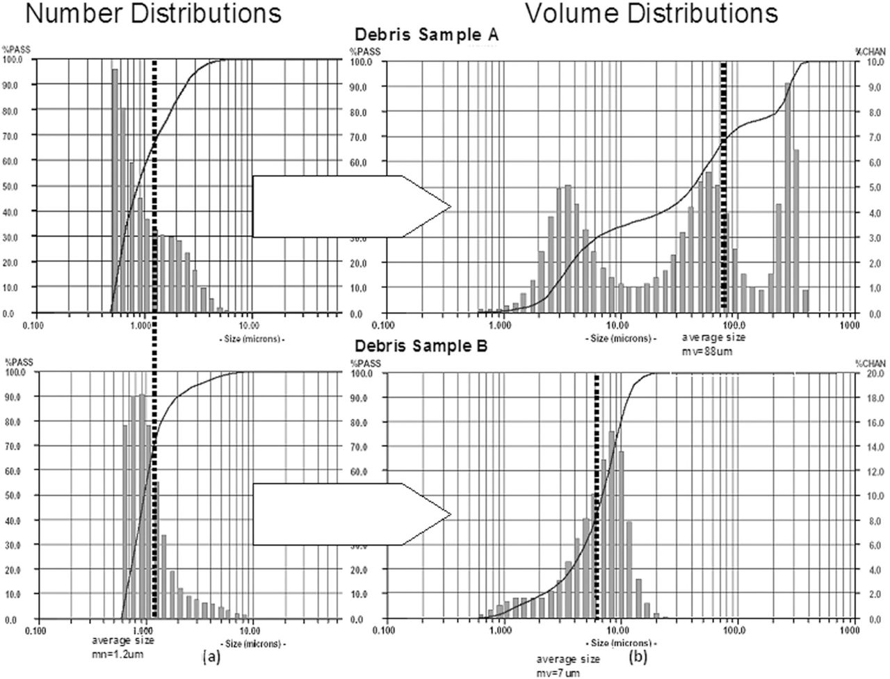
These analyses of (a) volume and (b) number distributions of two debris samples demonstrate how similar number distributions can result from very different actual size distributions as evident in (a) the volume distributions. Note: The x-axis represents increasing particle diameter and the y-axis is (a) percentage of total number of particles in each size range and (b) the percentage of total mass of each sample that is of that size range. (Courtesy of BioEngineering Solutions Inc.)
- Fig. 2
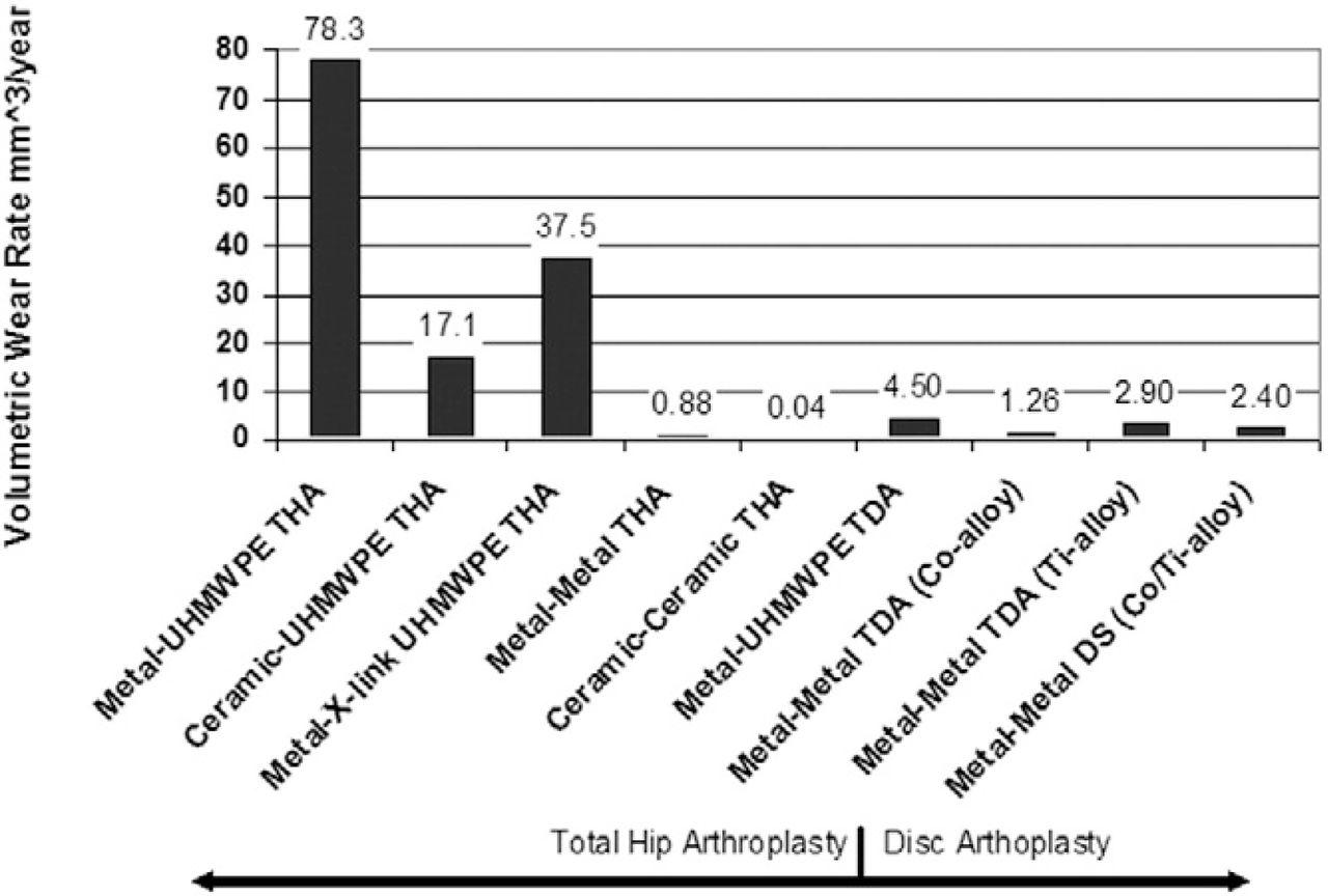
A graphical comparison of data showing the amount of wear debris generated from different types of total joint arthplasties demonstrating that there is relatively less (10×) polymeric debris generated by a total disc arthroplasty with a metal-on-polymer articulation. However, this difference is not apparent with metal-on-metal articulating implants. Note: Figure References: Metal-Poly:24Ceramic-Poly:25, Metal-X-linked Poly: 22 Metal-X-linked Poly: 26, Metal-X-linked Poly: 27, Metal-X-linked Poly: 28, Metal-Metal: 29, Ceramic-Ceramic: 30, Metal-UHMWPE TDA: 17, Metal-Metal TDA: 18.
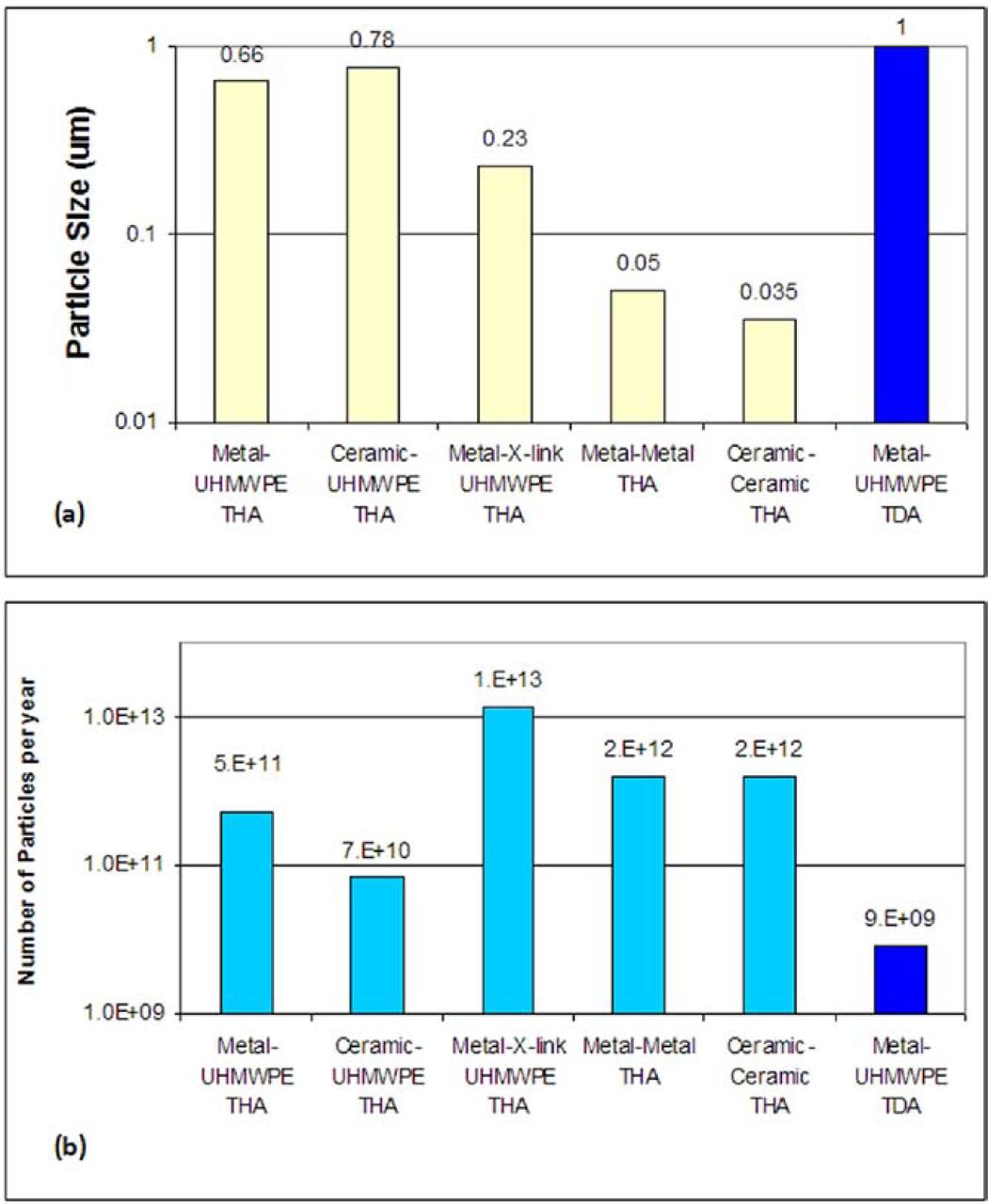
- Fig. 3
A graphical comparison of data showing (a) the reported size of implant debris generated from different types of total joint arthplasties and (b) the amount of particles per year this results in using the gravimetric data shown in Fig. 1. The smaller size of the particles reported in highly crosslinked polymer particles (copared to traditional poly) combined with the modest reductions in wear rate results in metal on Metal-X-linked Poly. Note: Fig. References: Metal-Poly:24Ceramic-Poly:25, Metal-X-linked Poly: 22 Metal-X-linked Poly: 26, Metal-X-linked Poly: 27, Metal-X-linked Poly: 28, Metal-Metal: 29, Ceramic-Ceramic: 30, Metal-UHMWPE TDA: 17, Metal-Metal TDA: 18.
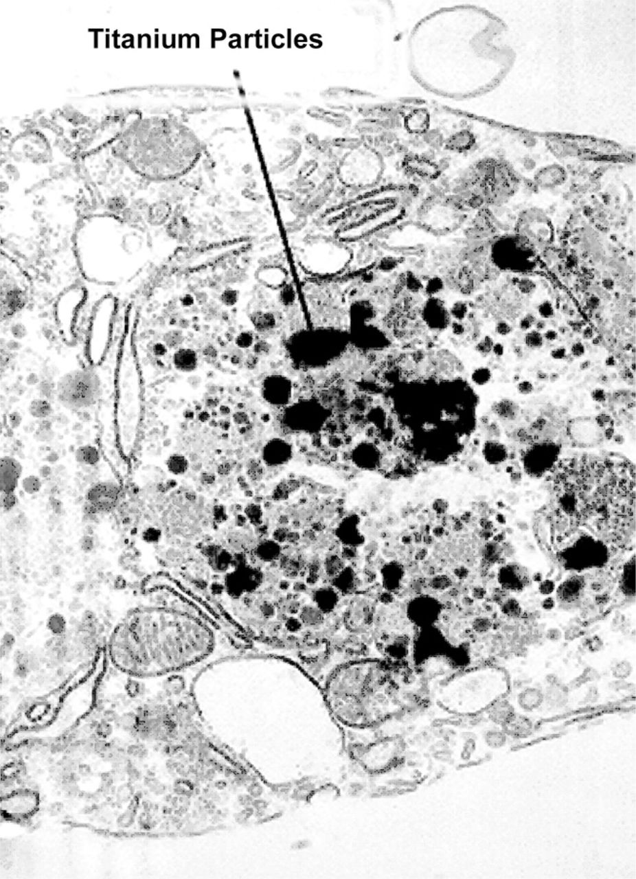
- Fig. 4
Transmission Electron Photomicrographs: (a) Macrophage containing phagocytized titanium particles. (b) Endothelial cell lining with embedded titanium debris. These specimens were obtained from a tissue sample overlying the posterolateral fusion mass (sixteen-week autograft + titanium) (TEM magnification = 20,000×) (courtesy of Bryan Cunningham).
- Fig. 5
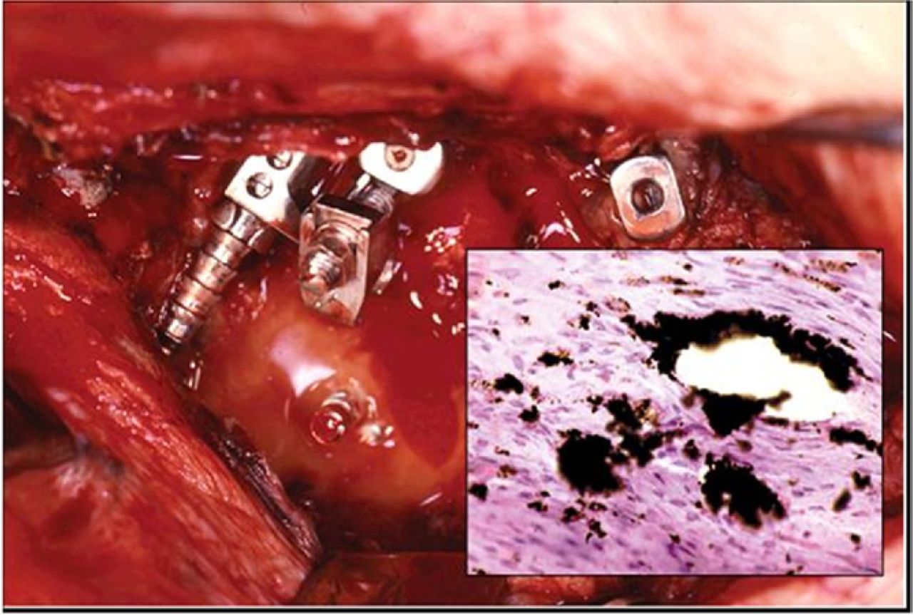
At surgical exploration the broken and dislodged instrumentation was accompanied by stainless steel particulate debris. Anteroposterior radiograph in a patient with breakage of a longitudinal rod connecting pedicle screws two years post-operatively. (Courtesy of Bryan Cunningham.)
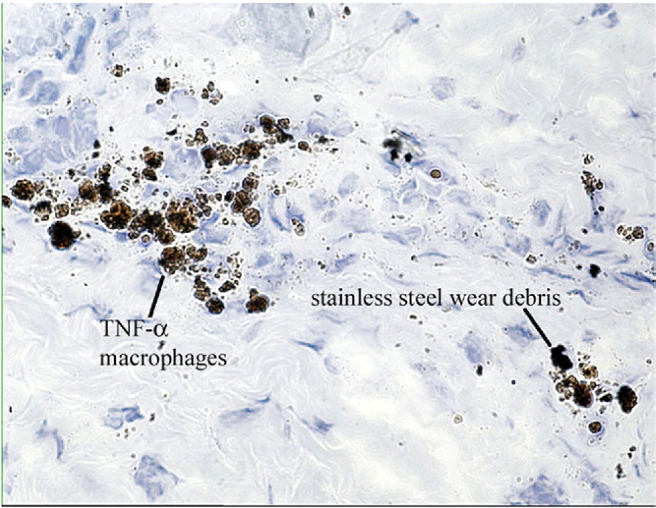
- Fig. 6
TNF-α Cytokine Expressing Macrophages: Membrane-bound or intracellular TNF-α, contained in the tissue layer overlying the posterolateral sites, produced yellow to brown chromagen label as shown in this sixteen-week autograft + stainless steel particles in a rabbit model. (Avitan-Biotin Complex horseradish peroxidase technique for TNF-α, magnification 40). (Courtesy of Bryan Cunningham.)
- Fig. 7
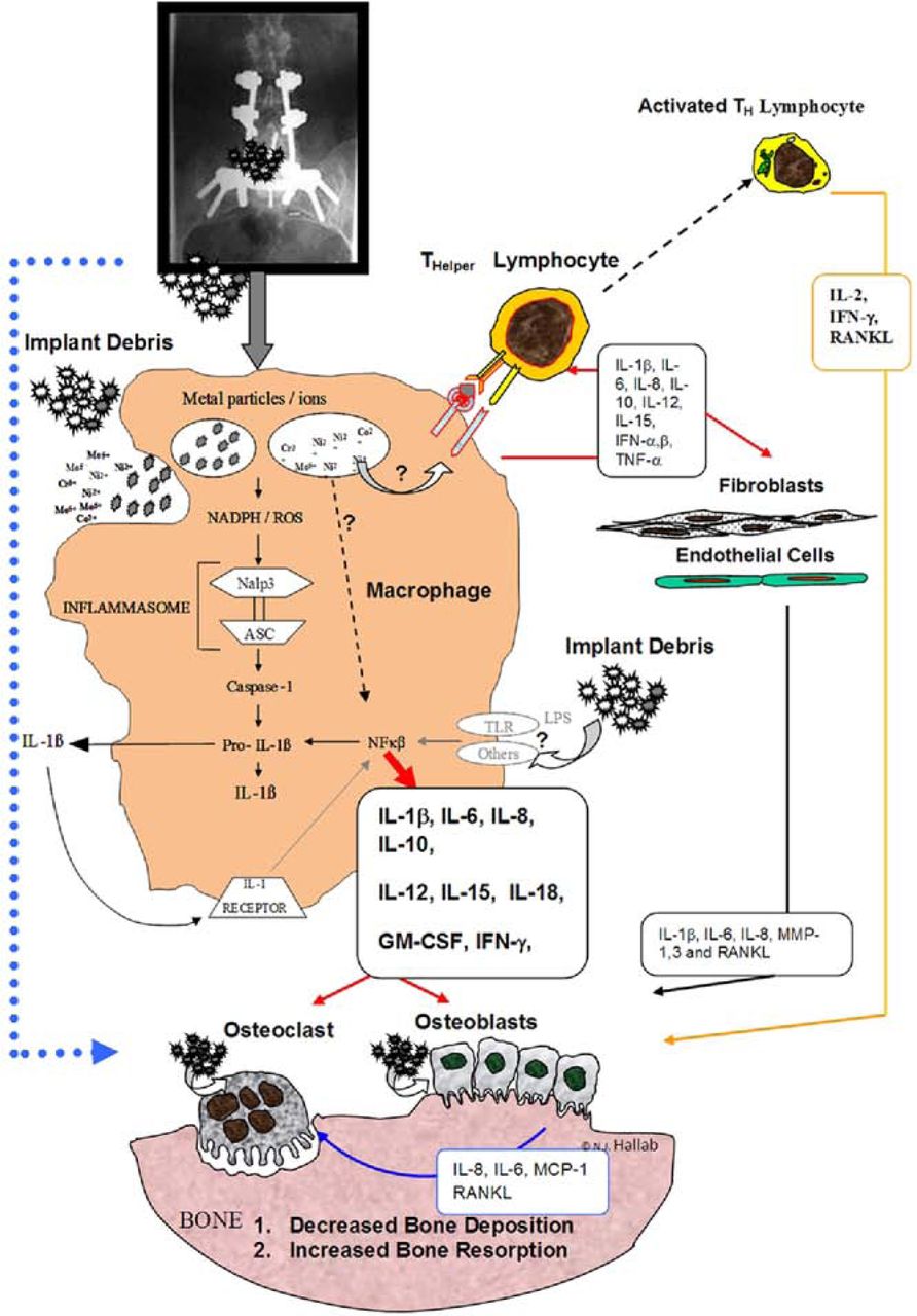
This schematic shows the numerous pro-inflammatory mediators produced by peri-implant tissue and immune cells reacting to implant debris, which can negatively affect bone turnover. The pro-inflammatory cytokines IL-1, IL-6, and TNF-alpha are thought to be some of the most potent cytokines in this cascade of signaling. The inflammasome pathway within cells such as macrophages has recently been reported to be central to implant debris mediated pro-inflammatory reactivity. Ingestion of the debris phagocytosis results in the release of pro-inflammatory cytokines that affect local cell types and induce a widening zone of soft-tissue damage and inflammation. (Courtesy of BioEngineering Solutions Inc)
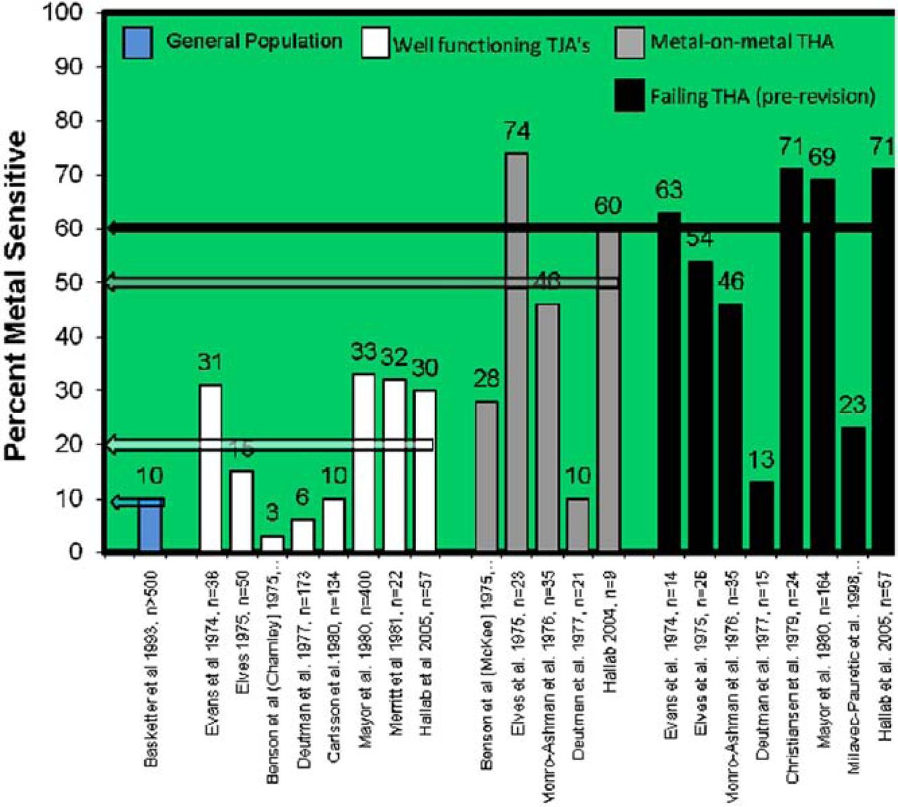
- Fig. 8
A compilation of investigations showing the averaged incidence percentages of metal sensitivity for nickel, cobalt or chromium among 1) the general population, 2) patients after receiving a metal containing TJA, 3) patients with metal-on-metal bearing arthroplasty and 4) patient populations with significant osteolysis or due to be revised. Note: Studies by Hallab et al used LTT to measure hypersensitivity, all other used dermal patch testing.


Tables
- Table 1
Approximate average concentrations of metal in human body fluids with and without total joint replacements. Note: where ranges were reported they are included here within parentheses, eg (1– 4). All concentrations are reported in ppb (ng/mL)
Concentrations of metal in body fluids (ng/mL or ppb) Co Cr Mo Ni Ti Al References Serum Normal <0.2–0.6109 (0.2–8.3)109 0.2110 (0.1–0.7)110 0.42110 (0.3–1.8)110 * 1.2–2.7109;111 (1.1–7.9) 1.2–2.2111 (1.1–6.4) <0.8111 <0.8 109, 111 TKA-M/P 0.2 (<0.02–1.15)112 0.1 (<0.02–0.6)112 * 0.4 (0.05–1.5)112 3.2 (<2.1–6.3)111 6.5 (2.1–9.4)111 <0.8 <0.8111 111, 112 TKA-F * * * * 135.6 (24.1–716.9)111 3.7 (0.8–6.2)111 0.9 (<0.8–2.6)111 111 THA-M/P 0.9 (<0.3–3.9)113 1.28 (0.1–2.4)113 * * 1.4–4.16, 2.6113(<1.1–11.17)109 ;113 1.2–1.7109 (0.2–2.46)109 * 40;109;113;114 THA-M-M 2.4 (0.6–7.9)113 3.5 (0.8–9.1)113 * * 1.9113 (1.1–6.0)113 * * 113 TDA 1.9–4.854;55 1.9–2.454;55 * * * * * 54;55 Synovial fluid Normal * * * * <0.1 (<0.1–7.6)115 7.3 (1.9–19)115 * 115;116 THA 5116, 0.2 (<0.02–1.15)112 3116, 0.1 (<0.02–0.6)112 21 ± 8116 4116, 0.4 (0.05–1.5)112 13–556116 109–654116 5–62116 112;116 THA-F 588 ± 427116 385 ± 232116 58 ± 53116 32 ± 16116 86 ± 35116 256 ± 271116 25 ± 19116 116 Whole blood Normal 0.1–1.2116 2–6116 0.5–1.8116 2.9–7.0116 17 ± 60116 12 ± 4116 5.8 ± 4116 116 THA 67 ± 62116 218 ± 233116 23 ± 31116 116 THA-F 20 ± 25116 110 ± 150116 12 ± 9116 29 ± 29116 602 ± 927116 237 ± 307116 55 ± 63116 116 Human tissue (µg/g) (roughly equivalent to: 10−1 to 10−2 mM) Liver Normal 100 890 14 120 <14 * * 117;118 TJA 560 680 22 15200 1130 * * 117;118 Psuedocapsule Normal <65 120 <9 50 150 * * 116;117;119;120 TJA 39400 460 121 5490 3820 * * 116;117;119;120 Lymphatic Normal * * * 10 690 * * 117 Tissue TJA * * * 390 690 * * 117 Abbreviations: Normal, subjects without any metallic prosthesis (not including dental); M/P, metal on polyarticulation; M/M, metal-on-metal articulation; F, subjects with a poorly functioning arthroplasty (loose or migratory osteolysis prior to surgical revision); THA, total hip arthroplasty; TKA, total knee arthroplasty; TJA, subjects with well functioning total joint arthroplasty (either knee or hip); TDA Spine, total disc arthroplasty (metal on metal).
↵* Data not available.
In this issue


Jump to section
Related Articles
Cited By...
- No citing articles found.