
Main menu

  • Home
  • Content
    • Current Issue
    • Advance Online Publication
    • Archive
  • About Us
    • About ISASS
    • About the Journal
    • Author Instructions
    • Editorial Board
    • Reviewer Guidelines & Publication Criteria
  • More
    • Advertise
    • Subscribe
    • Alerts
    • Feedback
  • Join Us
  • Reprints & Permissions
  • Sponsored Content
  • Other Publications
    • ijss

User menu

  • My alerts

Search

  • Advanced search
International Journal of Spine Surgery
  • My alerts
International Journal of Spine Surgery

Advanced Search

  • Home
  • Content
    • Current Issue
    • Advance Online Publication
    • Archive
  • About Us
    • About ISASS
    • About the Journal
    • Author Instructions
    • Editorial Board
    • Reviewer Guidelines & Publication Criteria
  • More
    • Advertise
    • Subscribe
    • Alerts
    • Feedback
  • Join Us
  • Reprints & Permissions
  • Sponsored Content
  • Follow ijss on Twitter
  • Visit ijss on Facebook
Research ArticleMinimally Invasive Surgery

Percutaneous Lumbar Interbody Fusion With an Expandable Titanium Cage Through Kambin’s Triangle: A Case Series With Initial Clinical and Radiographic Results

Timothy Y. Wang, Vikram A. Mehta, Mostafa Gabr, Eric W. Sankey, Alexia Bwensa, C. Rory Goodwin, Isaac O. Karikari, John H. Chi and Muhammad M. Abd-El-Barr
International Journal of Spine Surgery December 2021, 15 (6) 1133-1141; DOI: https://doi.org/10.14444/8144
Timothy Y. Wang
1 Department of Neurosurgery, Division of Spine, Duke University Medical Center, Durham, North Carolina, 27701, USA
  • Find this author on Google Scholar
  • Find this author on PubMed
  • Search for this author on this site
Vikram A. Mehta
1 Department of Neurosurgery, Division of Spine, Duke University Medical Center, Durham, North Carolina, 27701, USA
  • Find this author on Google Scholar
  • Find this author on PubMed
  • Search for this author on this site
Mostafa Gabr
1 Department of Neurosurgery, Division of Spine, Duke University Medical Center, Durham, North Carolina, 27701, USA
  • Find this author on Google Scholar
  • Find this author on PubMed
  • Search for this author on this site
Eric W. Sankey
1 Department of Neurosurgery, Division of Spine, Duke University Medical Center, Durham, North Carolina, 27701, USA
  • Find this author on Google Scholar
  • Find this author on PubMed
  • Search for this author on this site
Alexia Bwensa
1 Department of Neurosurgery, Division of Spine, Duke University Medical Center, Durham, North Carolina, 27701, USA
  • Find this author on Google Scholar
  • Find this author on PubMed
  • Search for this author on this site
C. Rory Goodwin
1 Department of Neurosurgery, Division of Spine, Duke University Medical Center, Durham, North Carolina, 27701, USA
  • Find this author on Google Scholar
  • Find this author on PubMed
  • Search for this author on this site
Isaac O. Karikari
1 Department of Neurosurgery, Division of Spine, Duke University Medical Center, Durham, North Carolina, 27701, USA
  • Find this author on Google Scholar
  • Find this author on PubMed
  • Search for this author on this site
John H. Chi
2 Department of Neurosurgery, Brigham and Women’s Hospital, Harvard Medical School, Boston, Massachusetts, 02115, USA
  • Find this author on Google Scholar
  • Find this author on PubMed
  • Search for this author on this site
Muhammad M. Abd-El-Barr
1 Department of Neurosurgery, Division of Spine, Duke University Medical Center, Durham, North Carolina, 27701, USA
  • Find this author on Google Scholar
  • Find this author on PubMed
  • Search for this author on this site
  • Article
  • Figures & Data
  • Info & Metrics
  • PDF
Loading

Article Figures & Data

Figures

  • Tables
  • Figure 1
    • Download figure
    • Open in new tab
    • Download powerpoint
    Figure 1

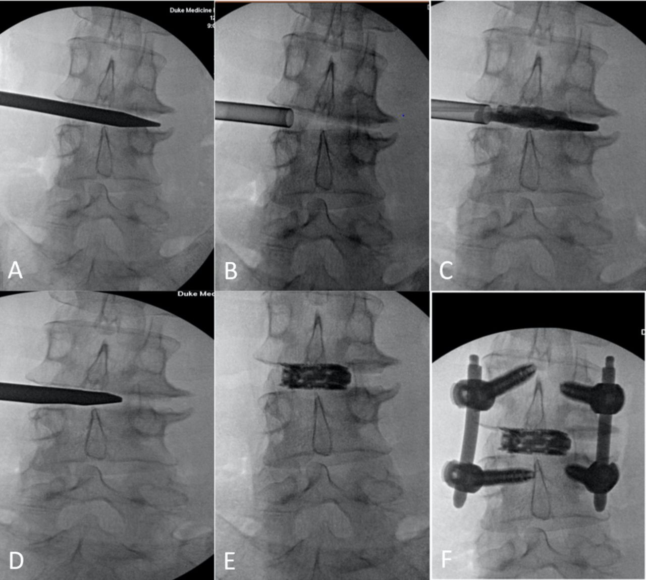
    Sequential fluoroscopic imaging of the percutaneous lumbar interbody fusion. (A) A blunt electromyography guided probe traverses the fascia, Kambin’s triangle, and is introduced into the disc space. (B) After sequential dilation, a working cannula is docked inside the disc space to protect surround structures. (C) After appropriate discectomy, a balloon is placed inside the disc space and inflated with radio-opaque material to confirm satisfactory discectomy. (D) After satisfactory end plate preparation, an introducer is placed at the center of the disc space and loaded with an expandable cage. (E) The cage is expanded as it is shown. (F) Percutaneous screws are placed to complete the procedure.

  • Figure 2
    • Download figure
    • Open in new tab
    • Download powerpoint
    Figure 2

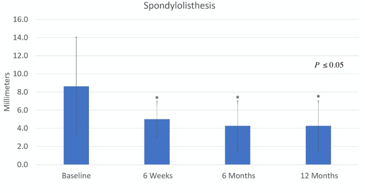
    Bar graph illustrates the correction of the grade 1 spondylolisthesis preoperatively compared to 6 weeks, 6 months, and 12 months postoperatively. The improvement in spondylolisthesis was significantly improved at 6 weeks, 6 months, and 12 months postoperatively (P < 0.05).

  • Figure 3
    • Download figure
    • Open in new tab
    • Download powerpoint
    Figure 3

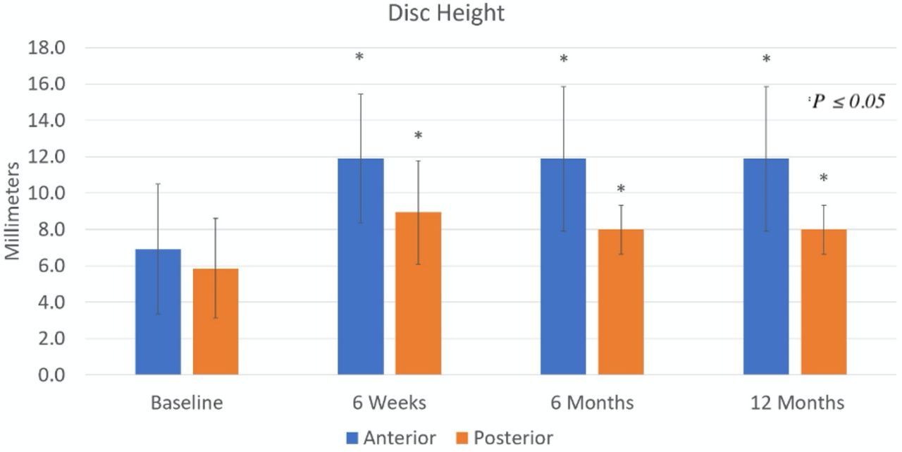
    Bar graph shows baseline disc height and its increase at 6 weeks, 6 months and 12 months postoperatively. The anterior and posterior disc height was significantly increased at 6 weeks, 6 months, and 12 months postoperatively (P ≤ 0.05).

  • Figure 4
    • Download figure
    • Open in new tab
    • Download powerpoint
    Figure 4

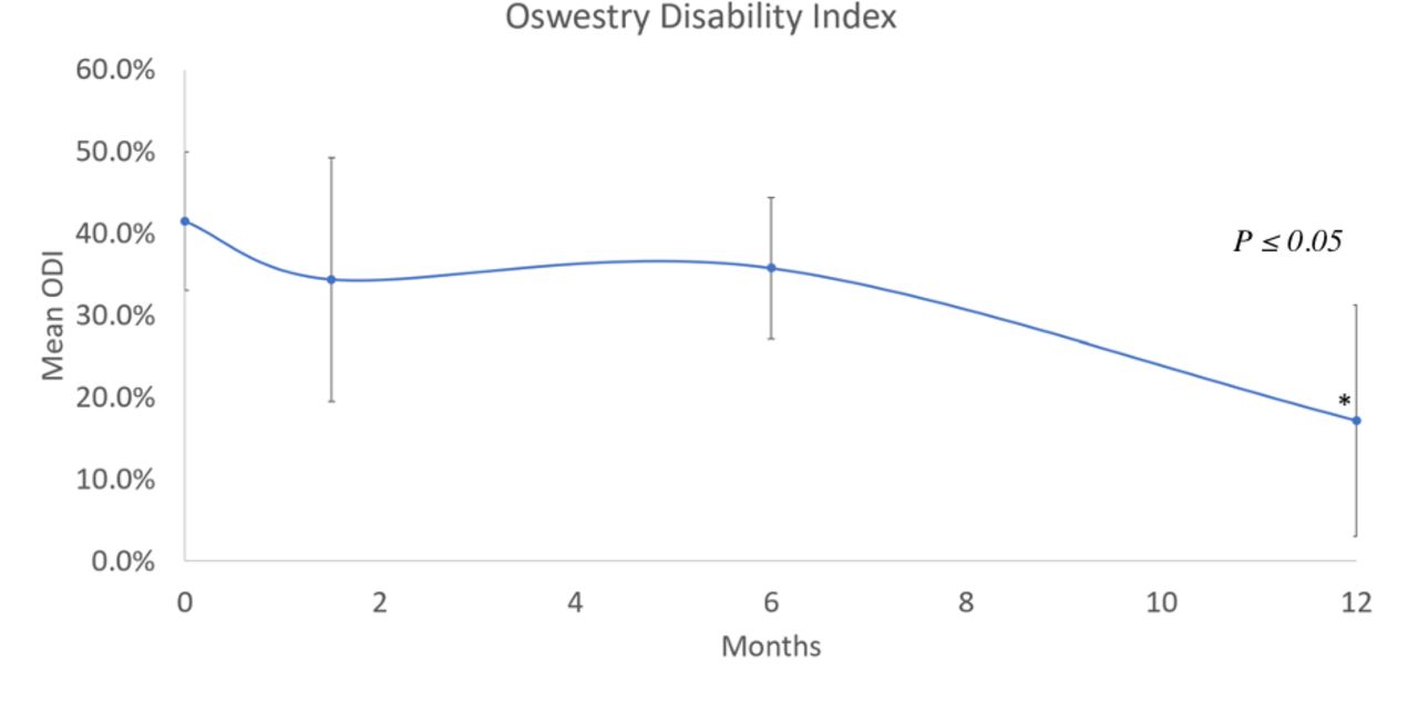
    Scatter plot demonstrating mean Oswestry Disability Index (ODI) scores of patients receiving percutaneous lumbar interbody fusion procedure over 1-year period. ODI scores were significantly improved at 1 year when compared to baseline (P ≤ 0.05).

  • Figure 5
    • Download figure
    • Open in new tab
    • Download powerpoint
    Figure 5

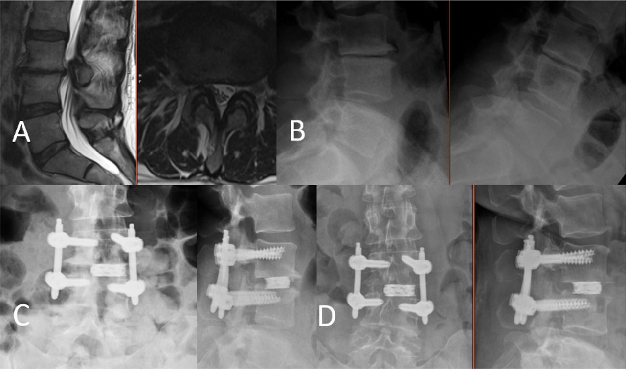
    Radiographical evaluation of the patient. (A) Preoperative lumbar sagittal and axial magnetic resonance imaging without contrast showing degenerative disc disease, height loss, and disc bulge at L3-4 with left neuroforaminal stenosis. (B) Preoperative flexion-extension lumbar x-rays showing a grade 1 spondylolisthesis at L3-4. (C) Anterior-posterior and lateral lumbar x-rays immediately after L3-4 percutaneous lumbar interbody fusion (percLIF) showing satisfactory placement of the interbody expandable cage and rigid pedicle screw and rod fixation. (D) Anterior-posterior and lateral lumbar x-rays 6 months after L3-4 percLIF showing appropriate hardware placement.

Tables

  • Figures
    • View popup
    Table 1

    Demographic data.

    VariableValue Count (%) or Mean (SD)
    N 16 (100%)
    Age (years)56.9 (11.4)
    Female13 (81.3%)
    BMI (kg/m2)30.8 (5.3)
    Grade 1 spondylolisthesis16 (100%)
    Hypertension7 (43.8%)
    Dyslipidemia2 (12.5%)
    Diabetes mellitus type 24 (25.0%)
    Coronary artery disease2 (25.0%)
    Rheumatoid arthritis0 (0.0%)
    Operative level
     L1-L20 (0%)
     L2-L31 (6.3%)
     L3-L43 (18.8%)
     L4-L59 (56.3%)
     L5-S13 (18.8%)
    • View popup
    Table 2

    Operative and hospitalization data.

    VariableValue Count (%) or Mean (SD)
    Operative time (minutes)149 (33.8)
    Estimated blood loss (milliliters)29.6 (12.4)
    Length of stay (nights)1.50 (.650)a
    • aTwo patients were considered outliers and omitted as they were outside the mean plus 2 standard deviations.

    • View popup
    Table 3

    Patient spondylolisthesis, anterior disc height, and posterior disc height.

    VariableBaseline, Mean (SD) (n = 16)6 Weeks, Mean (SD) (n = 16)6 Months, Mean (SD) (n = 10)12 Months, Mean (SD) (n = 6) P Value
    Spondylolisthesis (mm)8.6 (5.4)5.0 (2.0)4.3 (2.7)3.7 (2.11)6 weeks: P = 0.005
    6 months: P = 0.036
    12 months: P = 0.044
    Anterior disc height (mm)6.9 (3.6)11.9 (3.5)11.9 (4.0)10.5 (3.0)6 weeks: P < 0.0002
    6 months: P = 0.003
    12 months: P = 0.040
    Posterior disc height (mm)5.9 (2.7)8.9 (2.8)8.0 (1.3)9.0 (4.1)6 weeks: P = 0.0005
    6 months: P = 0.018
    12 months: P = 0.048
PreviousNext
Back to top

In this issue

International Journal of Spine Surgery
Vol. 15, Issue 6
1 Dec 2021
  • Table of Contents
  • Index by author

Print
Download PDF
Article Alerts
Sign In to Email Alerts with your Email Address
Email Article

Thank you for your interest in spreading the word on International Journal of Spine Surgery.

NOTE: We only request your email address so that the person you are recommending the page to knows that you wanted them to see it, and that it is not junk mail. We do not capture any email address.

Enter multiple addresses on separate lines or separate them with commas.
Percutaneous Lumbar Interbody Fusion With an Expandable Titanium Cage Through Kambin’s Triangle: A Case Series With Initial Clinical and Radiographic Results
(Your Name) has sent you a message from International Journal of Spine Surgery
(Your Name) thought you would like to see the International Journal of Spine Surgery web site.
CAPTCHA
This question is for testing whether or not you are a human visitor and to prevent automated spam submissions.
Citation Tools
Percutaneous Lumbar Interbody Fusion With an Expandable Titanium Cage Through Kambin’s Triangle: A Case Series With Initial Clinical and Radiographic Results
Timothy Y. Wang, Vikram A. Mehta, Mostafa Gabr, Eric W. Sankey, Alexia Bwensa, C. Rory Goodwin, Isaac O. Karikari, John H. Chi, Muhammad M. Abd-El-Barr
International Journal of Spine Surgery Dec 2021, 15 (6) 1133-1141; DOI: 10.14444/8144

Citation Manager Formats

  • BibTeX
  • Bookends
  • EasyBib
  • EndNote (tagged)
  • EndNote 8 (xml)
  • Medlars
  • Mendeley
  • Papers
  • RefWorks Tagged
  • Ref Manager
  • RIS
  • Zotero
Share
Percutaneous Lumbar Interbody Fusion With an Expandable Titanium Cage Through Kambin’s Triangle: A Case Series With Initial Clinical and Radiographic Results
Timothy Y. Wang, Vikram A. Mehta, Mostafa Gabr, Eric W. Sankey, Alexia Bwensa, C. Rory Goodwin, Isaac O. Karikari, John H. Chi, Muhammad M. Abd-El-Barr
International Journal of Spine Surgery Dec 2021, 15 (6) 1133-1141; DOI: 10.14444/8144
Twitter logo Facebook logo Mendeley logo
  • Tweet Widget
  • Facebook Like
  • Google Plus One

Jump to section

  • Article
    • Abstract
    • Introduction
    • Methods
    • Results
    • Discussion
    • Conclusion
    • Footnotes
    • References
  • Figures & Data
  • Info & Metrics
  • PDF

Related Articles

  • No related articles found.
  • PubMed
  • Google Scholar

Cited By...

  • No citing articles found.
  • Google Scholar

More in this TOC Section

  • Comparative Review of Lateral and Oblique Lumbar Interbody Fusion: Technique, Outcomes, and Complications
  • A Spine Surgeon’s Learning Curve With the Minimally Invasive L5 to S1 Lateral ALIF Surgical Approach: Perioperative Outcomes and Technical Considerations
  • Fully Navigated Single-Position Prone Lateral Lumbar Interbody Fusion: A Detailed Technical Report and Description of 15 Cases
Show more Minimally Invasive Surgery

Similar Articles

Keywords

  • expandable cage
  • Kambin’s triangle
  • lumbar interbody fusion
  • minimally invasive surgery
  • percutaneous spinal fusion

Content

  • Current Issue
  • Latest Content
  • Archive

More Information

  • About IJSS
  • About ISASS
  • Privacy Policy

More

  • Subscribe
  • Alerts
  • Feedback

Other Services

  • Author Instructions
  • Join ISASS
  • Reprints & Permissions

© 2025 International Journal of Spine Surgery

International Journal of Spine Surgery Online ISSN: 2211-4599

Powered by HighWire