


Article Figures & Data
Figures
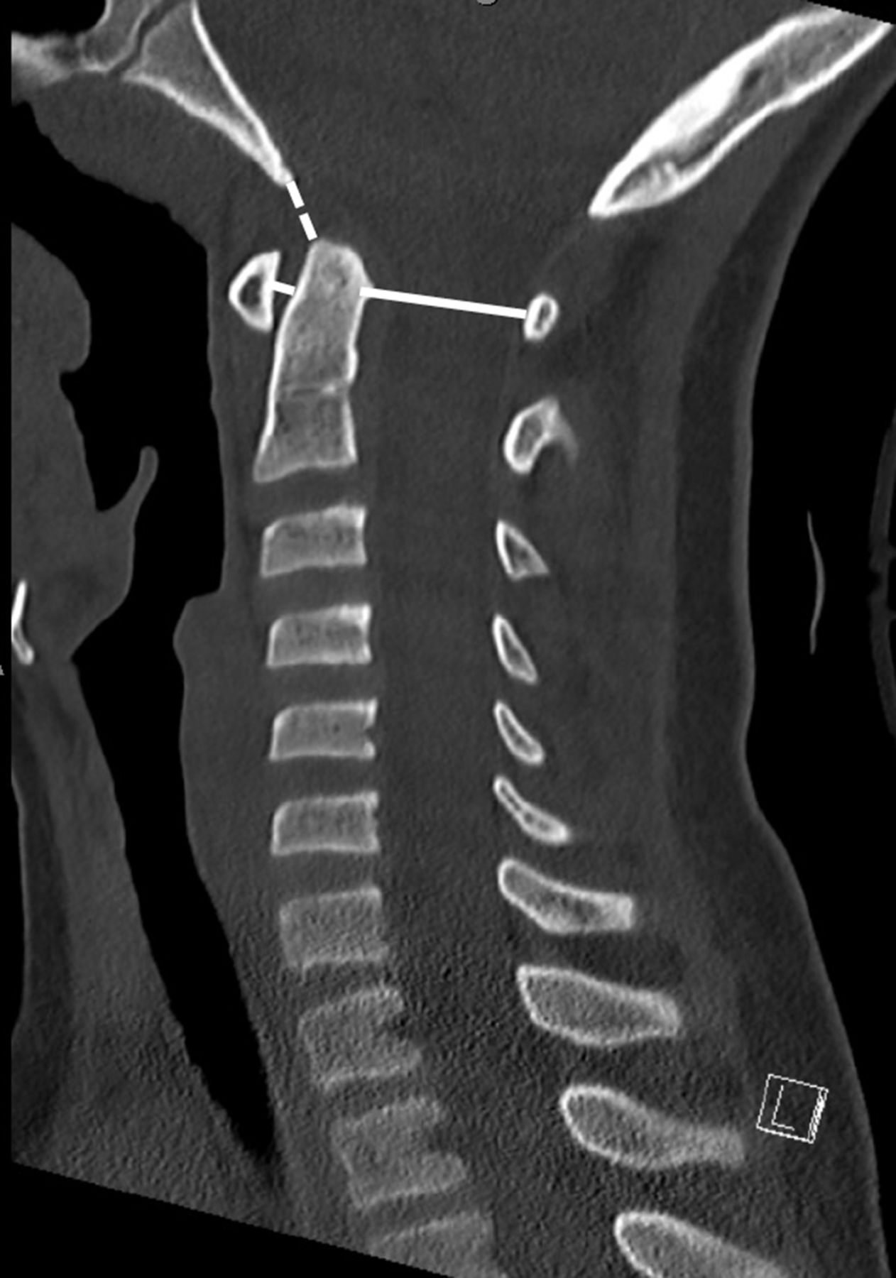
- Figure 1
On the midsagittal image, the basion-dens interval (dashed line) is measured from the tip of the basion to the apex of the dens. The anterior atlanto-dens interval is measured between the posterior aspect of the anterior C1 arch and the anterior aspect of the odontoid process—the measurement is taken at the midpoint of C1. The posterior atlanto-dens interval is measured from the posterior aspect of the odontoid process to the anterior aspect of the posterior arch of C1.
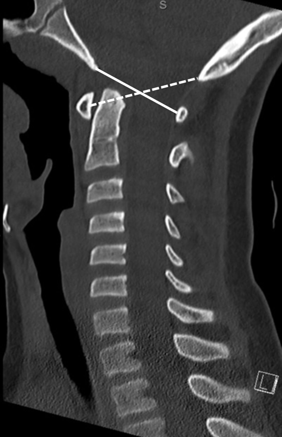
- Figure 2
On the midsagittal image, the basion-C1 distance (B-C1) (solid line) is measured from the tip of the basion to the anterior aspect of the posterior arch of C1. The opisthion-C1 distance (Op-C1) (dashed line) is measured from the tip of the opisthion to the posterior aspect of the anterior arch of C1. The same C1 points were used for measuring the atlanto-dens interval and posterior atlanto-dens interval. To calculate the Powers ratio, B-C1 was divided by Op-C1 as well described.
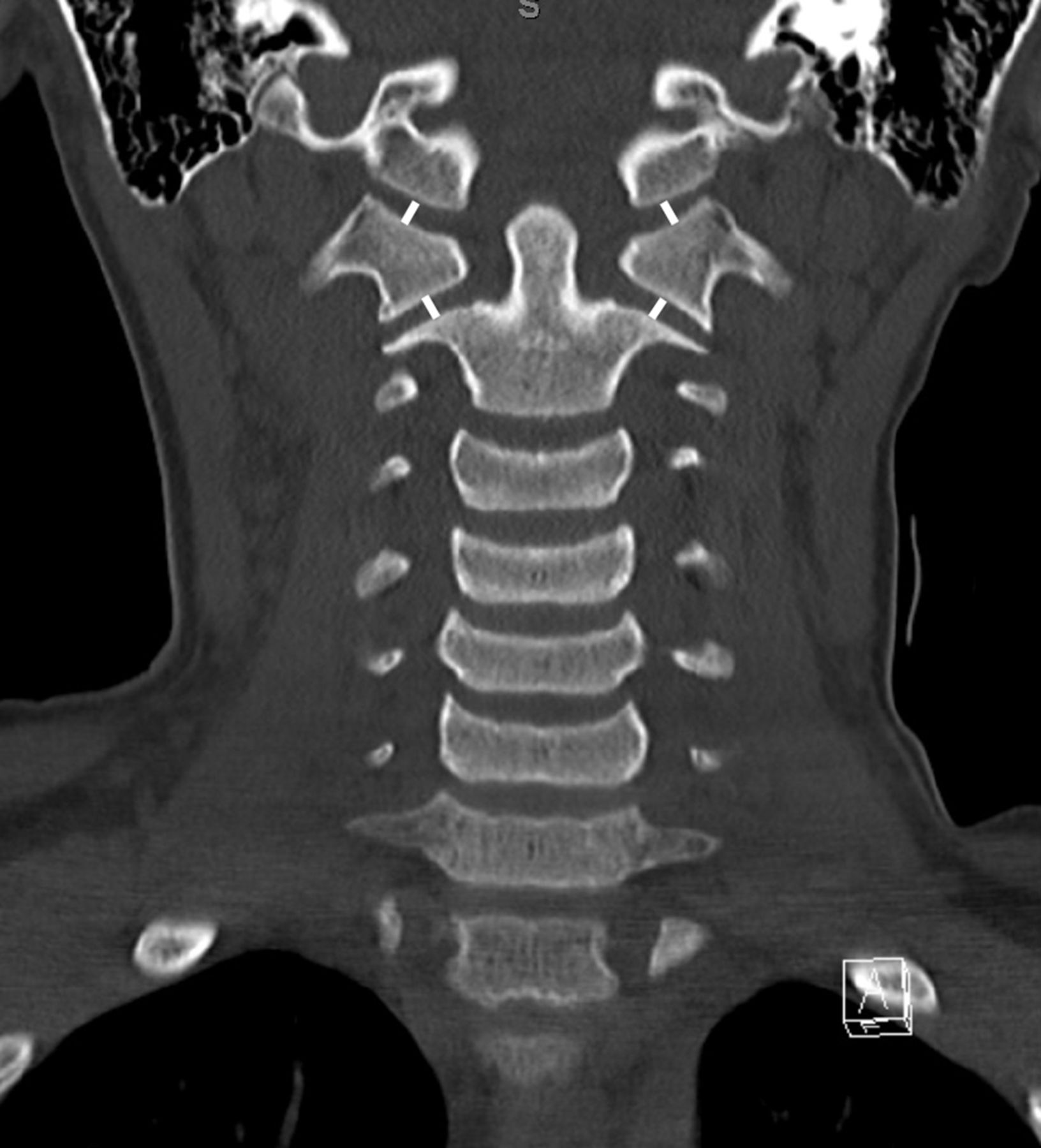
- Figure 3
On the midcoronal image, the condylar-C1-interval is measured with a line in the middle of the joint drawn perpendicular to the joint surfaces; the lateral mass interval is drawn similarly with a line in the middle of the joint drawn perpendicular to the joint surfaces.
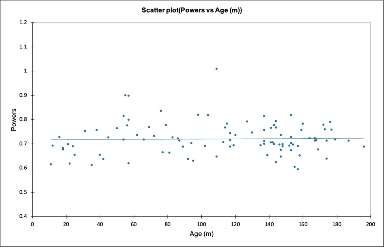
- Figure 4
Scatter plot of the Powers ratio (y) plotted against age in months (x). Of all the measurements, the Powers ratio remained least variable across the age groups.

Tables
Variable Mean SD Minimum Maximum Age 111.0 51.0 11 196 BDI 7.1 1.7 3.6 12.2 ADI 2.8 0.8 0.8 4.8 PADI 18.7 2.1 14.1 23.2 B-C1 29.2 2.8 21.4 35.3 Op-C1 40.7 4.2 30.4 48.8 Powers ratio 0.72 0.10 0.59 1.01 CCI-r 2.8 0.8 1.1 5 CCI-l 2.8 0.7 0.9 5 LMI-r 3.2 0.6 1.9 4.8 LMI-l 3.3 0.5 1.7 4.6 ADI, atlanto-dens interval; B-C1, basion-to-C1 distance; BDI, basion-dens interval; CCI, condylar-to-cervical spine interval; l, left; LMI, lateral mass interval; Op-C1, opisthion-to-C1 distance; PADI, posterior atlanto-dens interval; r, right.
Variable Female Male P Value Age 114 108 0.56 BDI 6.7 7.4 0.06 ADI 2.3 3.1 <0.001 PADI 17.8 19.1 0.003 B-C1 28.2 29.8 0.008 Op-C1 39.0 41.7 0.001 Powers ratio 0.72 0.71 0.46 CCI-r 2.5 3.0 0.002 CCI-l 2.4 3.1 <0.001 LMI-r 2.9 3.4 <0.001 LMI-l 3.1 3.4 0.002 Statistically significant findings appear in boldface.
ADI, atlanto-dens interval; B-C1, basion-to-C1 distance; BDI, basion-dens interval; CCI, condylar-to-cervical spine interval; l, left; LMI, lateral mass interval; Op-C1, opisthion-to-C1 distance; PADI, posterior atlanto-dens interval; r, right.
Age, mo ≤24 25–48 49–72 73–96 97–120 121–144 145–168 >168 P Value n 8 7 12 11 13 16 18 15 BDI 7.6 (1.0) 7.7 (1.9) 8.5 (2.0) 7.9 (1.2) 7.5 (1.3) 6.9 (1.4) 6.0 (1.3) 6.3 (1.8) 0.003 ADI 2.3 (0.9) 2.9 (0.8) 3.2 (0.7) 3.4 (0.9) 2.9 (0.7) 2.8 (0.7) 2.7 (0.6) 2.2 (0.5) 0.002 PADI 17.1 (2.5) 18.7 (2.3) 17.9 (1.8) 17.6 (1.7) 19.1 (1.6) 18.4 (2.1) 19.1 (2.0) 19.8 (2.2) 0.129 B-C1 24.2 (2.0) 27.6 (3.5) 29.1 (2.4) 27.7 (2.0) 30.1 (1.4) 30.3 (2.2) 30.1 (2.3) 30.9 (2.2) <0.001 Op-C1 36.0 (3.8) 40.4 (4.7) 38.0 (3.6) 39.5 (3.6) 40.2 (3.6) 41.6 (3.3) 43.4 (3.6) 42.8 (3.3) <0.001 Powers ratio 0.67 (0.04) 0.69 (0.06) 0.77 (0.08) 0.71 (0.06) 0.76 (0.09) 0.73 (0.05) 0.70 (0.06) 0.72 (0.04) 0.017 CCI-r 3.0 (1.0) 3.5 (1.1) 3.2 (0.5) 3.0 (0.5) 2.9 (0.8) 2.9 (0.7) 2.7 (0.8) 2.2 (0.7) 0.020 CCI-l 3.1 (0.9) 3.2 (0.4) 3.3 (0.5) 2.9 (0.5) 3.0 (0.8) 2.9 (0.6) 2.6 (0.7) 2.1 (0.7) 0.004 LMI-r 3.0 (0.5) 3.4 (0.2) 3.4 (0.5) 3.5 (0.8) 3.3 (0.6) 3.3 (0.7) 3.0 (0.6) 3.0 (0.5) 0.150 LMI-l 2.9 (0.4) 3.5 (0.4) 3.4 (0.4) 3.4 (0.3) 3.4 (0.7) 3.4 (0.5) 3.1 (0.6) 3.1 (0.4) 0.031 Mean (SD) values are presented for each age cohort. All measurements are in mm except for the Powers ratio. Statistically significant results are in boldface.
ADI, atlanto-dens interval; B-C1, basion-to-C1 distance; BDI, basion-dens interval; CCI, condylar-to-cervical spine interval; l, left; LMI, lateral mass interval; Op-C1, opisthion-to-C1 distance; PADI, posterior atlanto-dens interval;; r, right.
BDI = 9.4 – 0.02 × age ADI = 3.13 – 0.003 × age PADI = 17.5 + 0.01 × age B-C1 = 25.9 + 0.03 × age Op-C1 = 36.2 + 0.04 × age CCI = 3.52 – 0.006 × age LMI = 3.38 – 0.001 × age
In this issue


Jump to section
Related Articles
Cited By...
- No citing articles found.